A-Level Biology (9700) starts at the cell and works outwards: membranes and transport, the mitotic cell cycle, nucleic acids and protein synthesis, then transport in plants and mammals, gas exchange, infectious disease and immunity. A2 adds respiration, photosynthesis, homeostasis, inheritance, selection and evolution.
Biology is called the memorising science, and that is exactly what catches people out. The command word does most of the work: "explain" wants a mechanism, "describe" does not. A perfect description scores nothing when the question said explain.
Answers are marked against specific scheme points. One vague sentence covering three ideas usually earns one mark, not three.
make temporary preparations of cellular material suitable for viewing with a light microscope
draw cells from microscope slides and photomicrographs
calculate magnifications of images and actual sizes of specimens from drawings, photomicrographs and electron micrographs (scanning and transmission)
use an eyepiece graticule and stage micrometer scale to make measurements and use the appropriate units, millimetre (mm), micrometre (µm) and nanometre (nm)
define resolution and magnification and explain the differences between these terms, with reference to light microscopy and electron microscopy
Source: Cambridge International syllabus · 出典: Cambridge International シラバス
English
Cells 细胞 are very small, so you cannot see them with your eyes alone. You use a microscope 显微镜 to make a bigger picture of them. The first kind you meet is the light microscope 光学显微镜. It shines light through a thin specimen 标本 (the material you look at) and uses glass lenses to enlarge the view.
Making a slide and drawing what you see
To look at living material, you make a temporary preparation 临时装片. You put a small, thin piece of material on a glass slide 载玻片, add a drop of stain 染色剂 (a coloured liquid that makes parts easier to see), then lower a thin cover slip 盖玻片 on top to flatten it and keep out air.
When you draw cells from a slide or a photograph, follow simple rules:
use a sharp pencil and clear, single lines (no shading).
draw only what you can really see, with the parts in the correct sizes.
label the parts with straight lines that do not cross.
Magnification and actual size
Magnification 放大倍数 tells you how many times bigger the image is than the real object. It has no unit. You find it with one equation:
$$\text{magnification} = \frac{\text{size of image}}{\text{actual size of object}}$$
You can rearrange this to find any one value from the other two:
$$\text{actual size} = \frac{\text{size of image}}{\text{magnification}}$$
The top and the bottom of the fraction must use the same unit. Cells are tiny, so you work in small units:
$1\ \text{mm} = 1000\ \text{micrometre}$ 微米 (µm)
$1\ \text{µm} = 1000\ \text{nanometre}$ 纳米 (nm)
so $1\ \text{mm} = 1\,000\,000\ \text{nm}$.
Worked example. In a photomicrograph at magnification $5000$, a chloroplast 叶绿体 measures $25\ \text{mm}$ across. Its actual size is
The same equation works for drawings, photomicrographs 显微照片 (photos taken through a light microscope) and electron micrographs 电子显微照片 (the most detailed photos, explained below). Always convert to the same unit first, then divide.
Eyepiece graticule and stage micrometer
To measure a real cell under the microscope, you use an eyepiece graticule 目镜测微尺 — a tiny scale inside the eyepiece. Its divisions have no fixed size, so first you must calibrate 校准 them (work out what one division is worth).
You calibrate using a stage micrometer 载物台测微尺 — a special slide with an accurate scale on it (often $1\ \text{mm}$ split into $100$ parts, so each part is $10\ \text{µm}$). You line up the two scales, count how many graticule divisions fit a known length, and divide. Once calibrated, you can swap in your specimen and measure it with the graticule.
Resolution and magnification
These two words are easy to mix up. The examiner gives marks for the difference.
Magnification is how many times bigger the image is than the object.
Resolution 分辨率 is the smallest distance between two points that still lets you see them as two separate points.
Making an image bigger does not always show more detail. Past a certain point you just get a bigger, blurry image. Resolution sets the real limit on detail.
A light microscope has lower resolution because light has a fairly long wavelength 波长. An electron microscope 电子显微镜 uses beams of electrons 电子 instead of light. Electrons have a much shorter wavelength, so the resolution is far higher and you can see very small structures inside the cell. There are two kinds: scanning 扫描 (shows the surface in 3D) and transmission 透射 (passes electrons through a thin slice to show inside detail).
日本語
Cells 细胞 are very small, so you cannot see them with your eyes alone. You use a microscope 显微镜 to make a bigger picture of them. The first kind you meet is the light microscope 光学显微镜. It shines light through a thin specimen 标本 (the material you look at) and uses glass lenses to enlarge the view.
A light microscope: light passes up through the specimen, then two lenses (the objective and the eyepiece) magnify it
Making a slide and drawing what you see
To look at living material, you make a temporary preparation 临时装片. You put a small, thin piece of material on a glass slide 载玻片, add a drop of stain 染色剂 (a coloured liquid that makes parts easier to see), then lower a thin cover slip 盖玻片 on top to flatten it and keep out air.
Making a temporary wet mount — lower the cover slip at an angle so no air bubbles are trapped
When you draw cells from a slide or a photograph, follow simple rules:
use a sharp pencil and clear, single lines (no shading).
draw only what you can really see, with the parts in the correct sizes.
label the parts with straight lines that do not cross.
Magnification and actual size
Magnification 放大倍数 tells you how many times bigger the image is than the real object. It has no unit. You find it with one equation:
Magnification = image size ÷ actual size
$$\text{magnification} = \frac{\text{size of image}}{\text{actual size of object}}$$
You can rearrange this to find any one value from the other two:
$$\text{actual size} = \frac{\text{size of image}}{\text{magnification}}$$
The top and the bottom of the fraction must use the same unit. Cells are tiny, so you work in small units:
$1\ \text{mm} = 1000\ \text{micrometre}$ 微米 (µm)
$1\ \text{µm} = 1000\ \text{nanometre}$ 纳米 (nm)
so $1\ \text{mm} = 1\,000\,000\ \text{nm}$.
Worked example. In a photomicrograph at magnification $5000$, a chloroplast 叶绿体 measures $25\ \text{mm}$ across. Its actual size is
The same equation works for drawings, photomicrographs 显微照片 (photos taken through a light microscope) and electron micrographs 电子显微照片 (the most detailed photos, explained below). Always convert to the same unit first, then divide.
Eyepiece graticule and stage micrometer
To measure a real cell under the microscope, you use an eyepiece graticule 目镜测微尺 — a tiny scale inside the eyepiece. Its divisions have no fixed size, so first you must calibrate 校准 them (work out what one division is worth).
You calibrate using a stage micrometer 载物台测微尺 — a special slide with an accurate scale on it (often $1\ \text{mm}$ split into $100$ parts, so each part is $10\ \text{µm}$). You line up the two scales, count how many graticule divisions fit a known length, and divide. Once calibrated, you can swap in your specimen and measure it with the graticule.
Calibrate the graticule by lining it up with the stage micrometer's known scale
Resolution and magnification
These two words are easy to mix up. The examiner gives marks for the difference.
Magnification is how many times bigger the image is than the object.
Resolution 分辨率 is the smallest distance between two points that still lets you see them as two separate points.
Making an image bigger does not always show more detail. Past a certain point you just get a bigger, blurry image. Resolution sets the real limit on detail.
A light microscope has lower resolution because light has a fairly long wavelength 波长. An electron microscope 电子显微镜 uses beams of electrons 电子 instead of light. Electrons have a much shorter wavelength, so the resolution is far higher and you can see very small structures inside the cell. There are two kinds: scanning 扫描 (shows the surface in 3D) and transmission 透射 (passes electrons through a thin slice to show inside detail).
Resolution: a light microscope blurs two very close points into one; an electron microscope, with its shorter wavelength, resolves them as two
Explore · 探索
Microscope decision lab · 顕微鏡選択実験室
Choose the right microscopy idea from what the student wants to see. · 学生が見たいものから、適切な観察方法を選択してください。
recognise organelles and other cell structures found in eukaryotic cells and outline their structures and functions, limited to: • cell surface membrane • nucleus, nuclear envelope and nucleolus • rough endoplasmic reticulum • smooth endoplasmic reticulum • Golgi body (Golgi apparatus or Golgi complex) • mitochondria (including the presence of small circular DNA) • ribosomes (80S in the cytoplasm and 70S in chloroplasts and mitochondria) • lysosomes • centrioles and microtubules • cilia • microvilli • chloroplasts (including the presence of small circular DNA) • cell wall • plasmodesmata • large permanent vacuole and tonoplast of plant cells
describe and interpret photomicrographs, electron micrographs and drawings of typical plant and animal cells
compare the structure of typical plant and animal cells
state that cells use ATP from respiration for energy-requiring processes
outline key structural features of a prokaryotic cell as found in a typical bacterium, including: • unicellular • generally 1–5 μm diameter • peptidoglycan cell walls • circular DNA • 70S ribosomes • absence of organelles surrounded by double membranes
compare the structure of a prokaryotic cell as found in a typical bacterium with the structures of typical eukaryotic cells in plants and animals
state that all viruses are non-cellular structures with a nucleic acid core (either DNA or RNA) and a capsid made of protein, and that some viruses have an outer envelope made of phospholipids
Source: Cambridge International syllabus · 出典: Cambridge International シラバス
English
Plant and animal cells are eukaryotic cells 真核细胞: their DNA is kept inside a nucleus 细胞核. Inside the cell are many small parts called organelles 细胞器, each with its own job. The jelly-like fluid around them is the cytoplasm 细胞质.
In a photomicrograph or electron micrograph you identify organelles by their shape, size and position; in a drawing you show their outlines and label them.
Organelle
Structure
Function
cell surface membrane 细胞膜
thin layer around the cell
controls what enters and leaves the cell
nucleus
large, surrounded by a nuclear envelope 核膜 (a double membrane with holes); contains a nucleolus 核仁
holds the DNA; controls the cell; the nucleolus makes ribosomes
rough endoplasmic reticulum 粗面内质网 (rough ER)
sheets of membrane with ribosomes on the surface
makes and transports proteins 蛋白质 (for example antibodies 抗体)
smooth endoplasmic reticulum 滑面内质网 (smooth ER)
sheets of membrane, no ribosomes
makes lipids 脂质
Golgi body 高尔基体
stack of flat membrane sacs
changes and packs proteins and lipids into vesicles 囊泡 for secretion 分泌
mitochondria 线粒体
oval, with a folded inner membrane; has small circular DNA
site of respiration 呼吸作用 — releases energy 能量 as ATP
ribosomes 核糖体
very small; $80\text{S}$ in the cytoplasm, $70\text{S}$ in chloroplasts and mitochondria
join amino acids 氨基酸 to synthesise 合成 proteins
lysosomes 溶酶体
small sacs of enzymes 酶
break down old organelles and waste
centrioles 中心粒 and microtubules 微管
small tubes made of protein
help move chromosomes and form the cell's "skeleton"
cilia 纤毛
tiny hairs on the cell surface that beat
move fluid or move the cell
microvilli 微绒毛
tiny folds of the cell surface membrane
increase surface area for absorption 吸收
chloroplasts (plants)
green, with stacked membranes; has small circular DNA
site of photosynthesis 光合作用
cell wall 细胞壁 (plants)
strong outer layer of cellulose 纤维素
supports and protects the cell; stops it bursting
plasmodesmata 胞间连丝 (plants)
tiny channels through the cell walls
link the cytoplasm of neighbouring cells
large permanent vacuole 液泡 (plants)
big sac of watery fluid, with a membrane called the tonoplast 液泡膜
stores water and keeps the cell firm
Cells use ATP made in respiration as their energy supply for every job that needs energy, such as making proteins, moving things and dividing.
Comparing plant and animal cells
Feature
Plant cell
Animal cell
cell wall
present (cellulose)
absent
chloroplasts
present
absent
large permanent vacuole
present
absent (only small, temporary ones)
centrioles
absent in most
present
shape
fixed and regular
rounder and more flexible
Both have a cell surface membrane, cytoplasm, a nucleus, mitochondria, ribosomes, ER and a Golgi body.
日本語
Plant and animal cells are eukaryotic cells 真核细胞: their DNA is kept inside a nucleus 细胞核. Inside the cell are many small parts called organelles 细胞器, each with its own job. The jelly-like fluid around them is the cytoplasm 细胞质.
A generalised animal cell and its organellesA plant cell also has a cell wall, chloroplasts and a large vacuole
In a photomicrograph or electron micrograph you identify organelles by their shape, size and position; in a drawing you show their outlines and label them.
Organelle
Structure
Function
cell surface membrane 细胞膜
thin layer around the cell
controls what enters and leaves the cell
nucleus
large, surrounded by a nuclear envelope 核膜 (a double membrane with holes); contains a nucleolus 核仁
holds the DNA; controls the cell; the nucleolus makes ribosomes
rough endoplasmic reticulum 粗面内质网 (rough ER)
sheets of membrane with ribosomes on the surface
makes and transports proteins 蛋白质 (for example antibodies 抗体)
smooth endoplasmic reticulum 滑面内质网 (smooth ER)
sheets of membrane, no ribosomes
makes lipids 脂质
Golgi body 高尔基体
stack of flat membrane sacs
changes and packs proteins and lipids into vesicles 囊泡 for secretion 分泌
mitochondria 线粒体
oval, with a folded inner membrane; has small circular DNA
site of respiration 呼吸作用 — releases energy 能量 as ATP
ribosomes 核糖体
very small; $80\text{S}$ in the cytoplasm, $70\text{S}$ in chloroplasts and mitochondria
join amino acids 氨基酸 to synthesise 合成 proteins
lysosomes 溶酶体
small sacs of enzymes 酶
break down old organelles and waste
centrioles 中心粒 and microtubules 微管
small tubes made of protein
help move chromosomes and form the cell's "skeleton"
cilia 纤毛
tiny hairs on the cell surface that beat
move fluid or move the cell
microvilli 微绒毛
tiny folds of the cell surface membrane
increase surface area for absorption 吸收
chloroplasts (plants)
green, with stacked membranes; has small circular DNA
site of photosynthesis 光合作用
cell wall 细胞壁 (plants)
strong outer layer of cellulose 纤维素
supports and protects the cell; stops it bursting
plasmodesmata 胞间连丝 (plants)
tiny channels through the cell walls
link the cytoplasm of neighbouring cells
large permanent vacuole 液泡 (plants)
big sac of watery fluid, with a membrane called the tonoplast 液泡膜
stores water and keeps the cell firm
Cells use ATP made in respiration as their energy supply for every job that needs energy, such as making proteins, moving things and dividing.
Comparing plant and animal cells
Feature
Plant cell
Animal cell
cell wall
present (cellulose)
absent
chloroplasts
present
absent
large permanent vacuole
present
absent (only small, temporary ones)
centrioles
absent in most
present
shape
fixed and regular
rounder and more flexible
Both have a cell surface membrane, cytoplasm, a nucleus, mitochondria, ribosomes, ER and a Golgi body.
Explore · 探索
Explore an animal cell · 動物細胞を探索する
Tap each numbered part to check you know its job — the same organelles as the table above. · 番号付きの各部分タップして機能を確認 — 上記の表と同じ小器官です。
A prokaryotic cell 原核细胞, such as a bacterium 细菌, is much smaller and simpler than a eukaryotic cell. Its key features are:
unicellular 单细胞 — it is a single cell.
generally $1$–$5\ \text{µm}$ across.
a cell wall made of peptidoglycan 肽聚糖 (not cellulose).
circular DNA lying free in the cytoplasm — there is no nucleus.
$70\text{S}$ ribosomes (smaller than the $80\text{S}$ ones in the cytoplasm of eukaryotes).
no organelles surrounded by a double membrane — so no nucleus, no mitochondria and no chloroplasts.
Comparing prokaryotic and eukaryotic cells
Feature
Prokaryotic cell
Eukaryotic cell
size
about $1$–$5\ \text{µm}$
about $10$–$100\ \text{µm}$
DNA
circular, free in cytoplasm
linear, inside a nucleus
nucleus
none
present
double-membrane organelles
none
mitochondria (and chloroplasts in plants)
ribosomes
$70\text{S}$
$80\text{S}$ (with $70\text{S}$ inside mitochondria and chloroplasts)
cell wall
peptidoglycan
cellulose (plants) or none (animals)
日本語
A prokaryotic cell 原核细胞, such as a bacterium 细菌, is much smaller and simpler than a eukaryotic cell. Its key features are:
unicellular 单细胞 — it is a single cell.
generally $1$–$5\ \text{µm}$ across.
a cell wall made of peptidoglycan 肽聚糖 (not cellulose).
circular DNA lying free in the cytoplasm — there is no nucleus.
$70\text{S}$ ribosomes (smaller than the $80\text{S}$ ones in the cytoplasm of eukaryotes).
no organelles surrounded by a double membrane — so no nucleus, no mitochondria and no chloroplasts.
A prokaryotic cell: the circular DNA (nucleoid) lies free, with no nucleus and no double-membrane organelles
Comparing prokaryotic and eukaryotic cells
Feature
Prokaryotic cell
Eukaryotic cell
size
about $1$–$5\ \text{µm}$
about $10$–$100\ \text{µm}$
DNA
circular, free in cytoplasm
linear, inside a nucleus
nucleus
none
present
double-membrane organelles
none
mitochondria (and chloroplasts in plants)
ribosomes
$70\text{S}$
$80\text{S}$ (with $70\text{S}$ inside mitochondria and chloroplasts)
cell wall
peptidoglycan
cellulose (plants) or none (animals)
Explore · 探索
Explore a bacterial cell · 細菌細胞を探索する
A prokaryote is smaller and simpler. Tap each part — notice there is no nucleus and no double-membrane organelles. · 原核生物は小さく単純です。各部分タップ — 核がなく二重膜小器官もないことに注意してください。
All viruses 病毒 are non-cellular 非细胞 — they are not made of cells at all. Each virus is built from just two or three parts:
a core of nucleic acid 核酸, which is either DNA or RNA (never both).
a protein coat around the core called a capsid 衣壳.
in some viruses, an outer envelope 包膜 made of phospholipids 磷脂.
A virus has no cytoplasm, no organelles and no ribosomes. It cannot respire or make its own proteins. It can only copy itself inside a living host 宿主 cell, so it sits at the edge of what we call "living".
日本語
All viruses 病毒 are non-cellular 非细胞 — they are not made of cells at all. Each virus is built from just two or three parts:
a core of nucleic acid 核酸, which is either DNA or RNA (never both).
a protein coat around the core called a capsid 衣壳.
in some viruses, an outer envelope 包膜 made of phospholipids 磷脂.
A real virus, magnified hugely. The rounded head is the protein capsid wrapped around the nucleic acid core; the tail injects that nucleic acid into a bacterium. Note the scale bar — the whole virus is about 100 nm, far smaller than any cellA generalised virus: nucleic acid inside a protein capsid, with a lipid envelope in some viruses
A virus has no cytoplasm, no organelles and no ribosomes. It cannot respire or make its own proteins. It can only copy itself inside a living host 宿主 cell, so it sits at the edge of what we call "living".
Explore · 探索
Explore a virus · ウイルスを探索する
A virus is non-cellular — just a few parts. Tap each one: there is no cytoplasm, no organelles and no ribosomes. · ウイルスは非細胞性 — 数個の部分のみ。各部分タップ: 細胞質も小器官もリボソームもありません。
Magnification $=$ image size $\div$ actual size — convert units first ($\text{mm} \leftrightarrow \mu\text{m} \leftrightarrow \text{nm}$), then rearrange for whichever is unknown.
Distinguish magnification (how many times larger) from resolution (smallest distance still seen as two points); electron microscopes resolve more because electrons have a shorter wavelength.
Give each organelle a structure + function pair (e.g. mitochondrion: folded inner membrane → aerobic respiration).
State the prokaryote vs eukaryote differences exactly: no nucleus, smaller (70S) ribosomes, no membrane-bound organelles, circular DNA.
Viruses are non-living — describe them only by capsid, genetic material and (sometimes) an envelope.
describe and carry out the Benedict’s test for reducing sugars, the iodine test for starch, the emulsion test for lipids and the biuret test for proteins
describe and carry out a semi-quantitative Benedict’s test on a reducing sugar solution by standardising the test and using the results (time to first colour change or comparison to colour standards) to estimate the concentration
describe and carry out a test to identify the presence of non-reducing sugars, using acid hydrolysis and Benedict’s solution
Source: Cambridge International syllabus · 出典: Cambridge International シラバス
English
Living things are built from four main kinds of large molecule 分子: carbohydrates 碳水化合物, lipids 脂质, proteins 蛋白质 and nucleic acids 核酸. You can use simple chemical tests to find out which kinds are present in a sample.
Test
What it finds
Method
Positive result
Benedict's test
reducing sugar 还原糖
add Benedict's solution and heat in a water bath
blue changes to green, yellow, orange, then brick-red precipitate 沉淀
iodine test
starch 淀粉
add orange-brown iodine 碘 solution
colour changes to blue-black
emulsion test
lipid
mix sample with ethanol, then pour into water
a white, cloudy emulsion 乳浊液 forms
biuret 双缩脲 test
protein
add biuret solution at room temperature
blue changes to purple
Semi-quantitative Benedict's test
The normal Benedict's test only tells you "yes or no". A semi-quantitative 半定量 test gives a rough amount. First you standardise 标准化 the test: you run it on solutions of known concentration 浓度 and record the result for each. Then you can estimate an unknown by either:
the time to the first colour change (more sugar changes colour faster), or
comparing the final colour to your set of colour standards.
Testing for non-reducing sugars
Some sugars, such as sucrose, are non-reducing sugar 非还原糖: they give a negative Benedict's test. To detect them:
Do a normal Benedict's test first. It stays blue (no reducing sugar).
Take a fresh sample and add dilute hydrochloric acid 盐酸, then heat. This acid hydrolysis breaks the sugar into smaller reducing sugars.
Cool, then neutralise 中和 the acid with sodium hydrogencarbonate.
Now do the Benedict's test again. A brick-red colour shows a non-reducing sugar was present.
Worked example. A solution gives a negative Benedict's test. It is then boiled with dilute hydrochloric acid, neutralised with sodium hydrogencarbonate, and re-tested with Benedict's - now it turns brick-red. What was present, and why is each step needed? The first negative result rules out a reducing sugar. Boiling with acid hydrolyses the glycosidic bond, splitting a non-reducing sugar such as sucrose into its reducing monosaccharides. The neutralising step is essential because Benedict's only works in alkaline conditions - skip it and the test fails even when sugar is present. The positive re-test therefore shows a non-reducing sugar was there all along. Quote the first, negative test as part of the answer: without it, the final red colour cannot tell a non-reducing sugar from a reducing one.
日本語
Living things are built from four main kinds of large molecule 分子: carbohydrates 碳水化合物, lipids 脂质, proteins 蛋白质 and nucleic acids 核酸. You can use simple chemical tests to find out which kinds are present in a sample.
Benedict's test: blue turns green, then orange, then brick-red as more reducing sugar is present
Test
What it finds
Method
Positive result
Benedict's test
reducing sugar 还原糖
add Benedict's solution and heat in a water bath
blue changes to green, yellow, orange, then brick-red precipitate 沉淀
iodine test
starch 淀粉
add orange-brown iodine 碘 solution
colour changes to blue-black
emulsion test
lipid
mix sample with ethanol, then pour into water
a white, cloudy emulsion 乳浊液 forms
biuret 双缩脲 test
protein
add biuret solution at room temperature
blue changes to purple
Semi-quantitative Benedict's test
The normal Benedict's test only tells you "yes or no". A semi-quantitative 半定量 test gives a rough amount. First you standardise 标准化 the test: you run it on solutions of known concentration 浓度 and record the result for each. Then you can estimate an unknown by either:
the time to the first colour change (more sugar changes colour faster), or
comparing the final colour to your set of colour standards.
Testing for non-reducing sugars
Some sugars, such as sucrose, are non-reducing sugar 非还原糖: they give a negative Benedict's test. To detect them:
Do a normal Benedict's test first. It stays blue (no reducing sugar).
Take a fresh sample and add dilute hydrochloric acid 盐酸, then heat. This acid hydrolysis breaks the sugar into smaller reducing sugars.
Cool, then neutralise 中和 the acid with sodium hydrogencarbonate.
Now do the Benedict's test again. A brick-red colour shows a non-reducing sugar was present.
Testing for a non-reducing sugar: hydrolyse it with acid first, then the second Benedict's test turns red
Worked example. A solution gives a negative Benedict's test. It is then boiled with dilute hydrochloric acid, neutralised with sodium hydrogencarbonate, and re-tested with Benedict's - now it turns brick-red. What was present, and why is each step needed? The first negative result rules out a reducing sugar. Boiling with acid hydrolyses the glycosidic bond, splitting a non-reducing sugar such as sucrose into its reducing monosaccharides. The neutralising step is essential because Benedict's only works in alkaline conditions - skip it and the test fails even when sugar is present. The positive re-test therefore shows a non-reducing sugar was there all along. Quote the first, negative test as part of the answer: without it, the final red colour cannot tell a non-reducing sugar from a reducing one.
Explore · 探索
Testing for a non-reducing sugar · 非還元糖の検査
Step through the trick: a non-reducing sugar stays blue, so you hydrolyse it with acid, neutralise, then re-test. · 手順を確認する:非還元糖は青色のままなので、酸で加水分解し、中和してから再検査する。
describe and draw the ring forms of α-glucose and β-glucose
define the terms monomer, polymer, macromolecule, monosaccharide, disaccharide and polysaccharide
state the role of covalent bonds in joining smaller molecules together to form polymers
state that glucose, fructose and maltose are reducing sugars and that sucrose is a non-reducing sugar
describe the formation of a glycosidic bond by condensation, with reference to disaccharides, including sucrose, and polysaccharides
describe the breakage of a glycosidic bond in polysaccharides and disaccharides by hydrolysis, with reference to the non-reducing sugar test
describe the molecular structure of the polysaccharides starch (amylose and amylopectin) and glycogen and relate their structures to their functions in living organisms
describe the molecular structure of the polysaccharide cellulose and outline how the arrangement of cellulose molecules contributes to the function of plant cell walls
state that triglycerides are non-polar hydrophobic molecules and describe the molecular structure of triglycerides with reference to fatty acids (saturated and unsaturated), glycerol and the formation of ester bonds
relate the molecular structure of triglycerides to their functions in living organisms
describe the molecular structure of phospholipids with reference to their hydrophilic (polar) phosphate heads and hydrophobic (non-polar) fatty acid tails
Source: Cambridge International syllabus · 出典: Cambridge International シラバス
English
Monomers, polymers and macromolecules
a monomer 单体 is a small molecule that is a single unit.
a polymer 聚合物 is a long molecule made of many monomers joined together.
a macromolecule 大分子 is any very large molecule.
Sugars come in three sizes:
a monosaccharide 单糖 is a single sugar unit, such as glucose 葡萄糖 and fructose.
a disaccharide 二糖 is two units joined, such as maltose and sucrose.
a polysaccharide 多糖 is many units joined into a polymer.
Glucose, fructose 果糖 and maltose 麦芽糖 are reducing sugars. Sucrose 蔗糖 is a non-reducing sugar.
Two ring forms of glucose
Glucose has six carbon atoms and forms a ring. There are two ring forms. They differ only at carbon 1:
in α-glucose, the –OH group on carbon 1 points down, below the ring.
in β-glucose, the –OH group on carbon 1 points up, above the ring.
This small difference decides which polysaccharide the glucose can build.
Joining and breaking sugars
Monomers are joined by strong covalent bonds 共价键. When two sugars join, a glycosidic bond 糖苷键 forms between them. This happens by condensation 缩合: a molecule of water is removed each time a bond forms.
The reverse is hydrolysis 水解: a water molecule is added to break a glycosidic bond. This is why the non-reducing sugar test needs acid and heat — they hydrolyse sucrose into glucose and fructose.
Storage polysaccharides: starch and glycogen
Starch is the energy 能量 store in plants. It is made of two polymers of α-glucose:
amylose 直链淀粉 — a long, unbranched chain that coils into a spiral.
amylopectin 支链淀粉 — a chain with many side branches.
Glycogen 糖原 is the energy store in animals. It is like amylopectin but has even more branches, so it can be broken down quickly when energy is needed.
These stores suit their job well: they are compact, they are insoluble 不溶 (so they do not leave the cell), and they do not change the water potential 水势 of the cell (so they do not pull water in by osmosis 渗透). The many branches give many ends, so glucose can be added or removed fast.
Cellulose
Cellulose 纤维素 is made of β-glucose. Because of the β form, every other glucose is flipped over, so the chains are long and straight. Many straight chains lie side by side and are held together by hydrogen bonds 氢键 into strong bundles called microfibrils 微纤丝. These give the plant cell wall 细胞壁 its strength and stop the cell bursting.
日本語
Monomers, polymers and macromolecules
a monomer 单体 is a small molecule that is a single unit.
a polymer 聚合物 is a long molecule made of many monomers joined together.
a macromolecule 大分子 is any very large molecule.
Sugars come in three sizes:
a monosaccharide 单糖 is a single sugar unit, such as glucose 葡萄糖 and fructose.
a disaccharide 二糖 is two units joined, such as maltose and sucrose.
a polysaccharide 多糖 is many units joined into a polymer.
Glucose, fructose 果糖 and maltose 麦芽糖 are reducing sugars. Sucrose 蔗糖 is a non-reducing sugar.
Two ring forms of glucose
Glucose has six carbon atoms and forms a ring. There are two ring forms. They differ only at carbon 1:
in α-glucose, the –OH group on carbon 1 points down, below the ring.
in β-glucose, the –OH group on carbon 1 points up, above the ring.
This small difference decides which polysaccharide the glucose can build.
The two ring forms differ only at carbon 1: the –OH points down in α, up in β
Joining and breaking sugars
Monomers are joined by strong covalent bonds 共价键. When two sugars join, a glycosidic bond 糖苷键 forms between them. This happens by condensation 缩合: a molecule of water is removed each time a bond forms.
The reverse is hydrolysis 水解: a water molecule is added to break a glycosidic bond. This is why the non-reducing sugar test needs acid and heat — they hydrolyse sucrose into glucose and fructose.
Condensation removes water to join monomers; hydrolysis adds water to split them
Storage polysaccharides: starch and glycogen
Starch is the energy 能量 store in plants. It is made of two polymers of α-glucose:
amylose 直链淀粉 — a long, unbranched chain that coils into a spiral.
amylopectin 支链淀粉 — a chain with many side branches.
Glycogen 糖原 is the energy store in animals. It is like amylopectin but has even more branches, so it can be broken down quickly when energy is needed.
These stores suit their job well: they are compact, they are insoluble 不溶 (so they do not leave the cell), and they do not change the water potential 水势 of the cell (so they do not pull water in by osmosis 渗透). The many branches give many ends, so glucose can be added or removed fast.
Real starch grains inside a potato, stained by iodine. Each grain is a dense, insoluble package of amylose and amylopectin — note the scale: the largest are only about 0.1 mm across
Cellulose
Cellulose 纤维素 is made of β-glucose. Because of the β form, every other glucose is flipped over, so the chains are long and straight. Many straight chains lie side by side and are held together by hydrogen bonds 氢键 into strong bundles called microfibrils 微纤丝. These give the plant cell wall 细胞壁 its strength and stop the cell bursting.
The shape fits the job: amylose coils and amylopectin/glycogen branch (for compact stores), while straight cellulose chains pack into strong fibres
Explore · 探索
Condensation and hydrolysis · 縮合反応と加水分解
Step through how two sugars join. Condensation removes one water to make the bond; hydrolysis is the reverse — adding water splits it again. · 2つの糖が結合する様子を確認します。脱水縮合では、結合を作るために水分子1つが失われます;加水分解はその逆反応で、水を加えると再び分解されます。
A triglyceride 甘油三酯 is the main fat or oil. It is non-polar 非极性 and hydrophobic 疏水 (it does not mix with water). It is made from one glycerol 甘油 molecule joined to three fatty acids 脂肪酸 by ester bonds 酯键. Each ester bond forms by condensation, so three water molecules are removed.
Fatty acids are of two kinds:
saturated 饱和 — no carbon–carbon double bonds 双键; these fats are usually solid.
unsaturated 不饱和 — one or more double bonds; these oils are usually liquid.
Triglycerides make a good long-term energy store: they release about twice as much energy per gram as carbohydrates, they are insoluble, and they store little extra mass because they hold no water. Under the skin they also give insulation 隔热 and protect the organs.
Phospholipids
A phospholipid 磷脂 is like a triglyceride, but one fatty acid is replaced by a phosphate 磷酸 group. This gives the molecule two ends with different behaviour:
a hydrophilic 亲水 ("water-loving") polar 极性 phosphate head.
two hydrophobic ("water-fearing") fatty acid tails.
This split personality is why phospholipids form the membranes around cells.
日本語
Triglycerides
A triglyceride 甘油三酯 is the main fat or oil. It is non-polar 非极性 and hydrophobic 疏水 (it does not mix with water). It is made from one glycerol 甘油 molecule joined to three fatty acids 脂肪酸 by ester bonds 酯键. Each ester bond forms by condensation, so three water molecules are removed.
One glycerol plus three fatty acid tails; a straight tail is saturated, a kinked one unsaturated
Fatty acids are of two kinds:
saturated 饱和 — no carbon–carbon double bonds 双键; these fats are usually solid.
unsaturated 不饱和 — one or more double bonds; these oils are usually liquid.
Triglycerides make a good long-term energy store: they release about twice as much energy per gram as carbohydrates, they are insoluble, and they store little extra mass because they hold no water. Under the skin they also give insulation 隔热 and protect the organs.
Phospholipids
A phospholipid 磷脂 is like a triglyceride, but one fatty acid is replaced by a phosphate 磷酸 group. This gives the molecule two ends with different behaviour:
a hydrophilic 亲水 ("water-loving") polar 极性 phosphate head.
two hydrophobic ("water-fearing") fatty acid tails.
This split personality is why phospholipids form the membranes around cells.
The hydrophilic heads face the water; the hydrophobic tails hide inside, forming a bilayer
Explore · 探索
Building a triglyceride · トリグリセリドの合成
Watch one glycerol join three fatty acids. Each ester bond forms by condensation, removing one water — three bonds, three waters. · 1つのグリセロールが3つの脂肪酸と結合する様子を見ます。各エステル結合は脱水縮合によって形成され、水分子1つが失われます——結合3つ、水分子3つ。
describe and draw the general structure of an amino acid and the formation and breakage of a peptide bond
explain the meaning of the terms primary structure, secondary structure, tertiary structure and quaternary structure of proteins
describe the types of interaction that hold protein molecules in shape: • hydrophobic interactions • hydrogen bonding • ionic bonding • covalent bonding, including disulfide bonds
state that globular proteins are generally soluble and have physiological roles and fibrous proteins are generally insoluble and have structural roles
describe the structure of a molecule of haemoglobin as an example of a globular protein, including the formation of its quaternary structure from two alpha (α) chains (α–globin), two beta (β) chains (β–globin) and a haem group
relate the structure of haemoglobin to its function, including the importance of iron in the haem group
describe the structure of a molecule of collagen as an example of a fibrous protein, and the arrangement of collagen molecules to form collagen fibres
relate the structures of collagen molecules and collagen fibres to their function
Source: Cambridge International syllabus · 出典: Cambridge International シラバス
English
Amino acids and the peptide bond
Proteins are polymers of amino acids 氨基酸. Every amino acid has the same general structure around a central carbon atom: an amino group 氨基 (–NH₂), a carboxyl group 羧基 (–COOH), a hydrogen atom, and a variable side chain 侧链 (the R group). The R group is different in each amino acid.
Two amino acids join by condensation. The bond formed between the amino group of one and the carboxyl group of the next is a peptide bond 肽键, and a water molecule is removed. Many amino acids joined this way make a polypeptide 多肽. Adding water (hydrolysis) breaks a peptide bond.
Four levels of protein structure
Level
What it means
primary structure 一级结构
the order of amino acids in the chain
secondary structure 二级结构
local shapes — the α-helix 螺旋 and the β-pleated sheet 折叠片 — held by hydrogen bonds
tertiary structure 三级结构
the whole chain folded into a precise 3-D shape
quaternary structure 四级结构
two or more polypeptide chains joined into one protein
The folded shape is held together by four kinds of interaction between R groups:
hydrophobic interactions 疏水作用 (non-polar R groups cluster away from water).
hydrogen bonding.
ionic bonds 离子键 (between charged R groups).
covalent bonding, including strong disulfide bonds 二硫键.
Globular and fibrous proteins
globular proteins 球状蛋白质 fold into a rounded shape, are usually soluble 可溶, and do jobs in the body (for example enzymes and haemoglobin).
fibrous proteins 纤维状蛋白质 form long strands, are usually insoluble, and give structure and support (for example collagen).
Haemoglobin — a globular protein
Haemoglobin 血红蛋白 carries oxygen 氧气 in red blood cells. It has a quaternary structure made of four polypeptide chains: two alpha (α-globin) chains and two beta (β-globin) chains. Each chain holds a haem group 血红素. At the centre of each haem group is an iron 铁 atom, and this is where one oxygen molecule binds. Four chains mean one haemoglobin molecule can carry four oxygen molecules.
Collagen — a fibrous protein
Collagen 胶原蛋白 gives strength to skin, tendons 肌腱, bone and blood vessel walls. One collagen molecule is three polypeptide chains wound tightly around each other in a triple strand, held by hydrogen bonds. Many of these molecules lie side by side, slightly staggered, and are cross-linked into thick fibres 纤维. The staggered, cross-linked arrangement makes collagen very strong when pulled.
日本語
Amino acids and the peptide bond
Proteins are polymers of amino acids 氨基酸. Every amino acid has the same general structure around a central carbon atom: an amino group 氨基 (–NH₂), a carboxyl group 羧基 (–COOH), a hydrogen atom, and a variable side chain 侧链 (the R group). The R group is different in each amino acid.
Two amino acids join by condensation. The bond formed between the amino group of one and the carboxyl group of the next is a peptide bond 肽键, and a water molecule is removed. Many amino acids joined this way make a polypeptide 多肽. Adding water (hydrolysis) breaks a peptide bond.
Every amino acid has an amino group, a carboxyl group and an R group; two join by a peptide bond
Four levels of protein structure
Level
What it means
primary structure 一级结构
the order of amino acids in the chain
secondary structure 二级结构
local shapes — the α-helix 螺旋 and the β-pleated sheet 折叠片 — held by hydrogen bonds
tertiary structure 三级结构
the whole chain folded into a precise 3-D shape
quaternary structure 四级结构
two or more polypeptide chains joined into one protein
The four levels: primary → secondary → tertiary → quaternary structure
The folded shape is held together by four kinds of interaction between R groups:
hydrophobic interactions 疏水作用 (non-polar R groups cluster away from water).
hydrogen bonding.
ionic bonds 离子键 (between charged R groups).
covalent bonding, including strong disulfide bonds 二硫键.
Globular and fibrous proteins
globular proteins 球状蛋白质 fold into a rounded shape, are usually soluble 可溶, and do jobs in the body (for example enzymes and haemoglobin).
fibrous proteins 纤维状蛋白质 form long strands, are usually insoluble, and give structure and support (for example collagen).
Haemoglobin — a globular protein
Haemoglobin 血红蛋白 carries oxygen 氧气 in red blood cells. It has a quaternary structure made of four polypeptide chains: two alpha (α-globin) chains and two beta (β-globin) chains. Each chain holds a haem group 血红素. At the centre of each haem group is an iron 铁 atom, and this is where one oxygen molecule binds. Four chains mean one haemoglobin molecule can carry four oxygen molecules.
A real haemoglobin molecule, worked out from X-ray data. Count them: two alpha chains (red), two beta chains (blue), and one green haem group held in each — so four oxygen molecules in total
Collagen — a fibrous protein
Collagen 胶原蛋白 gives strength to skin, tendons 肌腱, bone and blood vessel walls. One collagen molecule is three polypeptide chains wound tightly around each other in a triple strand, held by hydrogen bonds. Many of these molecules lie side by side, slightly staggered, and are cross-linked into thick fibres 纤维. The staggered, cross-linked arrangement makes collagen very strong when pulled.
Explore · 探索
The four levels of protein structure · タンパク質構造の4段階
Build a protein up one level at a time: sequence → local shapes → a folded 3-D shape → several chains joined. · タンパク質を段階的に構築する:配列 → 局所形状 → 折りたたまれた3D構造 → 複数の鎖が結合。
explain how hydrogen bonding occurs between water molecules and relate the properties of water to its roles in living organisms, limited to solvent action, high specific heat capacity and latent heat of vaporisation
Source: Cambridge International syllabus · 出典: Cambridge International シラバス
English
Water is a small molecule, but its two O–H bonds are polar: the oxygen end is slightly negative and the hydrogen ends are slightly positive. So one water molecule attracts its neighbours, forming weak hydrogen bonds between them. These hydrogen bonds explain water's useful properties:
solvent action — water is a good solvent 溶剂, so many substances dissolve in it. This lets reactions happen and lets substances be carried around the body.
high specific heat capacity 比热容 — water needs a lot of energy to warm up, so its temperature stays steady. This protects living things from quick temperature changes.
latent heat of vaporisation 汽化潜热 — water needs a lot of energy to evaporate 蒸发. So when water evaporates (for example as sweat dries), it carries away a lot of heat and cools the body.
日本語
Water is a small molecule, but its two O–H bonds are polar: the oxygen end is slightly negative and the hydrogen ends are slightly positive. So one water molecule attracts its neighbours, forming weak hydrogen bonds between them. These hydrogen bonds explain water's useful properties:
solvent action — water is a good solvent 溶剂, so many substances dissolve in it. This lets reactions happen and lets substances be carried around the body.
high specific heat capacity 比热容 — water needs a lot of energy to warm up, so its temperature stays steady. This protects living things from quick temperature changes.
latent heat of vaporisation 汽化潜热 — water needs a lot of energy to evaporate 蒸发. So when water evaporates (for example as sweat dries), it carries away a lot of heat and cools the body.
Water is polar (δ− oxygen, δ+ hydrogens), so its molecules attract each other by hydrogen bonds — the reason for all the properties above
Explore · 探索
Why water is polar · 水が極性分子である理由
Tap each part. Oxygen pulls the shared electrons closer, so it is slightly negative and the hydrogens slightly positive — and these opposite charges form hydrogen bonds. · 各部分をクリック。酸素が共有電子対を引き寄せるため、わずかに負電荷となり、水素はわずかに正電荷となる。これらの逆電荷が水素結合を形成する。
state that enzymes are globular proteins that catalyse reactions inside cells (intracellular enzymes) or are secreted to catalyse reactions outside cells (extracellular enzymes)
explain the mode of action of enzymes in terms of an active site, enzyme–substrate complex, lowering of activation energy and enzyme specificity, including the lock-and-key hypothesis and the induced-fit hypothesis
investigate the progress of enzyme-catalysed reactions by measuring rates of formation of products using catalase and rates of disappearance of substrate using amylase
outline the use of a colorimeter for measuring the progress of enzyme-catalysed reactions that involve colour changes
Source: Cambridge International syllabus · 出典: Cambridge International シラバス
English
Enzyme action: lock and key
Enzymes 酶 are globular proteins 球状蛋白质 — a type of protein 蛋白质 with a rounded, soluble shape. They are biological catalysts: they catalyse 催化 (speed up) the chemical reactions in living things, and they are not used up, so each enzyme works again and again.
Enzymes work in two places:
intracellular 细胞内 enzymes work inside the cell 细胞 that made them. An example is catalase 过氧化氢酶, which breaks down harmful hydrogen peroxide.
extracellular 细胞外 enzymes are secreted 分泌 (sent out) to work outside the cell. An example is amylase 淀粉酶, which is released into the gut to digest 消化starch 淀粉.
Each enzyme has a special pocket called the active site 活性位点. The molecule it acts on is its substrate 底物. The substrate fits into the active site to form an enzyme–substrate complex 酶底物复合物. The reaction then happens, and the products 产物 leave, freeing the active site for the next substrate.
Specificity
An enzyme is specific: it usually works on only one substrate. This is because the shape of the active site is complementary 互补 to (fits) the shape of that substrate and no other. We call this specificity 专一性.
Two ideas explain how the substrate fits:
the lock-and-key hypothesis 锁钥学说 — the active site is a fixed shape, and only a substrate with the matching shape fits, like a key in a lock.
the induced-fit hypothesis 诱导契合学说 — the active site is not quite the right shape at first. When the substrate binds, the active site changes shape a little to wrap around it tightly. This idea fits the evidence better.
Lowering activation energy
Every reaction needs a small "push" of energy to start, called the activation energy 活化能. An enzyme lowers the activation energy. This lets the reaction go quickly at the cell's normal temperature 温度, instead of needing high heat.
Step through the cycle. The enzyme binds its substrate, the reaction happens, the products leave — and the same enzyme is free to go again. · サイクルを順に確認する。酵素が基質に結合し、反応が起こり、生成物が離脱する。そして同じ酵素が再び自由になって反復できる。
measure how fast product is made. With catalase, oxygen gas is a product, so you collect the gas and measure its volume over time.
measure how fast substrate disappears. With amylase, you remove samples and use the iodine test; the blue-black colour fades as the starch is used up.
A colorimeter 比色计 makes this exact. It shines light through the tube and measures how much light is absorbed, so a colour change becomes a number you can plot.
The rate of reaction 反应速率 is steepest at the start (most substrate present), so the initial rate (the slope at time zero) is the fairest value to compare.
Worked example. In the first $20$ seconds of a catalase reaction, $16\ \text{cm}^3$ of oxygen is collected. Estimate the rate of reaction.
$$\text{rate} = \frac{\text{volume of product}}{\text{time}} = \frac{16}{20} = 0.8\ \text{cm}^3\,\text{s}^{-1}.$$
Because the reaction is fastest at the start, measuring over this short early interval gives a value close to the initial rate; averaging over a longer time would include the slower later stages and underestimate it.
Factors that affect enzyme activity · 酵素活性に影響を与える要因
Syllabus · シラバス
English
investigate and explain the effects of the following factors on the rate of enzyme-catalysed reactions: • temperature • pH (using buffer solutions) • enzyme concentration • substrate concentration • inhibitor concentration
explain that the maximum rate of reaction ($V_{\text{max}}$) is used to derive the Michaelis–Menten constant ($K_{\text{m}}$), which is used to compare the affinity of different enzymes for their substrates
explain the effects of reversible inhibitors, both competitive and non-competitive, on enzyme activity
investigate the difference in activity between an enzyme immobilised in alginate and the same enzyme free in solution, and state the advantages of using immobilised enzymes
Source: Cambridge International syllabus · 出典: Cambridge International シラバス
English
Temperature
As temperature rises, molecules gain more kinetic energy 动能 and collide 碰撞 more often, so the rate rises. But above the optimum temperature 最适温度 the enzyme begins to denature 变性: the heat breaks the bonds holding its shape, so the active site changes and no longer fits the substrate. The rate then falls quickly.
pH
Each enzyme has an optimum pH. If the pH moves too far from it, the enzyme denatures and the rate drops. To study pH fairly, you keep it steady with a buffer solution 缓冲液.
Enzyme concentration
With plenty of substrate, more enzyme means more active sites, so the rate goes up in proportion to enzyme concentration.
Substrate concentration
At first, adding more substrate speeds the reaction. But once every active site is busy, adding more makes no difference — the rate levels off at a maximum.
Inhibitor concentration
An inhibitor 抑制剂 is a molecule that slows an enzyme. The more inhibitor present, the lower the rate.
How temperature changes enzyme activity · 温度変化が酵素活性に与える影響
Drag the temperature slider. Activity rises to an optimum, then crashes as the enzyme denatures and its active site loses shape. · 温度スライダーをドラッグ。活性は最適温度まで上昇しますが、酵素が変性して活性部位の形状を失うと急落します。
V_max and the Michaelis–Menten constant · V_max とマイケリス・メンテン定数
English
The levelling-off rate, when all active sites are full, is the maximum rate, written $V_{\text{max}}$.
The Michaelis–Menten constant 米氏常数 ($K_{\text{m}}$) is the substrate concentration that gives half of $V_{\text{max}}$. It tells you about the enzyme's affinity 亲和力 (pulling power) for its substrate:
a low$K_{\text{m}}$ means the enzyme reaches half-speed at a low substrate concentration, so it has a high affinity.
a high$K_{\text{m}}$ means a low affinity.
So $K_{\text{m}}$ lets you compare how strongly different enzymes hold their substrates.
Add more substrate: the rate climbs, then plateaus at $V_{max}$ once every active site is busy. $K_m$ is the substrate concentration that gives half of $V_{max}$. · 基質を増やす: 速度は上昇し、すべての活性部位が飽和すると$V_{max}$で限速します。$K_m$は$V_{max}$の半分を与える基質濃度です。
3.2
Reversible inhibitors · 可逆的阻害剤
English
Some inhibitors are reversible 可逆: they can leave the enzyme again. There are two types.
Type
Where it binds
Effect of adding more substrate
Effect on $V_{\text{max}}$ and $K_{\text{m}}$
competitive inhibitor 竞争性抑制剂
in the active site (it has a similar shape to the substrate)
more substrate out-competes it, so its effect is reduced
An immobilised enzyme 固定化酶 is fixed in place — for example, trapped inside small beads of alginate 海藻酸盐 — instead of floating free in solution. The substrate solution flows past the beads.
A free enzyme usually works a little faster, because the substrate can reach it easily. But immobilised enzymes have big practical advantages:
the enzyme is not washed away, so it can be used again and again.
the product is pure — it is not mixed with enzyme.
the enzyme is more stable, so it survives changes in temperature and pH better.
the process can run continuously, with substrate flowing in and product flowing out.
Explain enzyme action with the induced-fit model (the active site moulds around the substrate) — now preferred to lock-and-key.
Above the optimum the enzyme denatures (hydrogen bonds and tertiary structure break) — write "denatures", never "dies" or "is killed".
Distinguish inhibitors: competitive binds the active site (overcome by more substrate, same $V_{max}$); non-competitive binds elsewhere (lower $V_{max}$).
Compare rates using the initial rate (tangent at time zero) — the fairest measure, before substrate becomes limiting.
describe the fluid mosaic model of membrane structure with reference to the hydrophobic and hydrophilic interactions that account for the formation of the phospholipid bilayer and the arrangement of proteins
describe the arrangement of cholesterol, glycolipids and glycoproteins in cell surface membranes
describe the roles of phospholipids, cholesterol, glycolipids, proteins and glycoproteins in cell surface membranes, with reference to stability, fluidity, permeability, transport (carrier proteins and channel proteins), cell signalling (cell surface receptors) and cell recognition (cell surface antigens – see 11.1.2)
outline the main stages in the process of cell signalling leading to specific responses: • secretion of specific chemicals (ligands) from cells • transport of ligands to target cells • binding of ligands to cell surface receptors on target cells
Source: Cambridge International syllabus · 出典: Cambridge International シラバス
English
Every cell is wrapped in a cell surface membrane 细胞膜. We describe its structure with the fluid mosaic model 流动镶嵌模型.
The phospholipid bilayer
The membrane is built mainly from phospholipids 磷脂. Each phospholipid has a hydrophilic 亲水 ("water-loving") head and two hydrophobic 疏水 ("water-fearing") tails. There is water on both sides of the membrane, so the phospholipids line up in two layers — a bilayer 双层 — with the heads facing the water outside and inside, and the tails hidden in the middle, away from water. This arrangement forms by itself because of those hydrophilic and hydrophobic interactions.
The model is called "fluid" because the phospholipids are not fixed: they slide past each other, so the membrane can move and bend. It is called a "mosaic" because many proteins 蛋白质 are dotted through it, like tiles in a picture.
What floats in the membrane
Part
Where it sits
Main roles
phospholipids
the two layers
form the basic barrier
proteins
through the membrane or on its surface
transport, support and signalling
carrier proteins 载体蛋白
span the membrane
carry specific molecules across
channel proteins 通道蛋白
span the membrane
form water-filled pores for ions to pass
cholesterol 胆固醇
between the phospholipid tails
controls fluidity 流动性 and adds strength
glycolipids 糖脂 and glycoproteins 糖蛋白
carbohydrate chains on the outer surface
cell recognition; some act as antigens 抗原
So the membrane molecules together give the membrane its stability 稳定性, its fluidity, its permeability 通透性 (control over what gets through), its transport jobs, its signalling jobs, and its cell recognition.
The membrane is partially permeable 半透膜: it lets some substances through easily but blocks others.
Tap each part of the fluid mosaic model — the bilayer plus the proteins and other molecules dotted through it. · 流動モザイクモデルの各部分——二重層、およびそこに点在するタンパク質や他の分子——をタップしてください。
4.1
Cell signalling · 細胞シグナル伝達
English
Cells talk to each other by cell signalling 细胞信号传递. The main stages are:
a cell secretes a signal chemical called a ligand 配体 (for example a hormone 激素).
the ligand is carried (often in the blood) to a target cell 靶细胞.
the ligand binds to a specific receptor 受体 on the target cell's surface membrane. The shape of the receptor matches that ligand only. Binding then triggers a particular response inside the target cell.
Moving substances across the membrane · 膜を越えた物質移動
Syllabus · シラバス
English
describe and explain the processes of simple diffusion, facilitated diffusion, osmosis, active transport, endocytosis and exocytosis
investigate simple diffusion and osmosis using plant tissue and non-living materials, including dialysis (Visking) tubing and agar
illustrate the principle that surface area to volume ratios decrease with increasing size by calculating surface areas and volumes of simple 3-D shapes (as shown in the Mathematical requirements)
investigate the effect of changing surface area to volume ratio on diffusion using agar blocks of different sizes
investigate the effects of immersing plant tissues in solutions of different water potentials, using the results to estimate the water potential of the tissues
explain the movement of water between cells and solutions in terms of water potential and explain the different effects of the movement of water on plant cells and animal cells (knowledge of solute potential and pressure potential is not expected)
Source: Cambridge International syllabus · 出典: Cambridge International シラバス
English
Active transport vs diffusionOsmosis: water crosses the membraneDiffusion: random motion, one-way flow
There are six processes. Some are passive 被动 (they need no energy 能量), and some are active (they use energy from ATP).
Simple diffusion
Diffusion 扩散 is the net movement of particles from where they are at a high concentration 浓度 to where they are at a low concentration, until they are spread evenly. Simple diffusion 简单扩散 is when particles pass straight through the bilayer, down the concentration gradient 浓度梯度. Only small or non-polar molecules can do this — such as oxygen 氧气 and carbon dioxide 二氧化碳. It is passive.
Facilitated diffusion
Charged ions 离子 and large polar molecules (such as glucose 葡萄糖) cannot cross the oily bilayer by themselves. In facilitated diffusion 易化扩散 they cross through channel proteins or carrier proteins, still moving down the concentration gradient. It is also passive.
Osmosis
Osmosis 渗透 is the diffusion of water across a partially permeable membrane, from a higher water potential 水势 to a lower water potential. It is passive.
Active transport
Active transport 主动运输 moves a substance against its concentration gradient — from low to high concentration. This needs carrier proteins and energy from ATP.
Endocytosis and exocytosis
These move large amounts of material in bulk, using ATP.
in endocytosis 胞吞作用, the membrane folds inwards around material and pinches off a vesicle 囊泡 to bring it into the cell.
in exocytosis 胞吐作用, a vesicle fuses with the membrane and releases its contents outside the cell.
Set the concentration on each side. Particles spread from high to low concentration until both sides are equal. · 両側の濃度を設定。粒子は高い方から低い方へ拡散し、両側が等しくなるまで続きます。
A cell takes in and removes substances across its surface. As an object gets bigger, its volume 体积 grows faster than its surface area 表面积. So the surface area to volume ratio gets smaller as size increases.
A large $L$ gives a small ratio. This is why small cells (and thin, flat shapes) exchange materials quickly, while large cells cannot rely on diffusion alone.
Worked example. Compare the surface area : volume ratio of a cube-shaped cell of side $4$ with one of side $10$.
For side $4$: surface area $= 6 \times 4^2 = 96$ and volume $= 4^3 = 64$, so the ratio is $96 : 64 = 1.5 : 1$. For side $10$: surface area $= 6 \times 10^2 = 600$ and volume $= 10^3 = 1000$, so the ratio is $600 : 1000 = 0.6 : 1$. The larger cell has the smaller ratio, so it exchanges materials across its surface more slowly for its size — which is why large organisms need specialised exchange surfaces such as lungs and gills.
You can show this with agar 琼脂 blocks of different sizes soaked in dye or acid: the smallest block, with the largest surface area to volume ratio, changes colour all the way through fastest. Diffusion across non-living materials can also be studied with dialysis tubing 透析袋 (Visking tubing).
Make the cube bigger: its volume grows faster than its surface, so the SA:V ratio falls — which is why exchange surfaces and cells stay small. · 立方体を大きくすると、体積は表面積よりも速く増大するため、SA:V比は低下する——これが交換面や細胞が小さい理由である。
Explore · 探索
Diffusion across the surface · 表面への拡散
Particles spread on their own from crowded to sparse. A small cell has a large surface-area-to-volume ratio, so substances diffuse in and out fast enough. · 粒子は密な場所から希薄な場所へ自ら広がる。小さな細胞は表面積対体積比が大きいため、物質は十分に速く出入りする。
Water potential measures how likely water is to leave a solution. Pure water has the highest water potential. Adding a solute 溶质 (a dissolved substance) lowers it. Water always moves by osmosis from a higher to a lower water potential.
To estimate the water potential of plant tissue, you place pieces in sucrose solutions of different water potentials. The solution that causes no change in mass or length has about the same water potential as the tissue.
Effect on plant cells
in a solution of higher water potential (for example distilled water 蒸馏水), water enters the cell. The cell swells and becomes turgid 膨胀, but the strong cell wall stops it bursting.
in a solution of lower water potential, water leaves. The cell contents shrink and the membrane pulls away from the cell wall — this is plasmolysis 质壁分离.
Effect on animal cells
Animal cells have no cell wall to protect them.
in a solution of higher water potential, water enters and the cell may burst. In a red blood cell this bursting is called haemolysis 溶血.
in a solution of lower water potential, water leaves and the cell shrinks.
Drag the concentrations. A water-potential gradient drives net movement — it stops only when the two sides match. · 濃度をドラッグしてください。水分ポテンシャル勾配は正味移動を駆動し、両側が等しくなるまで停止しない。
Describe the membrane as a fluid mosaic: a phospholipid bilayer with proteins, cholesterol and glycoproteins.
Sort each process: diffusion, facilitated diffusion and osmosis are passive (down a gradient); active transport and endo/exocytosis need ATP and can go against it.
For osmosis always use water potential ($\Psi$): water moves from high (less negative) to low (more negative)$\Psi$; pure water is $0$, the highest.
State the outcome by cell type: plant cell turgid/plasmolysed; animal cell lyses/crenates — link it to the water-potential gradient.
describe the structure of a chromosome, limited to: • DNA • histone proteins • sister chromatids • centromere • telomeres
explain the importance of mitosis in the production of genetically identical daughter cells during: • growth of multicellular organisms • replacement of damaged or dead cells • repair of tissues by cell replacement • asexual reproduction
outline the mitotic cell cycle, including: • interphase (growth in G_1 and G_2 phases and DNA replication in S phase) • mitosis • cytokinesis
outline the role of telomeres in preventing the loss of genes from the ends of chromosomes during DNA replication
outline the role of stem cells in cell replacement and tissue repair by mitosis
explain how uncontrolled cell division can result in the formation of a tumour
Source: Cambridge International syllabus · 出典: Cambridge International シラバス
English
Mitosis: one cell into two
A chromosome 染色体 is one very long molecule of DNA wound tightly around special proteins 蛋白质 called histones 组蛋白. Winding the DNA like this lets a huge length fit inside the nucleus and keeps it tidy.
Before a cell divides, its DNA is copied (this copying is called replication 复制). After copying, each chromosome is made of two identical copies joined together. These two copies are the sister chromatids 姐妹染色单体, and they are held together at a point called the centromere 着丝粒. The tips of each chromosome are capped by telomeres 端粒, which protect the ends.
日本語
Mitosis: one cell into two
A chromosome 染色体 is one very long molecule of DNA wound tightly around special proteins 蛋白质 called histones 组蛋白. Winding the DNA like this lets a huge length fit inside the nucleus and keeps it tidy.
Before a cell divides, its DNA is copied (this copying is called replication 复制). After copying, each chromosome is made of two identical copies joined together. These two copies are the sister chromatids 姐妹染色单体, and they are held together at a point called the centromere 着丝粒. The tips of each chromosome are capped by telomeres 端粒, which protect the ends.
After replication a chromosome is two sister chromatids joined at the centromere, with telomeres at the tips
Mitosis 有丝分裂 is a type of nuclear division that makes two daughter cells 子细胞 that are genetically identical — they carry exactly the same genes 基因 as the parent cell and as each other.
This matters for:
growth of multicellular 多细胞organisms 生物体 (making more cells).
replacement of damaged or dead cells.
repair of tissues 组织 by making new cells.
asexual reproduction 无性生殖 (one parent makes identical offspring).
日本語
Mitosis 有丝分裂 is a type of nuclear division that makes two daughter cells 子细胞 that are genetically identical — they carry exactly the same genes 基因 as the parent cell and as each other.
This matters for:
growth of multicellular 多细胞 organisms 生物体 (making more cells).
replacement of damaged or dead cells.
repair of tissues 组织 by making new cells.
asexual reproduction 无性生殖 (one parent makes identical offspring).
Onion root tip at metaphase: mitosis produces identical body cells for growth and repair
Explore · 探索
Why mitosis matters · 有糸分裂が重要な理由
Classify real cases by the role mitosis is playing. · 実際のケースを、有糸分裂が果たす役割に基づいて分類せよ。
The cell cycle 细胞周期 is the full life of a cell from one division to the next. It has three parts:
interphase 间期 — the longest part. The cell grows in the G₁ phase, copies its DNA in the S phase (replication), and grows again and prepares to divide in the G₂ phase.
mitosis — the nucleus divides into two identical nuclei.
cytokinesis 胞质分裂 — the rest of the cell splits, giving two separate daughter cells.
The mitotic index
The mitotic index is the fraction of cells in a sample that are in mitosis. You find it by counting cells under a microscope:
$$\text{mitotic index} = \frac{\text{number of cells in mitosis}}{\text{total number of cells}}.$$
Worked example. In a root-tip sample, $8$ of the $50$ cells seen are in mitosis. Find the mitotic index.
$$\text{mitotic index} = \frac{8}{50} = 0.16.$$
A high mitotic index means many cells are dividing quickly — normal in a growing root tip, but in adult animal tissue it can be a warning sign of uncontrolled growth.
日本語
The cell cycle 细胞周期 is the full life of a cell from one division to the next. It has three parts:
interphase 间期 — the longest part. The cell grows in the G₁ phase, copies its DNA in the S phase (replication), and grows again and prepares to divide in the G₂ phase.
mitosis — the nucleus divides into two identical nuclei.
cytokinesis 胞质分裂 — the rest of the cell splits, giving two separate daughter cells.
Most of the cycle is interphase (G₁, S, G₂); mitosis (M) and cytokinesis are a short part
The mitotic index
The mitotic index is the fraction of cells in a sample that are in mitosis. You find it by counting cells under a microscope:
$$\text{mitotic index} = \frac{\text{number of cells in mitosis}}{\text{total number of cells}}.$$
Worked example. In a root-tip sample, $8$ of the $50$ cells seen are in mitosis. Find the mitotic index.
$$\text{mitotic index} = \frac{8}{50} = 0.16.$$
A high mitotic index means many cells are dividing quickly — normal in a growing root tip, but in adult animal tissue it can be a warning sign of uncontrolled growth.
Explore · 探索
The cell cycle · 細胞周期
Step around the cycle. Most of it is interphase (grow, copy DNA, grow); mitosis and cytokinesis are short, then it repeats. · サイクルの各段階を踏む。大部分は間期(増大、DNA複製、増大)であり、有糸分裂と細胞質分裂は短く、その後繰り返される。
Explore · 探索
The cell cycle · 細胞周期
Step through the cycle. Most of a cell's life is interphase (G1, S, G2); mitosis and cytokinesis are the short dividing phase. · サイクルをステップバイステップで確認。細胞生命の大部分は間期(G1, S, G2)であり、有糸分裂と細胞質分裂は短い分裂期です。
When DNA is replicated, the copying cannot reach the very end of the molecule, so a little is lost each time. Telomeres are short, repeated lengths of DNA at the ends that carry no genes. Because the telomeres are shortened instead, no important genes are lost during replication.
The telomeres (which carry no genes) shorten a little at each division, so the genes in the middle are never lost
5.1
Stem cells
English
A stem cell 干细胞 is an unspecialised cell that can keep dividing by mitosis and can differentiate 分化 (change) into different specialised cell types. Stem cells are the source of new cells for replacing lost cells and repairing tissues.
日本語
A stem cell 干细胞 is an unspecialised cell that can keep dividing by mitosis and can differentiate 分化 (change) into different specialised cell types. Stem cells are the source of new cells for replacing lost cells and repairing tissues.
One unspecialised stem cell can differentiate into many specialised cell types
The cell cycle is normally tightly controlled, so cells divide only when needed. If this control is lost, a cell may divide again and again without stopping. This uncontrolled division produces a lump of cells called a tumour 肿瘤.
日本語
The cell cycle is normally tightly controlled, so cells divide only when needed. If this control is lost, a cell may divide again and again without stopping. This uncontrolled division produces a lump of cells called a tumour 肿瘤.
Normally cells stop dividing when they should; if that control is lost, they keep dividing into a tumour
describe the behaviour of chromosomes in plant and animal cells during the mitotic cell cycle and the associated behaviour of the nuclear envelope, the cell surface membrane and the spindle (names of the main stages of mitosis are expected: prophase, metaphase, anaphase and telophase)
interpret photomicrographs, diagrams and microscope slides of cells in different stages of the mitotic cell cycle and identify the main stages of mitosis
Source: Cambridge International syllabus · 出典: Cambridge International シラバス
English
Mitosis runs through four stages. You should be able to recognise them in photomicrographs and slides.
Stage
What happens
prophase 前期
chromosomes coil up and become visible as two sister chromatids; the nuclear envelope 核膜 breaks down; a spindle 纺锤体 of fibres forms across the cell
metaphase 中期
chromosomes line up along the middle (the equator 赤道); spindle fibres attach to each centromere
anaphase 后期
the centromeres split; the sister chromatids are pulled to opposite ends (poles) of the cell
telophase 末期
a set of chromosomes reaches each pole; a new nuclear envelope forms around each set, making two nuclei
Cytokinesis then follows. In an animal cell the cell surface membrane pinches inwards to split the cell; in a plant cell a new wall forms across the middle. The result is two genetically identical daughter cells.
日本語
Mitosis runs through four stages. You should be able to recognise them in photomicrographs and slides.
Stage
What happens
prophase 前期
chromosomes coil up and become visible as two sister chromatids; the nuclear envelope 核膜 breaks down; a spindle 纺锤体 of fibres forms across the cell
metaphase 中期
chromosomes line up along the middle (the equator 赤道); spindle fibres attach to each centromere
anaphase 后期
the centromeres split; the sister chromatids are pulled to opposite ends (poles) of the cell
telophase 末期
a set of chromosomes reaches each pole; a new nuclear envelope forms around each set, making two nuclei
The four stages: prophase, metaphase, anaphase, telophaseA real onion root tip: in a growing tip many cells are caught dividing
Cytokinesis then follows. In an animal cell the cell surface membrane pinches inwards to split the cell; in a plant cell a new wall forms across the middle. The result is two genetically identical daughter cells.
Explore · 探索
The stages of mitosis · 有糸分裂の段階
Drag through prophase, metaphase, anaphase and telophase to watch the chromosomes line up and then separate into two identical cells. · 前期、中期、後期、末期を通じてドラッグ&ドロップを行い、染色体が整列してから2つの同一細胞に分かれる様子を見る。
Explore · 探索
The stages of mitosis · 有糸分裂の段階
Step through PMAT. Watch the chromosomes condense, line up, split to the poles, then reform two nuclei. · PMATを順にステップアップし、染色体が凝縮し、整列し、極へ分裂し、2つの核が再形成される様子を見る。
Nucleotides — the building blocks · ニュクレオチド — 構成要素
Syllabus · シラバス
English
describe the structure of nucleotides, including the phosphorylated nucleotide ATP (structural formulae are not expected)
state that the bases adenine and guanine are purines with a double ring structure, and that the bases cytosine, thymine and uracil are pyrimidines with a single ring structure (structural formulae for bases are not expected)
describe the structure of a DNA molecule as a double helix, including: • the importance of complementary base pairing between the 5′ to 3′ strand and the 3′ to 5′ strand (antiparallel strands) • differences in hydrogen bonding between C–G and A–T base pairs • linking of nucleotides by phosphodiester bonds
describe the semi-conservative replication of DNA during the S phase of the cell cycle, including: • the roles of DNA polymerase and DNA ligase (knowledge of other enzymes in DNA replication in cells and different types of DNA polymerase is not expected) • the differences between leading strand and lagging strand replication as a consequence of DNA polymerase adding nucleotides only in a 5′ to 3′ direction
describe the structure of an RNA molecule, using the example of messenger RNA (mRNA)
Source: Cambridge International syllabus · 出典: Cambridge International シラバス
English
DNA replication: unzip and copy
Nucleic acids 核酸 (DNA and RNA) are polymers of small units called nucleotides 核苷酸. Each nucleotide is made of three parts joined together:
a phosphate 磷酸 group,
a sugar (a 5-carbon sugar),
a nitrogen-containing base 碱基.
ATP is a special nucleotide. It has the base adenine 腺嘌呤, the sugar ribose 核糖, and three phosphate groups. Breaking off the last phosphate releases energy 能量 for the cell.
There are five bases, in two groups:
purines 嘌呤 have a double ring (two rings): adenine and guanine 鸟嘌呤.
pyrimidines 嘧啶 have a single ring (one ring): cytosine 胞嘧啶, thymine 胸腺嘧啶 and uracil 尿嘧啶.
A DNA molecule is two strands twisted together into a double helix 双螺旋.
Each strand 链 has a backbone of alternating sugar (here the sugar is deoxyribose 脱氧核糖) and phosphate. The sugar of one nucleotide is joined to the phosphate of the next by a phosphodiester bond 磷酸二酯键.
The two strands are held together by their bases, which meet in the middle. The pairing is exact — this is complementary base pairing 碱基互补配对:
A always pairs with T, held by twohydrogen bonds 氢键.
C always pairs with G, held by three hydrogen bonds (so a C–G base pair 碱基对 is harder to separate).
The two strands run in opposite directions: one goes 5′ to 3′ while the other goes 3′ to 5′. We say they are antiparallel 反平行.
The flat ladder above is twisted into a spiral. This space-filling model, where every atom is a ball, shows the real shape of the double helix:
DNA replication 复制 (copying) happens during the S phase of the cell cycle. It is semi-conservative 半保留复制: each new molecule keeps one old strand and one new strand. The steps are:
the double helix unwinds and the hydrogen bonds break, so the two strands separate.
each old strand acts as a template. Free nucleotides pair with the exposed bases by complementary base pairing.
the enzyme 酶DNA polymerase 聚合酶 joins the new nucleotides into a strand. It can only add nucleotides in the 5′ to 3′ direction.
Because of that 5′ to 3′ rule, the two new strands are made differently:
the leading strand 前导链 is built continuously, following the unwinding.
the lagging strand 后随链 is built in short pieces, working away from the unwinding point. The enzyme DNA ligase 连接酶 then joins these pieces together.
Step through copying DNA. The helix unwinds, each old strand templates a new one, and you end with two identical molecules. · DNAの複製工程を追う。らせんが解开され、古い鎖それぞれが新しい鎖の鋳型となり、2つの同一分子が完成する。
6.1
RNA
English
RNA is also made of nucleotides, but it is a single strand, its sugar is ribose, and it uses uracil in place of thymine. The most important type here is messenger RNA (mRNA), which carries a copy of a gene's instructions out of the nucleus to be used.
state that a polypeptide is coded for by a gene and that a gene is a sequence of nucleotides that forms part of a DNA molecule
describe the principle of the universal genetic code in which different triplets of DNA bases either code for specific amino acids or correspond to start and stop codons
describe how the information in DNA is used during transcription and translation to construct polypeptides, including the roles of: • RNA polymerase • messenger RNA (mRNA) • codons • transfer RNA (tRNA) • anticodons • ribosomes
state that the strand of a DNA molecule that is used in transcription is called the transcribed or template strand and that the other strand is called the non-transcribed strand
explain that, in eukaryotes, the RNA molecule formed following transcription (primary transcript) is modified by the removal of non-coding sequences (introns) and the joining together of coding sequences (exons) to form mRNA
state that a gene mutation is a change in the sequence of base pairs in a DNA molecule that may result in an altered polypeptide
explain that a gene mutation is a result of substitution or deletion or insertion of nucleotides in DNA and outline how each of these types of mutation may affect the polypeptide produced
Source: Cambridge International syllabus · 出典: Cambridge International シラバス
English
Protein synthesis: DNA to protein
A gene 基因 is a sequence of DNA nucleotides that codes for one polypeptide 多肽.
The code is read in triplets 三联体 — groups of three bases. Each triplet either codes for one specific amino acid 氨基酸, or acts as a start or stop signal. The code is universal: nearly all living things use the same triplets for the same amino acids.
There are 64 possible triplets but only 20 common amino acids, so most amino acids are coded by more than one triplet.
Protein synthesis: transcription and translation · タンパク質合成:転写と翻訳
English
Transcription (in the nucleus)
The DNA gene is copied into mRNA. This is transcription 转录.
the strand of DNA that is copied is the template strand 模板链; the partner strand is the non-transcribed strand 非转录链.
the enzyme RNA polymerase joins RNA nucleotides that pair with the template bases (with uracil pairing to adenine).
in eukaryotes the first RNA made (the primary transcript 初级转录本) contains coding parts called exons 外显子 and non-coding parts called introns 内含子. The introns are cut out and the exons joined together to form the finished mRNA.
Translation (at the ribosome)
The mRNA leaves the nucleus and attaches to a ribosome 核糖体. Building the polypeptide from the mRNA code is translation 翻译.
the mRNA is read in codons 密码子 (each codon is one triplet of mRNA bases).
molecules of transfer RNA (tRNA) bring amino acids to the ribosome. Each tRNA has an anticodon 反密码子 that pairs with a matching codon.
as the codons are read in order, the ribosome joins the amino acids with peptide bonds 肽键, building the polypeptide.
Worked example. A DNA template strand reads TAC GGA CTT. Give the mRNA codons, and say how long the polypeptide is. Transcribe by complementary base pairing, remembering that RNA uses uracil in place of thymine: TAC gives AUG, GGA gives CCU, CTT gives GAA. So the mRNA reads AUG CCU GAA. AUG is the start codon (methionine), so the polypeptide is three amino acids long - two once the start methionine is removed. Two errors cost most of the marks here: pairing A with T instead of U when writing mRNA, and transcribing the coding strand instead of the template strand. The mRNA is complementary to the template, and identical to the coding strand apart from U replacing T.
** worked example.** DNAのテンプレート鎖の配列が TAC GGA CTT です。mRNAのコドンを与え、ポリペプチドの長さを答えよ。相補的塩基対合により転写し、RNAはチミンの代わりにウラシルを使用することを忘れないでください:TAC は AUG に、GGA は CCU に、CTT は GAA に変換されます。したがってmRNAの配列は AUG CCU GAA です。AUG は開始コドン(メチオニン)なので、ポリペプチドは3個のアミノ酸から成ります - 開始メチオニンを除くと2個です。ここでの大部分の点数を失う2つの誤りは、mRNAを書く際にTとペアにするのではなくUとペアにするべきところをTとペアにしたこと、そしてテンプレート鎖ではなくコード鎖を転写してしまったことです。mRNAはテンプレート鎖に相補的であり、TがUに置き換わったものを除けばコード鎖と同じです。
Explore · 探索
From gene to protein · 遺伝子からタンパク質へ
Step through the central dogma: the DNA template is transcribed into mRNA by base pairing (A→U, T→A, G→C, C→G), then read in codons and translated into a chain of amino acids. · 中央ドグマを順を追って確認する:DNA鋳型は塩基対形成(A→U, T→A, G→C, C→G)によりmRNAに転写され、コドン単位で読み取られ、アミノ酸鎖に翻訳される。
Explore · 探索
From gene to protein · 遺伝子からタンパク質へ
Step through how a gene becomes a protein. Transcription copies DNA into mRNA; translation reads the mRNA to build the polypeptide. · 遺伝子がタンパク質になるプロセスをステップバイステップで確認。転写でDNAをmRNAにコピー、翻訳でmRNAを読み取ってポリアミノ酸鎖を合成します。
A gene mutation 突变 is a change in the base sequence of a DNA molecule. It may change the polypeptide made. There are three types:
substitution 替换 — one base is swapped for a different base. This changes at most one amino acid, and sometimes none (because most amino acids have more than one triplet).
deletion 缺失 — a base is removed.
insertion 插入 — an extra base is added.
A deletion or insertion shifts how every later triplet is read, so it usually changes many amino acids after that point and has a large effect on the polypeptide.
Source: Cambridge International syllabus · 出典: Cambridge International シラバス
English
Plants move substances through two transport tissues 组织:
xylem 木质部 carries water and dissolved mineral ions 矿物离子 up from the roots.
phloem 韧皮部 carries dissolved foods (called assimilates 同化物, mainly sugars) to wherever they are needed.
In a transverse section 横切面 (a cut straight across) of a dicotyledonous plant 双子叶植物:
in the stem, xylem and phloem sit together in bundles near the outside, with xylem on the inside of each bundle.
in the root, the xylem is in the centre, often in a star shape, with phloem between the arms.
in the leaf, both are found in the veins.
When you draw a plan diagram, you draw only the outlines of the tissues, not the single cells.
Xylem vessels
Xylem water-carrying tubes are called vessels 导管. They are made of dead, empty cells joined end to end, with the end walls gone, so they form one long open pipe. Their walls are thickened and waterproofed with lignin 木质素. So the structure suits the job: the hollow, open tube with no contents lets water flow fast, and the lignin gives strength and support.
Phloem sieve tubes and companion cells
Phloem food-carrying tubes are called sieve tubes 筛管. They are living cells joined end to end, but their end walls are not gone — they become sieve plates 筛板 with many holes that sap flows through. To leave room for flow, a sieve tube cell loses most of its contents and has no nucleus.
Beside each sieve tube is a companion cell 伴胞. It keeps its nucleus 细胞核 and has many mitochondria 线粒体. It does the living work for the sieve tube and loads sugars into it.
This is what a real vascular bundle 维管束 looks like under the microscope, in a stained section of a young sunflower stem:
Tap each part. Xylem is a dead, hollow pipe for water; phloem is a living tube for sugars, helped by its companion cell. · 各部分をクリックしてください。木質部は水を運ぶ死んだ空洞の管であり、篩部は糖を運ぶ生きた管で、その補完細胞の助けを得ています。
state that some mineral ions and organic compounds can be transported within plants dissolved in water
describe the transport of water from the soil to the xylem through the: • apoplast pathway, including reference to lignin and cellulose • symplast pathway, including reference to the endodermis, Casparian strip and suberin
explain that transpiration involves the evaporation of water from the internal surfaces of leaves followed by diffusion of water vapour to the atmosphere
explain how hydrogen bonding of water molecules is involved with movement of water in the xylem by cohesion-tension in transpiration pull and by adhesion to cellulose in cell walls
make annotated drawings of transverse sections of leaves from xerophytic plants to explain how they are adapted to reduce water loss by transpiration
state that assimilates dissolved in water, such as sucrose and amino acids, move from sources to sinks in phloem sieve tubes
explain how companion cells transfer assimilates to phloem sieve tubes, with reference to proton pumps and cotransporter proteins
explain mass flow in phloem sieve tubes down a hydrostatic pressure gradient from source to sink
Source: Cambridge International syllabus · 出典: Cambridge International シラバス
English
Water enters a root hair cell 根毛细胞 by osmosis 渗透, because the root hair has a lower water potential than the soil water. Water then crosses the root to the xylem by two pathways:
the apoplast pathway 质外体途径 — water moves through the cell walls 细胞壁 (made of cellulose 纤维素) and the spaces between cells, without entering the cytoplasm. This is fast.
the symplast pathway 共质体途径 — water moves through the cytoplasm of cells, passing from cell to cell through the plasmodesmata 胞间连丝.
At a ring of cells called the endodermis 内皮层, the apoplast pathway is blocked by the Casparian strip 凯氏带, a waterproof band of suberin 木栓质. This forces all the water through the cell membranes, which lets the plant control what enters the xylem.
Follow water in from the soil. It crosses the root two ways, is forced through a membrane at the endodermis, then enters the xylem. · 土壌からの水を追跡する。根を2通りで横切り、内皮層の膜要通过され、その後木質部に入る。
7.2
Transpiration and the movement of water up the xylem · 蒸散と木部を登る水の移動
English
Transpiration 蒸腾作用 is the loss of water vapour from a plant. Water evaporates (turns to vapour) from the wet cell surfaces inside the leaf — this is evaporation 蒸发. The water vapour 水蒸气 then diffuses 扩散 out through the stomata 气孔 into the atmosphere 大气.
This loss at the top pulls water up the xylem in a continuous column. It works because of hydrogen bonding between water molecules:
water molecules attract each other through hydrogen bonds 氢键, so they stick together. This sticking is cohesion 内聚力, and it lets the whole column be pulled up under tension 张力 (the cohesion–tension idea).
water molecules also stick to the cellulose of the cell walls. This is adhesion 附着力, which helps hold the column in place.
Worked example. A plant cell with a water potential of $-800\ \text{kPa}$ is placed in a solution of water potential $-400\ \text{kPa}$. Which way does water move, and what happens to the cell? Water always moves down a water potential gradient - from the less negative (higher) value to the more negative (lower) one. The solution at $-400$ is higher than the cell at $-800$, so water moves into the cell. The cell swells, the protoplast presses on the cell wall, the pressure potential rises, and the cell becomes turgid. Two traps: $-400$ is greater than $-800$, which is the sign error that reverses half of all answers; and pure water is the maximum at 0, so every solution is negative and a water potential can never rise above zero.
Step through how water is pulled up a tree — evaporation at the top creates a tension that drags the whole cohesive column upward. · 水が木に引き上げられる仕組みを段階的に説明してください — 上部での蒸発が張力を生み、全体 cohesion した水柱を上へ引っ張ります。
A xerophyte 旱生植物 is a plant adapted 适应 to live where water is scarce. Its leaves reduce water loss by transpiration in several ways: a thick waxy cuticle 角质层, stomata sunk in pits, hairs that trap moist air, and leaves that can roll up. You should be able to draw a labelled leaf section showing these features.
Translocation: moving assimilates in the phloem · 輸送:篩部内の同化物の移動
English
Assimilates such as sucrose 蔗糖 and amino acids 氨基酸 are carried in the phloem from a source 源 to a sink 库.
a source is where the assimilate is made or released (for example a photosynthesising leaf).
a sink is where it is used or stored (for example a growing root).
Loading at the source
Companion cells load sucrose into the sieve tubes against its concentration gradient. They use proton pumps 质子泵 to pump hydrogen ions out, then cotransporter proteins 协同运输蛋白 bring sucrose back in together with those ions. This is a form of active transport 主动运输.
Loading sucrose lowers the water potential 水势 inside the sieve tube, so water follows by osmosis. This raises the hydrostatic pressure 静水压 there.
Mass flow
At the sink, sucrose is removed, so the water potential rises, water leaves, and the pressure falls. The result is a pressure difference between source (high) and sink (low). Sap flows from high to low pressure down this gradient. This pressure-driven flow is called mass flow 集流.
庫では蔗糖が除去されるためポテンシャルが上昇し、水が出て圧力が低下する。その結果、源(高圧)と庫(低圧)の間に圧力差が生じる。この勾配に沿って高圧から低圧へ sap が流れる。この圧力による流れを量流という。
源での蔗糖 Loading により圧力が上昇し、随后 sap は量流によって庫へ流れる
Explore · 探索
Translocation by mass flow · 質量流による移動
Step through how sugar moves. Loading at the source pulls water in and raises the pressure, pushing sap to the sink. · 糖が動く仕組みを段階的に説明してください。源でのロードが水を引き込み圧力を高め、 sap をシンクへ押します。
State the tissue for each direction: xylem carries water up (dead, lignified), phloem carries assimilates both ways (living sieve tubes + companion cells).
Explain water movement by cohesion-tension: transpiration pulls a continuous water column held by cohesion (H-bonds) and adhesion.
List the factors affecting transpiration rate (light, temperature, humidity, air movement) and how a potometer measures uptake.
For xerophytes, link each adaptation (thick cuticle, sunken stomata, rolled leaves, hairs) to reduced water loss.
state that the mammalian circulatory system is a closed double circulation consisting of a heart, blood and blood vessels including arteries, arterioles, capillaries, venules and veins
describe the functions of the main blood vessels of the pulmonary and systemic circulations, limited to pulmonary artery, pulmonary vein, aorta and vena cava
recognise arteries, veins and capillaries from microscope slides, photomicrographs and electron micrographs and make plan diagrams showing the structure of arteries and veins in transverse section (TS) and longitudinal section (LS)
explain how the structure of muscular arteries, elastic arteries, veins and capillaries are each related to their functions
recognise and draw red blood cells, monocytes, neutrophils and lymphocytes from microscope slides, photomicrographs and electron micrographs
state that water is the main component of blood and tissue fluid and relate the properties of water to its role in transport in mammals, limited to solvent action and high specific heat capacity
state the functions of tissue fluid and describe the formation of tissue fluid in a capillary network
Source: Cambridge International syllabus · 出典: Cambridge International シラバス
English
Mammals have a closed double circulation. "Closed" means the blood stays inside blood vessels 血管 the whole time. "Double" means the blood passes through the heart 心脏 twice for each full trip around the body. This gives two linked loops, so we call it a double circulation 双循环 and the whole thing a circulatory system 循环系统:
the pulmonary circulation 肺循环 carries blood from the heart to the lungs and back.
the systemic circulation 体循环 carries blood from the heart to the rest of the body and back.
Blood travels in this order: arteries 动脉 → arterioles 小动脉 → capillaries 毛细血管 → venules 小静脉 → veins 静脉.
The main vessels are:
the pulmonary artery 肺动脉 — carries blood low in oxygen 氧气 from the heart to the lungs.
the pulmonary vein 肺静脉 — carries oxygen-rich blood from the lungs back to the heart.
the aorta 主动脉 — the big artery that carries oxygen-rich blood from the heart to the body.
the vena cava 腔静脉 — the big vein that returns oxygen-poor blood from the body to the heart.
How the vessels suit their jobs
Vessel
Structure
Function
artery
thick wall of muscle 肌肉 and elastic 弹性 fibres; narrow lumen 管腔 (the space inside)
carries blood at high pressure away from the heart; elastic walls stretch and recoil to smooth the flow; muscle controls the flow
capillary
wall just one cell thick; very narrow
short distance for exchange of substances between blood and cells
vein
thin wall; wide lumen; has valves 瓣膜
returns blood at low pressure to the heart; valves stop blood flowing backwards
Blood cells
You should recognise: red blood cells 红细胞 (which carry oxygen), and three white blood cells — monocytes 单核细胞, neutrophils 中性粒细胞 and lymphocytes 淋巴细胞.
Water, plasma and tissue fluid
Water is the main part of blood. It is a good solvent 溶剂, so it carries dissolved substances, and it has a high specific heat capacity 比热容, so the blood's temperature stays steady.
At the start of a capillary, the high blood pressure pushes liquid (but not the cells or large proteins) out of the plasma 血浆 and through the capillary wall. This liquid around the cells is tissue fluid 组织液. It supplies the cells with oxygen and glucose and carries waste away. Most of it returns to the capillary at the far end, where the pressure is lower.
日本語
Mammals have a closed double circulation. "Closed" means the blood stays inside blood vessels 血管 the whole time. "Double" means the blood passes through the heart 心脏 twice for each full trip around the body. This gives two linked loops, so we call it a double circulation 双循环 and the whole thing a circulatory system 循环系统:
the pulmonary circulation 肺循环 carries blood from the heart to the lungs and back.
the systemic circulation 体循环 carries blood from the heart to the rest of the body and back.
Blood travels in this order: arteries 动脉 → arterioles 小动脉 → capillaries 毛细血管 → venules 小静脉 → veins 静脉.
Double circulation: the pulmonary loop goes to the lungs, the systemic loop to the body; blood passes through the heart twice
The main vessels are:
the pulmonary artery 肺动脉 — carries blood low in oxygen 氧气 from the heart to the lungs.
the pulmonary vein 肺静脉 — carries oxygen-rich blood from the lungs back to the heart.
the aorta 主动脉 — the big artery that carries oxygen-rich blood from the heart to the body.
the vena cava 腔静脉 — the big vein that returns oxygen-poor blood from the body to the heart.
How the vessels suit their jobs
Vessel
Structure
Function
artery
thick wall of muscle 肌肉 and elastic 弹性 fibres; narrow lumen 管腔 (the space inside)
carries blood at high pressure away from the heart; elastic walls stretch and recoil to smooth the flow; muscle controls the flow
capillary
wall just one cell thick; very narrow
short distance for exchange of substances between blood and cells
vein
thin wall; wide lumen; has valves 瓣膜
returns blood at low pressure to the heart; valves stop blood flowing backwards
An artery has a thick wall and narrow lumen; a vein a thin wall, wide lumen and valves; a capillary is one cell thick
Blood cells
You should recognise: red blood cells 红细胞 (which carry oxygen), and three white blood cells — monocytes 单核细胞, neutrophils 中性粒细胞 and lymphocytes 淋巴细胞.
A stained blood smear: many small red cells, plus a lymphocyte (centre) and neutrophils (lobed nucleus) — the white cells are larger and have a nucleus
Water, plasma and tissue fluid
Water is the main part of blood. It is a good solvent 溶剂, so it carries dissolved substances, and it has a high specific heat capacity 比热容, so the blood's temperature stays steady.
At the start of a capillary, the high blood pressure pushes liquid (but not the cells or large proteins) out of the plasma 血浆 and through the capillary wall. This liquid around the cells is tissue fluid 组织液. It supplies the cells with oxygen and glucose and carries waste away. Most of it returns to the capillary at the far end, where the pressure is lower.
High pressure at the arterial end pushes fluid out to form tissue fluid; most returns at the venous end
Explore · 探索
Double circulation
Follow one red blood cell around the loop. It passes through the heart twice each circuit — once to the lungs, once to the body.
describe the role of red blood cells in transporting oxygen and carbon dioxide with reference to the roles of: • haemoglobin • carbonic anhydrase • the formation of haemoglobinic acid • the formation of carbaminohaemoglobin
describe the chloride shift and explain the importance of the chloride shift
describe the role of plasma in the transport of carbon dioxide
describe and explain the oxygen dissociation curve of adult haemoglobin
explain the importance of the oxygen dissociation curve at partial pressures of oxygen in the lungs and in respiring tissues
describe the Bohr shift and explain the importance of the Bohr shift
Source: Cambridge International syllabus · 出典: Cambridge International シラバス
English
Carrying oxygen
Oxygen is carried by haemoglobin 血红蛋白 in the red blood cells. We show how well haemoglobin holds oxygen with the oxygen dissociation curve 氧解离曲线. This S-shaped graph plots the saturation 饱和度 (how full of oxygen the haemoglobin is) against the partial pressure 分压 of oxygen:
where the partial pressure of oxygen is high (in the lungs), haemoglobin loads up and becomes almost fully saturated.
where it is low (in respiring tissues), haemoglobin unloads its oxygen for the cells to use.
The Bohr shift
When tissues are very active, they release more carbon dioxide 二氧化碳, which lowers the pH. This makes haemoglobin release oxygen more easily, so the curve moves to the right. This helpful change is the Bohr shift 波尔位移: oxygen is given up exactly where it is most needed.
Carrying carbon dioxide
A little carbon dioxide dissolves straight into the plasma, but most is carried after a reaction inside the red blood cells:
the enzyme 酶carbonic anhydrase 碳酸酐酶 speeds up the reaction of carbon dioxide with water to make carbonic acid.
the carbonic acid splits into hydrogen ions and hydrogencarbonate ions 碳酸氢根离子.
the hydrogencarbonate ions move out into the plasma. This is the main way carbon dioxide is carried.
to keep the charge balanced, chloride ions 氯离子 move into the red blood cells. This movement is the chloride shift 氯转移.
the hydrogen ions join haemoglobin to form haemoglobinic acid 血红蛋白酸; this mops up the hydrogen ions and keeps the pH steady.
Some carbon dioxide also joins haemoglobin directly to form carbaminohaemoglobin 氨甲酰血红蛋白.
Worked example. At the lungs the partial pressure of oxygen is about $12\ \text{kPa}$ and haemoglobin is about 98% saturated; in an exercising muscle it is about $3\ \text{kPa}$ and saturation falls to about 40%. How much oxygen is unloaded, and why is the curve S-shaped? Subtract the saturations: $98 - 40 =$58% of the haemoglobin's oxygen is released in the muscle. The S shape comes from cooperative binding - the first oxygen to bind changes haemoglobin's shape so the next ones bind more easily, which is why the middle of the curve is so steep. That steepness is the point: a small fall in partial pressure in a respiring tissue causes a large release of oxygen. The extra carbon dioxide in an exercising muscle lowers the pH and shifts the curve right, so even more is unloaded at the same partial pressure - that is the Bohr shift doing its job.
日本語
Carrying oxygen
Oxygen is carried by haemoglobin 血红蛋白 in the red blood cells. We show how well haemoglobin holds oxygen with the oxygen dissociation curve 氧解离曲线. This S-shaped graph plots the saturation 饱和度 (how full of oxygen the haemoglobin is) against the partial pressure 分压 of oxygen:
where the partial pressure of oxygen is high (in the lungs), haemoglobin loads up and becomes almost fully saturated.
where it is low (in respiring tissues), haemoglobin unloads its oxygen for the cells to use.
The Bohr shift
When tissues are very active, they release more carbon dioxide 二氧化碳, which lowers the pH. This makes haemoglobin release oxygen more easily, so the curve moves to the right. This helpful change is the Bohr shift 波尔位移: oxygen is given up exactly where it is most needed.
Haemoglobin loads oxygen in the lungs and unloads it in the tissues; the Bohr shift moves the curve right so more is released
Carrying carbon dioxide
A little carbon dioxide dissolves straight into the plasma, but most is carried after a reaction inside the red blood cells:
the enzyme 酶 carbonic anhydrase 碳酸酐酶 speeds up the reaction of carbon dioxide with water to make carbonic acid.
the carbonic acid splits into hydrogen ions and hydrogencarbonate ions 碳酸氢根离子.
the hydrogencarbonate ions move out into the plasma. This is the main way carbon dioxide is carried.
to keep the charge balanced, chloride ions 氯离子 move into the red blood cells. This movement is the chloride shift 氯转移.
the hydrogen ions join haemoglobin to form haemoglobinic acid 血红蛋白酸; this mops up the hydrogen ions and keeps the pH steady.
Some carbon dioxide also joins haemoglobin directly to form carbaminohaemoglobin 氨甲酰血红蛋白.
Most CO₂ travels as hydrogencarbonate ions; the chloride shift keeps the charge balanced and haemoglobin mops up the H⁺
Worked example. At the lungs the partial pressure of oxygen is about $12\ \text{kPa}$ and haemoglobin is about 98% saturated; in an exercising muscle it is about $3\ \text{kPa}$ and saturation falls to about 40%. How much oxygen is unloaded, and why is the curve S-shaped? Subtract the saturations: $98 - 40 =$58% of the haemoglobin's oxygen is released in the muscle. The S shape comes from cooperative binding - the first oxygen to bind changes haemoglobin's shape so the next ones bind more easily, which is why the middle of the curve is so steep. That steepness is the point: a small fall in partial pressure in a respiring tissue causes a large release of oxygen. The extra carbon dioxide in an exercising muscle lowers the pH and shifts the curve right, so even more is unloaded at the same partial pressure - that is the Bohr shift doing its job.
Explore · 探索
How blood carries oxygen
Haemoglobin picks up oxygen where there is lots of it (the lungs) and releases it where there is little (the tissues).
describe the external and internal structure of the mammalian heart
explain the differences in the thickness of the walls of the: • atria and ventricles • left ventricle and right ventricle
describe the cardiac cycle, with reference to the relationship between blood pressure changes during systole and diastole and the opening and closing of valves
explain the roles of the sinoatrial node, the atrioventricular node and the Purkyne tissue in the cardiac cycle (knowledge of nervous and hormonal control is not expected)
Source: Cambridge International syllabus · 出典: Cambridge International シラバス
English
Structure
The heart has four chambers. The two upper chambers are the atria 心房 (singular: atrium); they have thin walls because they only push blood down into the chambers below. The two lower chambers are the ventricles 心室; they have thick muscular walls because they pump blood out of the heart.
The left ventricle wall is thicker than the right ventricle wall, because the left side must pump blood all the way round the body, while the right side only pumps to the nearby lungs.
The cardiac cycle
One heartbeat is the cardiac cycle 心动周期. It has two parts: systole 收缩期 (when heart muscle contracts) and diastole 舒张期 (when it relaxes and fills). When a chamber contracts, the pressure inside rises; this pressure change opens and closes the valves so that blood flows one way only.
Controlling the heartbeat
The heart sets its own rhythm:
the sinoatrial node 窦房结 in the right atrium is the pacemaker. It sends out a wave of electrical excitation that spreads across the atria and makes them contract.
the atrioventricular node 房室结 picks up the wave, holds it back for a moment (so the atria empty first), then passes it on.
the Purkyne tissue 浦肯野组织 carries the wave down and through the ventricle walls, so the ventricles contract from the bottom upwards and push blood out.
日本語
Structure
The heart has four chambers. The two upper chambers are the atria 心房 (singular: atrium); they have thin walls because they only push blood down into the chambers below. The two lower chambers are the ventricles 心室; they have thick muscular walls because they pump blood out of the heart.
The left ventricle wall is thicker than the right ventricle wall, because the left side must pump blood all the way round the body, while the right side only pumps to the nearby lungs.
The four chambers, the valves and the main vessels; the left ventricle wall is the thickest
The cardiac cycle
One heartbeat is the cardiac cycle 心动周期. It has two parts: systole 收缩期 (when heart muscle contracts) and diastole 舒张期 (when it relaxes and fills). When a chamber contracts, the pressure inside rises; this pressure change opens and closes the valves so that blood flows one way only.
The ventricle pressure spikes in systole and pushes blood into the aorta; a valve opens or closes whenever two pressure curves cross
Controlling the heartbeat
The heart sets its own rhythm:
the sinoatrial node 窦房结 in the right atrium is the pacemaker. It sends out a wave of electrical excitation that spreads across the atria and makes them contract.
the atrioventricular node 房室结 picks up the wave, holds it back for a moment (so the atria empty first), then passes it on.
the Purkyne tissue 浦肯野组织 carries the wave down and through the ventricle walls, so the ventricles contract from the bottom upwards and push blood out.
The SAN sets the rhythm; the wave passes to the AVN, then the Purkyne tissue makes the ventricles contract bottom-up
Explore · 探索
Explore the heart
Tap each part. The right side pumps blood to the lungs; the thicker-walled left side pumps it round the whole body.
Explore · 探索
The cardiac cycle
Step through one heartbeat — atria contract, then ventricles contract, then everything relaxes and refills.
Explore · 探索
The cardiac cycle
Step through one heartbeat. Pressure changes open and close the valves so blood always flows one way.
describe the structure of the human gas exchange system, limited to: • lungs • trachea • bronchi • bronchioles • alveoli • capillary network
describe the distribution in the gas exchange system of cartilage, ciliated epithelium, goblet cells, squamous epithelium of alveoli, smooth muscle and capillaries
recognise cartilage, ciliated epithelium, goblet cells, squamous epithelium of alveoli, smooth muscle and capillaries in microscope slides, photomicrographs and electron micrographs
recognise trachea, bronchi, bronchioles and alveoli in microscope slides, photomicrographs and electron micrographs and make plan diagrams of transverse sections of the walls of the trachea and bronchus
describe the functions of ciliated epithelial cells, goblet cells and mucous glands in maintaining the health of the gas exchange system
describe the functions in the gas exchange system of cartilage, smooth muscle, elastic fibres and squamous epithelium
describe gas exchange between air in the alveoli and blood in the capillaries
Source: Cambridge International syllabus · 出典: Cambridge International シラバス
English
Your body needs to take in oxygen and get rid of carbon dioxide. This swap happens in the gas exchange 气体交换 system. Air follows this path into the body:
down the trachea 气管 (the windpipe),
into two bronchi 支气管 (one to each lung),
into many smaller bronchioles 细支气管,
and finally into tiny air sacs called alveoli 肺泡, deep in the lungs 肺.
Each alveolus is wrapped in a network of capillaries 毛细血管, so air and blood are brought very close together.
Tap each part. Air branches from the trachea down to the tiny alveoli, where gas exchange happens. · 各部分をクリックしてください。空気は気管から微小な肺胞まで枝分かれし、そこでガス交換が行われます。
The tissues of the airways and what they do · 気道组织及其功能
English
Tissue
Where it is
Function
cartilage 软骨
C-shaped rings in the trachea and bronchi
holds the airway open so it cannot collapse when you breathe in
ciliated epithelium 纤毛上皮
lining the trachea and bronchi
tiny hairs called cilia 纤毛 beat to sweep mucus 黏液 up towards the throat, away from the lungs
goblet cells 杯状细胞 and mucous glands 黏液腺
in the lining of the airways
make the mucus, which traps dust and microbes 微生物 that you breathe in
smooth muscle 平滑肌
in the walls of bronchi and bronchioles
contracts to make the airway narrower
elastic fibres 弹性纤维
in the airway and alveolus walls
stretch when you breathe in, then spring back to help push air out
squamous epithelium 扁平上皮
the very thin, flat lining of the alveoli
gives a very short distance for gases to cross
The cilia, goblet cells and mucous glands work together to keep the lungs clean and healthy: the mucus traps dirt and microbes, and the cilia carry it away to be swallowed.
The alveoli are excellent surfaces for exchanging gases, because they have:
a very large total surface area (millions of tiny sacs),
very thin walls — the squamous epithelium of the alveolus and the capillary wall are each only one cell thick, so the distance to cross is tiny,
a rich blood supply from the capillary network,
a moist lining, so gases dissolve before crossing.
Gases move by diffusion 扩散 down their concentration gradients 浓度梯度:
oxygen 氧气 is at a high concentration in the alveolar air and a low concentration in the blood, so it diffuses from the air into the blood.
carbon dioxide 二氧化碳 is at a high concentration in the blood and a low concentration in the alveolar air, so it diffuses from the blood into the air to be breathed out.
Under the microscope, real lung tissue looks like a fine pink lace. The many open spaces are the alveoli, and the thin pink lines between them are the walls where gas exchange happens:
Breathing keeps fresh air in the alveoli, and the flowing blood keeps carrying gases away. Both of these keep the concentration gradients steep, so gas exchange stays fast.
Worked example. Use Fick's law to explain why the alveoli allow such rapid gas exchange. Fick's law makes the rate of diffusion proportional to
so a fast rate needs all three terms working for it. Surface area: millions of alveoli give a huge total area, roughly $70\ \text{m}^2$. Diffusion distance: the alveolar epithelium and the capillary endothelium are each one flattened cell thick, so oxygen crosses in under $1\ \mu\text{m}$. Concentration difference: ventilation constantly refreshes the air while the circulation constantly carries oxygenated blood away, so a steep gradient is maintained. Tie each adaptation to the term of the equation it serves - listing "big surface area, thin walls, good blood supply" without linking them to Fick's law is the weaker answer.
Tap each part. Oxygen and carbon dioxide swap across a wall just one cell thick, between the air and the blood. · 各部分をクリックしてください。酸素と二酸化炭素は、空気と血液の間にある1層細胞だけの壁を介して交換されます。
Link each alveolar feature to fast diffusion: large surface area, thin (one-cell) walls, moist surface, good blood supply — a "surface area, short distance, steep gradient" answer.
Match airway tissue to function: cartilage (holds airways open), ciliated + goblet cells (trap and sweep mucus), smooth muscle and elastic fibres.
Frame answers with Fick's law ideas: rate $\propto$ surface area $\times$ concentration difference $\div$ distance.
state that infectious diseases are caused by pathogens and are transmissible
state the name and type of pathogen that causes each of the following diseases: • cholera – caused by the bacterium Vibrio cholerae • malaria – caused by the protoctists Plasmodium falciparum, Plasmodium malariae, Plasmodium ovale and Plasmodium vivax • tuberculosis (TB) – caused by the bacteria Mycobacterium tuberculosis and Mycobacterium bovis • HIV/AIDS – caused by the human immunodeficiency virus (HIV)
explain how cholera, malaria, TB and HIV are transmitted
discuss the biological, social and economic factors that need to be considered in the prevention and control of cholera, malaria, TB and HIV (details of the life cycle of the malarial parasite are not expected)
Source: Cambridge International syllabus · 出典: Cambridge International シラバス
English
An infectious disease 传染病 is caused by a pathogen 病原体 — an organism that lives in or on a host and causes harm. Infectious diseases are transmissible: the pathogen can be transmitted 传播 (passed) from one person to another.
You need to know four diseases, the pathogen that causes each, and how each spreads.
Disease
Pathogen and type
How it spreads
cholera 霍乱
the bacterium 细菌Vibrio cholerae
drinking water or food contaminated 污染 with faeces 粪便 (human waste)
malaria 疟疾
the protoctist 原生生物Plasmodium
the bite of an infected mosquito 蚊子, which acts as a vector 媒介 (a carrier of the pathogen); also through infected blood
tuberculosis (TB) 结核病
the bacterium Mycobacterium
tiny airborne droplets 飞沫 from coughs and sneezes; spreads fast where people are crowded
HIV/AIDS
the virus 病毒 HIV (which leads to AIDS 艾滋病)
unprotected sex, infected blood (for example shared needles), and from mother to baby
HIV infects and destroys certain white blood cells, so it slowly weakens the body's immune system 免疫系统.
Preventing and controlling these diseases · これらの疾患の予防と管理
English
Control has biological, social and economic sides — the science of the pathogen, people's behaviour and education, and the money and resources available. Examples:
cholera: provide clean water and proper sewage treatment; good hygiene; vaccines 疫苗 in some areas.
malaria: sleep under nets; remove pools of still water where mosquitoes breed; spray insecticides 杀虫剂; take anti-malarial drugs.
TB: find and treat infected people with a long course of antibiotics; give the BCG vaccine; reduce overcrowding; trace contacts of patients.
HIV: use condoms; use clean needles; test donated blood; educate people. There is no cure and no vaccine yet, but drugs can slow the virus down.
In every case, cost (economic), people's willingness to change behaviour (social) and the supply of drugs or vaccines (biological) all affect how well a disease can be controlled.
Source: Cambridge International syllabus · 出典: Cambridge International シラバス
English
An antibiotic 抗生素 is a drug that kills bacteria or stops them growing. For example, penicillin 青霉素 stops bacteria from building their cell walls 细胞壁. As the bacterium grows, its weak wall cannot hold it, so the cell takes in water and bursts.
Antibiotics do not work against viruses. A virus has no cell wall and no chemical reactions of its own to attack — it simply uses the machinery of the host cell. So there is no antibiotic target in a virus.
Sometimes a mutation 突变 makes a bacterium resistant to an antibiotic, which means the antibiotic no longer kills it. This resistance 耐药性 is a serious problem:
when an antibiotic is used, the non-resistant bacteria die, but any resistant ones survive and multiply. Over time, more and more bacteria carry the resistance.
some infections then become very hard, or impossible, to treat.
Steps to slow resistance down:
only use antibiotics when they are really needed (not for viral illnesses such as colds).
always finish the full course, so no bacteria are left alive.
use the correct antibiotic for the infection.
reduce the heavy use of antibiotics in farming.
Worked example. A patient with influenza is prescribed an antibiotic. Explain why it will not help, and why it is actively harmful. Antibiotics work by attacking structures or processes that bacteria have and human cells do not - penicillin, for instance, blocks cell wall synthesis, so the growing bacterium bursts under osmotic pressure. A virus has no cell wall, no ribosomes of its own and no metabolism: it replicates inside the host's cells using the host's machinery, so there is simply no bacterial target for the drug to attack. Taking it anyway exposes the patient's harmless resident bacteria to the antibiotic, killing the susceptible ones and selecting for resistant ones - which is how resistance spreads through a population. Name the target that is missing: "viruses are not alive" is not the reason and earns nothing.
** worked example. インフルエンザ患者に抗生物質が処方された場合、なぜそれが効果を示さず、むしろ有害になるのかを説明せよ。抗生物質は、バクテリアにはあるがヒトの細胞にはない構造やプロセスを標的として作用する。例えばペニシリンは細胞壁**の合成を阻害するため、増殖中のバクテリアは浸透圧によって破裂する。一方、ウイルスには細胞壁も、独自のリボソームも、代謝機構もない。宿主細胞内で宿主のメカニズムを用いて増殖するため、薬が攻撃すべきバクテリアの標的は存在しない。にもかかわらず服用すると、患者体内的の無害な常在菌が抗生物質にさらされ、感受性のある菌は死滅し、耐性を獲得した菌だけが選択的に生存・繁殖する。これが集団内での耐性菌の拡散経路である。欠如している標的を答えよ。「ウイルスは生き物ではない」という記述は正解とみなされず、得点も与えられない。
Explore · 探索
How resistance evolves · 耐性が進化する過程
Step through natural selection in fast-forward. The antibiotic kills the rest, so only the resistant bacteria are left to breed. · 自然選択を早送りで確認する。抗生物質が残りを殺すため、耐性のある細菌だけが繁殖する。
describe the mode of action of phagocytes (macrophages and neutrophils)
explain what is meant by an antigen (see 4.1.3) and state the difference between self antigens and non-self antigens
describe the sequence of events that occurs during a primary immune response with reference to the roles of: • macrophages • B-lymphocytes, including plasma cells • T-lymphocytes, limited to T-helper cells and T-killer cells
explain the role of memory cells in the secondary immune response and in long-term immunity
Source: Cambridge International syllabus · 出典: Cambridge International シラバス
English
Your immune system 免疫系统 is the set of cells that defends the body against pathogens 病原体. The first cells to act are phagocytes 吞噬细胞, a group of white blood cells that includes macrophages 巨噬细胞 and neutrophils 中性粒细胞.
A phagocyte destroys a pathogen by phagocytosis 吞噬作用: it surrounds the pathogen, takes it inside in a vesicle, and digests it with enzymes. After this, a macrophage displays parts of the pathogen on its own surface, ready to alert other immune cells.
This false-coloured electron micrograph captures the moment in real life: a phagocyte reaching out to grab and engulf rod-shaped bacteria.
Step through how a phagocyte deals with a pathogen — engulf it, digest it, then display its antigen to call in the rest of the immune system. · 食胞体が病原体を処理する手順を確認する——包み込み、消化し、その後免疫系を呼び出すためにその抗原を示す。
Step through the first time the body meets a pathogen — slow at first, but it leaves memory cells behind. · 病原体との初接触をステップバイステップで確認 — 最初は遅いですが、記憶細胞を残します。
11.1
Memory cells and long-term immunity · 記憶細胞と長期免疫
English
Memory cells stay in the body for years after the infection is over. If the same pathogen enters again, the memory cells start a secondary immune response that is much faster and larger than the first. The pathogen is destroyed before it can make you ill. This is what we mean by long-term immunity.
relate the molecular structure of antibodies to their functions
outline the hybridoma method for the production of monoclonal antibodies
outline the principles of using monoclonal antibodies in the diagnosis of disease and in the treatment of disease
describe the differences between active immunity and passive immunity and between natural immunity and artificial immunity
explain that vaccines contain antigens that stimulate immune responses to provide long-term immunity
explain how vaccination programmes can help to control the spread of infectious diseases
日本語
抗体の分子構造と機能の関係を述べよ
クローン抗体の製造のためのハイブリドーマ法を概説せよ
疾病の診断および治療におけるクローン抗体の使用の原則を概説せよ
能動的免疫と受動的免疫の違い、および自然免疫と人工免疫の違いを記述せよ
ワクチンには抗原が含まれており、免疫応答を刺激して長期免疫を提供することを説明せよ
ワクチン接種プログラムが感染症の拡大を抑制するのにどう役立つかを説明せよ
Source: Cambridge International syllabus · 出典: Cambridge International シラバス
English
An antibody is a Y-shaped protein made by plasma cells. The two tips of the Y are antigen-binding sites 抗原结合位点. Each site has a special shape, called the variable region 可变区, that fits one antigen only, like a lock and key.
Antibodies help in several ways: they stick to antigens, clump pathogens together so they are easier to deal with, mark pathogens so phagocytes find them, and block harmful toxins.
Tap each part. The variable tips bind one specific antigen; the constant stem is the same in every antibody. · 各部分をクリックせよ。可変端部は特定の抗原1つに結合し、一定領域はすべての抗体で同じである。
A monoclonal antibody 单克隆抗体 is a single type of antibody, all identical. They are made by the hybridoma 杂交瘤 method:
an animal is given an antigen, so it makes B-lymphocytes that produce the wanted antibody.
these B-lymphocytes are fused with tumour cells, which divide endlessly.
the fused cell (the hybridoma) both makes the antibody and divides without stopping, producing large amounts of one identical antibody.
Monoclonal antibodies are used in the diagnosis 诊断 of disease (to detect a specific molecule, as in a pregnancy test) and in treatment (to carry drugs to specific target cells, such as cancer cells).
Immunity can be active or passive, and natural or artificial.
active immunity 主动免疫 — your own body meets an antigen and makes its own antibodies and memory cells. It is slow to start but long-lasting.
passive immunity 被动免疫 — ready-made antibodies are given to you from outside. It works at once but does not last, because there are no memory cells.
natural immunity 天然免疫 — gained in a natural way (active: after an infection; passive: antibodies passed from mother to baby).
artificial immunity 人工免疫 — gained on purpose (active: your body is made to respond to a safe dose of antigen; passive: you are injected with ready-made antibodies).
A vaccine 疫苗 contains antigens — often a dead or weakened pathogen, or part of one. The antigens trigger a primary immune response and make memory cells, so you gain long-term immunity without becoming ill.
Vaccination 疫苗接种 programmes can control the spread of a disease across a population. If enough people are vaccinated, the pathogen cannot pass easily from person to person. This protects even the people who are not vaccinated, an effect called herd immunity 群体免疫.
Worked example. Classify each as active or passive, natural or artificial: (a) a baby receives antibodies in breast milk; (b) a child is vaccinated against measles; (c) someone recovers from chickenpox; (d) a patient bitten by a snake is given antivenom. Ask two questions each time. Did the person make the antibodies themselves? If yes it is active; if they were handed ready-made ones, it is passive. Did it happen by chance or deliberately? Naturally, or artificially. So (a) is natural passive - ready-made antibodies by a natural route; (b) is artificial active - the vaccine's antigens make the child produce their own; (c) is natural active - the infection made them produce their own; (d) is artificial passive - ready-made antibodies given deliberately. Only active immunity makes memory cells, which is exactly why passive immunity acts immediately but is short-lived.
** worked example.** 次を能動/受動、自然/人工のいずれかに分類せよ:(a) 乳児が母乳から抗体を受ける;(b) 子供が麻疹ワクチンを受ける;(c) 某人が水疱瘡から回復する;(d) 蛇に噛まれた患者に抗血清を受ける。毎回2つの質問を行う。その人は自身で抗体を作ったか? はいなら能動性、既成のものを渡されたら受動性である。偶然か意図的か? 自然か人工か。したがって(a)は自然受動性—自然な経路での既成抗体;(b)は人工能動性—ワクチンの抗原が子供自身に抗体を作らせる;(c)は自然能動性—感染が自身での抗体生産を促した;(d)は人工受動性—意図的に渡された既成抗体。能動性免疫のみが記憶細胞を作り、これが受動性免疫が即効性はあるものの短命である理由そのものである。
Explore · 探索
How a vaccine works · ワクチンの仕組み
Step through it. A vaccine triggers a primary response and memory cells, so the real pathogen meets a fast, strong defence. · 順を追って確認しよう。ワクチンは一次応答と記憶細胞を誘導するため、実際の病原体は迅速かつ強力な防衛に遭遇する。
outline the need for energy in living organisms, as illustrated by active transport, movement and anabolic reactions, such as those occurring in DNA replication and protein synthesis
describe the features of ATP that make it suitable as the universal energy currency
state that ATP is synthesised by: • transfer of phosphate in substrate-linked reactions • chemiosmosis in membranes of mitochondria and chloroplasts
explain the relative energy values of carbohydrates, lipids and proteins as respiratory substrates
state that the respiratory quotient (RQ) is the ratio of the number of molecules of carbon dioxide produced to the number of molecules of oxygen taken in, as a result of respiration
calculate RQ values of different respiratory substrates from equations for respiration
describe and carry out investigations, using simple respirometers, to determine the RQ of germinating seeds or small invertebrates (e.g. blowfly larvae)
ATP is the molecule that carries energy to where it is needed. It is made from ADP and a phosphate group; when it loses that phosphate again, it releases a small, usable burst of energy. ATP suits this job well, so we call it the universal energy currency:
it releases energy quickly, in small amounts that match a cell's needs.
it is easily made and re-made, again and again.
it is small and soluble, so it moves easily around the cell.
ATP is made in two ways: by direct transfer of a phosphate group in phosphorylation 磷酸化 reactions, and by chemiosmosis 化学渗透 across the membranes of mitochondria 线粒体 and chloroplasts.
日本語
ATP is the molecule that carries energy to where it is needed. It is made from ADP and a phosphate group; when it loses that phosphate again, it releases a small, usable burst of energy. ATP suits this job well, so we call it the universal energy currency:
it releases energy quickly, in small amounts that match a cell's needs.
it is easily made and re-made, again and again.
it is small and soluble, so it moves easily around the cell.
Respiration adds a phosphate to make ATP; the cell breaks it off again to release energy
ATP is made in two ways: by direct transfer of a phosphate group in phosphorylation 磷酸化 reactions, and by chemiosmosis 化学渗透 across the membranes of mitochondria 线粒体 and chloroplasts.
Explore · 探索
The ATP cycle · ATPサイクル
Step around the loop. ATP is split to release a small burst of energy, then rebuilt by respiration — over and over. · ループの周りを回れ。ATPは分解されて少量のエネルギー放出 occurs、その後呼吸によって再合成され—繰り返される。
A respiratory substrate 呼吸底物 is a molecule that is broken down to release energy. Per gram, lipids release the most energy (they have the most hydrogen), proteins are next, and carbohydrates the least.
The respiratory quotient 呼吸商 (RQ) compares the gases exchanged:
$$\text{RQ} = \frac{\text{molecules of carbon dioxide produced}}{\text{molecules of oxygen taken in}}$$
You can work out the RQ from a respiration equation. Carbohydrates give an RQ of about 1.0, lipids about 0.7 and proteins about 0.9.
Worked example. Find the RQ for the aerobic respiration of glucose: $\text{C}_6\text{H}_{12}\text{O}_6 + 6\text{O}_2 \rightarrow 6\text{CO}_2 + 6\text{H}_2\text{O}$.
This is why an RQ near $1.0$ suggests the organism is respiring carbohydrate; a lower RQ (about $0.7$) suggests it is using lipid, which needs more oxygen per molecule of $\text{CO}_2$.
A respirometer 呼吸计 measures the oxygen 氧气 taken in by living things, such as germinating 萌发 seeds or small invertebrates 无脊椎动物, and is used to find their RQ.
日本語
A respiratory substrate 呼吸底物 is a molecule that is broken down to release energy. Per gram, lipids release the most energy (they have the most hydrogen), proteins are next, and carbohydrates the least.
Lipids release the most energy per gram, then proteins, then carbohydrates
The respiratory quotient 呼吸商 (RQ) compares the gases exchanged:
$$\text{RQ} = \frac{\text{molecules of carbon dioxide produced}}{\text{molecules of oxygen taken in}}$$
You can work out the RQ from a respiration equation. Carbohydrates give an RQ of about 1.0, lipids about 0.7 and proteins about 0.9.
Worked example. Find the RQ for the aerobic respiration of glucose: $\text{C}_6\text{H}_{12}\text{O}_6 + 6\text{O}_2 \rightarrow 6\text{CO}_2 + 6\text{H}_2\text{O}$.
This is why an RQ near $1.0$ suggests the organism is respiring carbohydrate; a lower RQ (about $0.7$) suggests it is using lipid, which needs more oxygen per molecule of $\text{CO}_2$.
A respirometer 呼吸计 measures the oxygen 氧气 taken in by living things, such as germinating 萌发 seeds or small invertebrates 无脊椎动物, and is used to find their RQ.
The soda lime absorbs the CO₂ given off, so the gas volume falls only by the oxygen used — and the bead moves in by that amount
Explore · 探索
Respiratory quotient lab · 呼吸係数の実験
RQ = CO2 produced / O2 used · RQ = 生成されたCO2 / 消費されたO2
Change oxygen use and see how RQ compares fuels. · 酸素消費量を変えてRQが燃料をどう比較するかを見る。
State where each of the four stages in aerobic respiration occurs in eukaryotic cells: • glycolysis in the cytoplasm • link reaction in the mitochondrial matrix • Krebs cycle in the mitochondrial matrix • oxidative phosphorylation on the inner membrane of mitochondria
outline glycolysis as phosphorylation of glucose and the subsequent splitting of fructose 1,6-bisphosphate (6C) into two triose phosphate molecules (3C), which are then further oxidised to pyruvate (3C), with the production of ATP and reduced NAD
explain that, when oxygen is available, pyruvate enters mitochondria to take part in the link reaction
describe the link reaction, including the role of coenzyme A in the transfer of acetyl (2C) groups
outline the Krebs cycle, explaining that oxaloacetate (4C) acts as an acceptor of the 2C fragment from acetyl coenzyme A to form citrate (6C), which is converted back to oxaloacetate in a series of small steps
explain that reactions in the Krebs cycle involve decarboxylation and dehydrogenation and the reduction of the coenzymes NAD and FAD
describe the role of NAD and FAD in transferring hydrogen to carriers in the inner mitochondrial membrane
explain that during oxidative phosphorylation: • hydrogen atoms split into protons and energetic electrons • energetic electrons release energy as they pass through the electron transport chain (details of carriers are not expected) • the released energy is used to transfer protons across the inner mitochondrial membrane • protons return to the mitochondrial matrix by facilitated diffusion through ATP synthase, providing energy for ATP synthesis (details of ATP synthase are not expected) • oxygen acts as the final electron acceptor to form water
describe the relationship between the structure and function of mitochondria using diagrams and electron micrographs
outline respiration in anaerobic conditions in mammals (lactate fermentation) and in yeast cells (ethanol fermentation)
explain why the energy yield from respiration in aerobic conditions is much greater than the energy yield from respiration in anaerobic conditions (a detailed account of the total yield of ATP from the aerobic respiration of glucose is not expected)
explain how rice is adapted to grow with its roots submerged in water, limited to the development of aerenchyma in roots, ethanol fermentation in roots and faster growth of stems
describe and carry out investigations using redox indicators, including DCPIP and methylene blue, to determine the effects of temperature and substrate concentration on the rate of respiration of yeast
describe and carry out investigations using simple respirometers to determine the effect of temperature on the rate of respiration
Source: Cambridge International syllabus · 出典: Cambridge International シラバス
English
Aerobic 有氧 respiration (with oxygen) has four stages, each in a set place in the cell:
Stage
Where it happens
glycolysis 糖酵解
the cytoplasm 细胞质
link reaction 连接反应
the matrix 基质 of the mitochondria
Krebs cycle 克雷布斯循环
the matrix of the mitochondria
oxidative phosphorylation 氧化磷酸化
the inner membrane of the mitochondria
Glycolysis
Glucose 葡萄糖 (6 carbons) is first phosphorylated, using 2 ATP, to form fructose bisphosphate (6C). This 6C molecule is split into two triose phosphate molecules (3C each). These are then oxidised 氧化 to pyruvate 丙酮酸 (3C). Glycolysis makes a net gain of 2 ATP and some reduced 还原 NAD (NAD is a coenzyme 辅酶, a helper molecule).
The link reaction
When oxygen is available, pyruvate enters the mitochondria. There each pyruvate loses a carbon dioxide 二氧化碳 and is turned into a 2-carbon acetyl 乙酰基 group. This group is carried by coenzyme A 辅酶A to form acetyl coenzyme A. Some carbon dioxide is released and NAD is reduced.
The Krebs cycle
The 2C acetyl group joins a 4-carbon molecule, oxaloacetate 草酰乙酸, to make a 6-carbon molecule, citrate 柠檬酸. Citrate is then changed back to oxaloacetate in a series of small steps, ready to accept the next acetyl group. During these steps:
decarboxylation 脱羧 removes carbon as carbon dioxide.
dehydrogenation 脱氢 removes hydrogen, which reduces the coenzymes NAD and FAD.
The reduced NAD and FAD then carry the hydrogen to the carriers in the inner mitochondrial membrane.
Oxidative phosphorylation
This stage makes most of the ATP:
the hydrogen atoms split into protons 质子 and energetic electrons 电子.
the electrons pass along the electron transport chain 电子传递链, releasing energy as they go.
this energy is used to pump protons across the inner membrane.
the protons flow back into the matrix through a channel called ATP synthase ATP合酶. This flow provides the energy to make ATP (this is chemiosmosis).
oxygen is the final electron acceptor: it joins with electrons and protons to form water.
The structure of mitochondria
The inner membrane is folded into cristae 嵴, giving a large surface for the electron transport chain and ATP synthase. The matrix inside holds the substances and helpers for the link reaction and the Krebs cycle.
日本語
Aerobic 有氧 respiration (with oxygen) has four stages, each in a set place in the cell:
Stage
Where it happens
glycolysis 糖酵解
the cytoplasm 细胞质
link reaction 连接反应
the matrix 基质 of the mitochondria
Krebs cycle 克雷布斯循环
the matrix of the mitochondria
oxidative phosphorylation 氧化磷酸化
the inner membrane of the mitochondria
The four stages and where each happens; reduced NAD and FAD carry hydrogen to the inner membrane where most ATP is made
Glycolysis
Glucose 葡萄糖 (6 carbons) is first phosphorylated, using 2 ATP, to form fructose bisphosphate (6C). This 6C molecule is split into two triose phosphate molecules (3C each). These are then oxidised 氧化 to pyruvate 丙酮酸 (3C). Glycolysis makes a net gain of 2 ATP and some reduced 还原 NAD (NAD is a coenzyme 辅酶, a helper molecule).
Glycolysis spends 2 ATP to start but makes 4, so the net gain is 2 ATP (plus reduced NAD) — and it needs no oxygen
The link reaction
When oxygen is available, pyruvate enters the mitochondria. There each pyruvate loses a carbon dioxide 二氧化碳 and is turned into a 2-carbon acetyl 乙酰基 group. This group is carried by coenzyme A 辅酶A to form acetyl coenzyme A. Some carbon dioxide is released and NAD is reduced.
The Krebs cycle
The 2C acetyl group joins a 4-carbon molecule, oxaloacetate 草酰乙酸, to make a 6-carbon molecule, citrate 柠檬酸. Citrate is then changed back to oxaloacetate in a series of small steps, ready to accept the next acetyl group. During these steps:
decarboxylation 脱羧 removes carbon as carbon dioxide.
dehydrogenation 脱氢 removes hydrogen, which reduces the coenzymes NAD and FAD.
The reduced NAD and FAD then carry the hydrogen to the carriers in the inner mitochondrial membrane.
Each turn releases carbon dioxide (decarboxylation) and reduced NAD and FAD (dehydrogenation)
Oxidative phosphorylation
This stage makes most of the ATP:
the hydrogen atoms split into protons 质子 and energetic electrons 电子.
the electrons pass along the electron transport chain 电子传递链, releasing energy as they go.
this energy is used to pump protons across the inner membrane.
the protons flow back into the matrix through a channel called ATP synthase ATP合酶. This flow provides the energy to make ATP (this is chemiosmosis).
oxygen is the final electron acceptor: it joins with electrons and protons to form water.
Electrons pump protons (H⁺) into the intermembrane space; they flow back through ATP synthase to make ATP (chemiosmosis)
The structure of mitochondria
The inner membrane is folded into cristae 嵴, giving a large surface for the electron transport chain and ATP synthase. The matrix inside holds the substances and helpers for the link reaction and the Krebs cycle.
Two real mitochondria, photographed with an electron microscope. The dark stripes crossing the inside are the cristae — all that folding is what makes the surface for the electron transport chain so large
Explore · 探索
Glycolysis · 糖解系
Step through it. Glucose is split in the cytoplasm into two pyruvate, for a small net gain of ATP and reduced NAD. · 手順を追って確認する。細胞質内でグルコースが2つのピルビンに分解され、少量の正味ATP産生および還元型NADが得られる。
Explore · 探索
The four stages of respiration · 呼吸の4段階
Step through where ATP comes from. Glucose is broken down in stages; most ATP is made at the last stage. · ATPの産生場所を確認。グルコースは段階的に分解され、最終段階で最も多くのATPが生成されます。
When there is no oxygen, only glycolysis can run. To keep glycolysis going, the cell must use up the reduced NAD. This happens by fermentation 发酵:
in mammals, pyruvate is turned into lactate 乳酸 (lactate fermentation). The lactate is later broken down when oxygen returns.
in yeast 酵母, pyruvate is turned into ethanol 乙醇 and carbon dioxide (ethanol fermentation).
Anaerobic 无氧 respiration gives far less energy than aerobic respiration. Aerobic respiration also runs the Krebs cycle and oxidative phosphorylation, which release a lot more ATP, while anaerobic respiration gains only the small amount from glycolysis.
日本語
When there is no oxygen, only glycolysis can run. To keep glycolysis going, the cell must use up the reduced NAD. This happens by fermentation 发酵:
in mammals, pyruvate is turned into lactate 乳酸 (lactate fermentation). The lactate is later broken down when oxygen returns.
in yeast 酵母, pyruvate is turned into ethanol 乙醇 and carbon dioxide (ethanol fermentation).
Without oxygen, pyruvate becomes lactate (mammals) or ethanol (yeast); this regenerates NAD for glycolysis
Anaerobic 无氧 respiration gives far less energy than aerobic respiration. Aerobic respiration also runs the Krebs cycle and oxidative phosphorylation, which release a lot more ATP, while anaerobic respiration gains only the small amount from glycolysis.
Explore · 探索
Why fermentation matters · 発酵の重要性
Step through it. Without oxygen, fermentation regenerates NAD so glycolysis can keep making a little ATP. · 手順を追って確認する。酸素がない場合、発酵はNADを再生することで、糖解系がわずかなATPの生成を続けられるようにする。
Rice can grow with its roots under water, where there is little oxygen. It is adapted in three ways: it develops aerenchyma 通气组织 (air-filled spaces) in the roots to carry air down; the roots use ethanol fermentation to keep making some ATP; and the stems grow faster to reach the air above the water.
日本語
Rice can grow with its roots under water, where there is little oxygen. It is adapted in three ways: it develops aerenchyma 通气组织 (air-filled spaces) in the roots to carry air down; the roots use ethanol fermentation to keep making some ATP; and the stems grow faster to reach the air above the water.
A root of a plant that grows in water, seen in cross-section. The big white gaps are the aerenchyma — connected air channels that let oxygen diffuse all the way down to roots sitting in oxygen-poor mud
A redox indicator 指示剂 such as DCPIP or methylene blue loses its colour when it gains hydrogen from respiring cells. The faster the colour is lost, the faster the yeast is respiring, so you can test the effect of temperature or substrate concentration. A respirometer can also be used to measure how temperature changes the rate of oxygen uptake.
日本語
A redox indicator 指示剂 such as DCPIP or methylene blue loses its colour when it gains hydrogen from respiring cells. The faster the colour is lost, the faster the yeast is respiring, so you can test the effect of temperature or substrate concentration. A respirometer can also be used to measure how temperature changes the rate of oxygen uptake.
State where each stage happens and its ATP yield: glycolysis (cytoplasm), link reaction + Krebs (matrix), oxidative phosphorylation (inner membrane, most ATP).
Explain the roles of NAD/FAD (carry hydrogen to the electron transport chain) and of oxygen (the final electron acceptor).
RQ$=$ CO2 produced $\div$ O2 used ($\approx 1.0$ carbohydrate, $0.7$ lipid); know how a respirometer measures it.
Anaerobic respiration gives lactate (animals) or ethanol + CO2 (yeast/plants) and far less ATP (glycolysis only).
describe the relationship between the structure of chloroplasts, as shown in diagrams and electron micrographs, and their function
explain that energy transferred as ATP and reduced NADP from the light-dependent stage is used during the light-independent stage (Calvin cycle) of photosynthesis to produce complex organic molecules
state that within a chloroplast, the thylakoids (thylakoid membranes and thylakoid spaces), which occur in stacks called grana, are the site of the light-dependent stage and the stroma is the site of the light-independent stage
describe the role of chloroplast pigments (chlorophyll a, chlorophyll b, carotene and xanthophyll) in light absorption in thylakoids
interpret absorption spectra of chloroplast pigments and action spectra for photosynthesis
describe and use chromatography to separate and identify chloroplast pigments (reference should be made to $R_f$ values in identification of chloroplast pigments)
state that cyclic photophosphorylation and non-cyclic photophosphorylation occur during the light-dependent stage of photosynthesis
explain that in cyclic photophosphorylation: • only photosystem I (PSI) is involved • photoactivation of chlorophyll occurs • ATP is synthesised
explain that in non-cyclic photophosphorylation: • photosystem I (PSI) and photosystem II (PSII) are both involved • photoactivation of chlorophyll occurs • the oxygen-evolving complex catalyses the photolysis of water • ATP and reduced NADP are synthesised
explain that during photophosphorylation: • energetic electrons release energy as they pass through the electron transport chain (details of carriers are not expected) • the released energy is used to transfer protons across the thylakoid membrane • protons return to the stroma from the thylakoid space by facilitated diffusion through ATP synthase, providing energy for ATP synthesis (details of ATP synthase are not expected)
outline the three main stages of the Calvin cycle: • rubisco catalyses the fixation of carbon dioxide by combination with a molecule of ribulose bisphosphate (RuBP), a 5C compound, to yield two molecules of glycerate 3-phosphate (GP), a 3C compound • GP is reduced to triose phosphate (TP) in reactions involving reduced NADP and ATP • RuBP is regenerated from TP in reactions that use ATP
state that Calvin cycle intermediates are used to produce other molecules, limited to GP to produce some amino acids and TP to produce carbohydrates, lipids and amino acids
Source: Cambridge International syllabus · 出典: Cambridge International シラバス
English
Photosynthesis 光合作用 happens inside the chloroplast 叶绿体. Its structure suits its two stages:
inside are stacks of flat sacs called thylakoids 类囊体. A stack of thylakoids is a granum (plural grana 基粒). The thylakoid membranes hold the light-trapping pigments and are the site of the first stage.
the fluid around the thylakoids is the stroma 基质, the site of the second stage.
日本語
Photosynthesis 光合作用 happens inside the chloroplast 叶绿体. Its structure suits its two stages:
inside are stacks of flat sacs called thylakoids 类囊体. A stack of thylakoids is a granum (plural grana 基粒). The thylakoid membranes hold the light-trapping pigments and are the site of the first stage.
the fluid around the thylakoids is the stroma 基质, the site of the second stage.
Leaves are green because their chloroplasts are full of the pigment chlorophyllThylakoids stack into grana (the first stage happens here); the stroma around them is where the second stage happensChloroplasts (the small green discs) inside the cells of an Elodea (pondweed) leaf, seen under a microscope
Explore · 探索
Explore the chloroplast · 葉緑体を調べる
Tap each part. The light-dependent stage runs on the thylakoid membranes (grana); the Calvin cycle runs in the stroma around them. · 各部分をクリックしなさい。光依存反応はチラコイド膜(粒状体)上で行われ、カルビン回路はその周囲のストロマで進行します。
the light-dependent stage 光反应阶段 happens in the thylakoids. It uses light energy to make ATP and reduced 还原 NADP.
the light-independent stage 暗反应阶段, also called the Calvin cycle 卡尔文循环, happens in the stroma. It uses the ATP and reduced NADP from the first stage to build complex organic molecules from carbon dioxide 二氧化碳.
日本語
Photosynthesis has two linked stages:
the light-dependent stage 光反应阶段 happens in the thylakoids. It uses light energy to make ATP and reduced 还原 NADP.
the light-independent stage 暗反应阶段, also called the Calvin cycle 卡尔文循环, happens in the stroma. It uses the ATP and reduced NADP from the first stage to build complex organic molecules from carbon dioxide 二氧化碳.
The two stages are linked: the first makes the ATP and reduced NADP that the second spends to turn CO₂ into sugars
A pigment 色素 is a coloured substance that absorbs light. The thylakoids hold several pigments that work together to trap as much light as possible:
chlorophyll 叶绿素 a and chlorophyll b (which absorb mainly red and blue light),
carotene 胡萝卜素 and xanthophyll 叶黄素 (which absorb other colours and pass the energy on).
We study them with two graphs. An absorption spectrum 吸收光谱 shows how much light each pigment absorbs at each wavelength. An action spectrum 作用光谱 shows how fast photosynthesis goes at each wavelength. The two graphs match closely, which shows the pigments drive photosynthesis.
You can separate the pigments by chromatography 色谱法: the pigments travel different distances up the paper. Each pigment is identified by its Rf value 比移值 (the distance the pigment moved divided by the distance the solvent moved).
日本語
A pigment 色素 is a coloured substance that absorbs light. The thylakoids hold several pigments that work together to trap as much light as possible:
chlorophyll 叶绿素 a and chlorophyll b (which absorb mainly red and blue light),
carotene 胡萝卜素 and xanthophyll 叶黄素 (which absorb other colours and pass the energy on).
We study them with two graphs. An absorption spectrum 吸收光谱 shows how much light each pigment absorbs at each wavelength. An action spectrum 作用光谱 shows how fast photosynthesis goes at each wavelength. The two graphs match closely, which shows the pigments drive photosynthesis.
The absorption spectrum and action spectrum match: blue and red light are used most, green light least
You can separate the pigments by chromatography 色谱法: the pigments travel different distances up the paper. Each pigment is identified by its Rf value 比移值 (the distance the pigment moved divided by the distance the solvent moved).
Each pigment travels its own distance up the paper; its Rf is the spot distance divided by the solvent distance
In the thylakoids, light is used to make ATP. This is photophosphorylation 光合磷酸化, and it comes in two forms.
In cyclic photophosphorylation 循环光合磷酸化:
only photosystem 光系统 I (PSI) is used.
photoactivation 光激活 of chlorophyll occurs (light boosts its electrons 电子 to a higher energy).
only ATP is made.
In non-cyclic photophosphorylation 非循环光合磷酸化:
both photosystem I (PSI) and photosystem II (PSII) are used.
photoactivation of chlorophyll occurs.
the oxygen-evolving complex 放氧复合体 carries out the photolysis 光解 (splitting by light) of water, which releases oxygen 氧气.
both ATP and reduced NADP are made.
In both forms, energy is released in the same way:
energetic electrons pass along an electron transport chain 电子传递链, releasing energy as they go.
this energy is used to pump protons 质子 across the thylakoid membrane.
the protons flow back into the stroma through ATP synthase ATP合酶, and this flow provides the energy to make ATP.
日本語
In the thylakoids, light is used to make ATP. This is photophosphorylation 光合磷酸化, and it comes in two forms.
In cyclic photophosphorylation 循环光合磷酸化:
only photosystem 光系统 I (PSI) is used.
photoactivation 光激活 of chlorophyll occurs (light boosts its electrons 电子 to a higher energy).
only ATP is made.
In non-cyclic photophosphorylation 非循环光合磷酸化:
both photosystem I (PSI) and photosystem II (PSII) are used.
photoactivation of chlorophyll occurs.
the oxygen-evolving complex 放氧复合体 carries out the photolysis 光解 (splitting by light) of water, which releases oxygen 氧气.
both ATP and reduced NADP are made.
In non-cyclic photophosphorylation, photolysis splits water; electrons flow PSII → PSI, making ATP and reduced NADP
In both forms, energy is released in the same way:
energetic electrons pass along an electron transport chain 电子传递链, releasing energy as they go.
this energy is used to pump protons 质子 across the thylakoid membrane.
the protons flow back into the stroma through ATP synthase ATP合酶, and this flow provides the energy to make ATP.
Explore · 探索
The light-dependent stage · 光依存反応
Step through it. Light excites electrons and splits water; the result is ATP, reduced NADP and oxygen. · 手順を追って確認します。光が電子を励起し、水を分解します;その結果、ATP、還元型NADP、および酸素が生成されます。
electron transport chain/ɪˈlektrɒn ˈtrænspɔːt tʃeɪn/
電子伝達系
proton/ˈprəʊtɒn/
陽子
ATP synthase/ˌeɪ tiː ˈpiː ˈsɪnθeɪs/
ATP合成酵素
13.1
The Calvin cycle
English
The light-independent stage builds sugars in three main steps:
fixation 固定 — the enzyme 酶 rubisco joins carbon dioxide to a 5-carbon molecule, RuBP (ribulose bisphosphate). This makes two molecules of a 3-carbon compound, GP (glycerate 3-phosphate).
reduction — GP is reduced to TP (triose phosphate) using reduced NADP and ATP from the light-dependent stage.
regeneration — most of the TP is used to regenerate 再生 the RuBP (using more ATP), so the cycle can keep running.
Some TP leaves the cycle to make useful molecules. GP can be used to make some amino acids 氨基酸, and TP can be used to make carbohydrates 碳水化合物, lipids 脂质 and amino acids.
日本語
The light-independent stage builds sugars in three main steps:
fixation 固定 — the enzyme 酶 rubisco joins carbon dioxide to a 5-carbon molecule, RuBP (ribulose bisphosphate). This makes two molecules of a 3-carbon compound, GP (glycerate 3-phosphate).
reduction — GP is reduced to TP (triose phosphate) using reduced NADP and ATP from the light-dependent stage.
regeneration — most of the TP is used to regenerate 再生 the RuBP (using more ATP), so the cycle can keep running.
Fixation adds CO₂ to RuBP; reduction makes TP using ATP and reduced NADP; regeneration remakes RuBP; some TP becomes sugars
Some TP leaves the cycle to make useful molecules. GP can be used to make some amino acids 氨基酸, and TP can be used to make carbohydrates 碳水化合物, lipids 脂质 and amino acids.
Explore · 探索
The Calvin cycle · カルビン回路
Step around the cycle. CO₂ is fixed onto RuBP, reduced to sugar using the light-stage's ATP and NADP, and RuBP is regenerated. · ループ内の行程。CO₂がRuBPに固定され、光反応で生成したATPとNADPを用いて糖に還元され、RuBPが再生される。
state that light intensity, carbon dioxide concentration and temperature are examples of limiting factors of photosynthesis
explain the effects of changes in light intensity, carbon dioxide concentration and temperature on the rate of photosynthesis
describe and carry out investigations using redox indicators, including DCPIP and methylene blue, and a suspension of chloroplasts to determine the effects of light intensity and light wavelength on the rate of photosynthesis
describe and carry out investigations using whole plants, including aquatic plants, to determine the effects of light intensity, carbon dioxide concentration and temperature on the rate of photosynthesis
Source: Cambridge International syllabus · 出典: Cambridge International シラバス
English
A limiting factor 限制因素 is the one in shortest supply that holds back the rate of photosynthesis. The three main ones are light intensity 光照强度, carbon dioxideconcentration 浓度, and temperature 温度.
raising light intensity speeds up photosynthesis, until some other factor becomes limiting.
raising carbon dioxide concentration speeds it up, until some other factor becomes limiting.
raising temperature speeds it up, but only to an optimum; too high and the enzymes denature.
You can measure the rate of the light-dependent stage with a redox indicator 指示剂 such as DCPIP or methylene blue and a suspension of chloroplasts: the dye loses its colour as the chloroplasts work, and you can test different light intensities or light wavelengths 波长. With a whole aquatic plant 水生植物 you can count the bubbles of oxygen given off to compare rates under different conditions.
Worked example. A plant photosynthesising steadily is suddenly deprived of carbon dioxide. What happens to the levels of GP and RuBP? Follow the cycle one step at a time. Carbon dioxide is fixed when RuBP combines with it, catalysed by rubisco, to make GP. Remove the carbon dioxide and that reaction stops, so GP is no longer being made - yet GP continues to be used up, reduced to TP using ATP and reduced NADP. So GP falls. Meanwhile RuBP is still being regenerated from TP but is no longer being consumed, so RuBP rises. The method never changes: find the reaction that stops, then ask of each substance whether it is still being made and still being used. Removing the light instead gives the mirror image: with no ATP or reduced NADP, GP cannot be reduced, so GP rises and RuBP falls.
日本語
A limiting factor 限制因素 is the one in shortest supply that holds back the rate of photosynthesis. The three main ones are light intensity 光照强度, carbon dioxideconcentration 浓度, and temperature 温度.
raising light intensity speeds up photosynthesis, until some other factor becomes limiting.
raising carbon dioxide concentration speeds it up, until some other factor becomes limiting.
raising temperature speeds it up, but only to an optimum; too high and the enzymes denature.
While the rate rises, light is the limiting factor; where it levels off, another factor (CO₂ or temperature) limits it
You can measure the rate of the light-dependent stage with a redox indicator 指示剂 such as DCPIP or methylene blue and a suspension of chloroplasts: the dye loses its colour as the chloroplasts work, and you can test different light intensities or light wavelengths 波长. With a whole aquatic plant 水生植物 you can count the bubbles of oxygen given off to compare rates under different conditions.
Worked example. A plant photosynthesising steadily is suddenly deprived of carbon dioxide. What happens to the levels of GP and RuBP? Follow the cycle one step at a time. Carbon dioxide is fixed when RuBP combines with it, catalysed by rubisco, to make GP. Remove the carbon dioxide and that reaction stops, so GP is no longer being made - yet GP continues to be used up, reduced to TP using ATP and reduced NADP. So GP falls. Meanwhile RuBP is still being regenerated from TP but is no longer being consumed, so RuBP rises. The method never changes: find the reaction that stops, then ask of each substance whether it is still being made and still being used. Removing the light instead gives the mirror image: with no ATP or reduced NADP, GP cannot be reduced, so GP rises and RuBP falls.
Explore · 探索
What limits photosynthesis · 光合成を制限する要因
Change light and CO₂. The rate is set by whichever factor is in shortest supply — raising the others won't help. · 光量とCO₂を変化させます。速度は最も不足している因子によって決まり、他の要因を上げても役立ちません。
Separate the two stages: light-dependent (thylakoid — photolysis, ATP, reduced NADP, O2) and the Calvin cycle (stroma — CO2 fixed by rubisco, GP → TP).
Read limiting-factor graphs: the plateau is where a different factor (light, CO2 or temperature) limits the rate.
Interpret chromatography with the $R_f$ value to identify pigments.
Link chloroplast structure to function (thylakoid stacks for the light reactions; stroma for the Calvin cycle).
explain what is meant by homeostasis and the importance of homeostasis in mammals
explain the principles of homeostasis in terms of internal and external stimuli, receptors, coordination systems (nervous system and endocrine system), effectors (muscles and glands) and negative feedback
state that urea is produced in the liver from the deamination of excess amino acids
describe the structure of the human kidney, limited to: • fibrous capsule • cortex • medulla • renal pelvis • ureter • branches of the renal artery and renal vein
Identify, in diagrams, photomicrographs and electron micrographs, the parts of a nephron and its associated blood vessels and structures, limited to: • glomerulus • Bowman’s capsule • proximal convoluted tubule • loop of Henle • distal convoluted tubule • collecting duct
describe and explain the formation of urine in the nephron, limited to: • the formation of glomerular filtrate by ultrafiltration in the Bowman’s capsule • selective reabsorption in the proximal convoluted tubule
relate the detailed structure of the Bowman’s capsule and proximal convoluted tubule to their functions in the formation of urine
describe the roles of the hypothalamus, posterior pituitary gland, antidiuretic hormone (ADH), aquaporins and collecting ducts in osmoregulation
describe the principles of cell signalling using the example of the control of blood glucose concentration by glucagon, limited to: • binding of hormone to cell surface receptor causing conformational change • activation of G-protein leading to stimulation of adenylyl cyclase • formation of the second messenger, cyclic AMP (cAMP) • activation of protein kinase A by cAMP leading to initiation of an enzyme cascade • amplification of the signal through the enzyme cascade as a result of activation of more and more enzymes by phosphorylation • cellular response in which the final enzyme in the pathway is activated, catalysing the breakdown of glycogen
explain how negative feedback control mechanisms regulate blood glucose concentration, with reference to the effects of insulin on muscle cells and liver cells and the effect of glucagon on liver cells
explain the principles of operation of test strips and biosensors for measuring the concentration of glucose in blood and urine, with reference to glucose oxidase and peroxidase enzymes
Source: Cambridge International syllabus · 出典: Cambridge International シラバス
English
Negative feedback: blood glucose
Homeostasis 稳态 means keeping the conditions inside the body steady, even when the outside changes. Keeping things like temperature, water and blood glucose steady lets enzymes and cells work properly all the time.
The principles of homeostasis
Most homeostasis follows the same plan:
a change (an internal or external stimulus 刺激) is detected by a receptor 受体.
a coordination system carries the message — either the nervous system 神经系统 (using nerve signals) or the endocrine system 内分泌系统 (using hormones 激素).
an effector 效应器 (a muscle or a gland 腺体) makes a response that corrects the change.
This works by negative feedback 负反馈: a change away from the normal level triggers a response that pushes it back towards normal.
日本語
Negative feedback: blood glucose
Homeostasis 稳态 means keeping the conditions inside the body steady, even when the outside changes. Keeping things like temperature, water and blood glucose steady lets enzymes and cells work properly all the time.
A blood glucose meter: homeostasis keeps blood glucose within narrow limits
The principles of homeostasis
Most homeostasis follows the same plan:
a change (an internal or external stimulus 刺激) is detected by a receptor 受体.
a coordination system carries the message — either the nervous system 神经系统 (using nerve signals) or the endocrine system 内分泌系统 (using hormones 激素).
an effector 效应器 (a muscle or a gland 腺体) makes a response that corrects the change.
Sweating cools the body — part of temperature homeostasis, with the skin as the effector
This works by negative feedback 负反馈: a change away from the normal level triggers a response that pushes it back towards normal.
Negative feedback: a receptor detects a change and an effector corrects it, returning to normal
Explore · 探索
Homeostasis · ホメオスタシス
negative feedback to a set point · 設定値へのネガティブフィードバック
Drag the disturbance. A change is corrected back to the set point — the basis of all homeostasis. · 攪乱をドラッグする。変化は設定値に戻される — これがすべてのホメオスタシスの基礎である。
The body cannot store extra amino acids. In the liver 肝脏, the process of deamination 脱氨基作用 removes the amino group from excess amino acids 氨基酸, and this is turned into urea 尿素. The urea is carried in the blood to the kidneys to be removed.
日本語
The body cannot store extra amino acids. In the liver 肝脏, the process of deamination 脱氨基作用 removes the amino group from excess amino acids 氨基酸, and this is turned into urea 尿素. The urea is carried in the blood to the kidneys to be removed.
The kidney 肾脏 cleans the blood and controls its water content. Its parts are:
an outer fibrous capsule,
an outer region, the cortex 皮质,
an inner region, the medulla 髓质,
a central space, the renal pelvis 肾盂, which collects urine,
the ureter 输尿管, which carries urine to the bladder,
branches of the renal artery (bringing blood in) and renal vein (taking blood out).
The nephron
Each kidney holds about a million tiny tubes called nephrons 肾单位. Along a nephron are: the glomerulus 肾小球 (a knot of capillaries), the Bowman's capsule 鲍曼囊 around it, the proximal convoluted tubule 近曲小管, the loop of Henle 亨利环, the distal convoluted tubule 远曲小管, and the collecting duct 集合管.
Making urine
Ultrafiltration 超滤 happens in the Bowman's capsule. The blood in the glomerulus is under high pressure, so water and small molecules (glucose 葡萄糖, ions 离子, urea) are pushed out into the capsule, forming the filtrate 滤液. Blood cells and large proteins are too big to pass, so they stay in the blood.
Selective reabsorption 选择性重吸收 happens in the proximal convoluted tubule. Useful substances are taken back into the blood. All the glucose and much of the water and ions are reabsorbed here. The tubule wall is well suited to this: its cells have microvilli 微绒毛 to give a large surface area, and many mitochondria 线粒体 to power active transport 主动运输.
日本語
The kidney 肾脏 cleans the blood and controls its water content. Its parts are:
an outer fibrous capsule,
an outer region, the cortex 皮质,
an inner region, the medulla 髓质,
a central space, the renal pelvis 肾盂, which collects urine,
the ureter 输尿管, which carries urine to the bladder,
branches of the renal artery (bringing blood in) and renal vein (taking blood out).
A section through the kidney: blood is cleaned in the cortex and medulla, and urine collects in the renal pelvis before leaving down the ureter
The nephron
Each kidney holds about a million tiny tubes called nephrons 肾单位. Along a nephron are: the glomerulus 肾小球 (a knot of capillaries), the Bowman's capsule 鲍曼囊 around it, the proximal convoluted tubule 近曲小管, the loop of Henle 亨利环, the distal convoluted tubule 远曲小管, and the collecting duct 集合管.
Ultrafiltration happens in the Bowman's capsule; selective reabsorption happens in the proximal convoluted tubule
Making urine
Ultrafiltration 超滤 happens in the Bowman's capsule. The blood in the glomerulus is under high pressure, so water and small molecules (glucose 葡萄糖, ions 离子, urea) are pushed out into the capsule, forming the filtrate 滤液. Blood cells and large proteins are too big to pass, so they stay in the blood.
Selective reabsorption 选择性重吸收 happens in the proximal convoluted tubule. Useful substances are taken back into the blood. All the glucose and much of the water and ions are reabsorbed here. The tubule wall is well suited to this: its cells have microvilli 微绒毛 to give a large surface area, and many mitochondria 线粒体 to power active transport 主动运输.
Ultrafiltration keeps blood cells and proteins in the blood
Explore · 探索
Explore the nephron · 腎単位を調べる
Tap each part. Filtration happens at the top; the long tubule then reabsorbs what the body needs, leaving urine. · 各部分タップしてください。上部で濾過が行われ、長い管が身体が必要な物質を再吸収し、尿が残ります。
Osmoregulation 渗透调节 controls the water content of the blood. It is run by the brain:
the hypothalamus 下丘脑 detects the water potential 水势 of the blood.
when the blood is too concentrated, the pituitary gland 垂体 releases antidiuretic hormone 抗利尿激素 (ADH).
ADH makes the collecting ducts more permeable to water by adding water channels called aquaporins 水通道蛋白.
more water is then reabsorbed back into the blood, so less, more concentrated urine is made. This is negative feedback.
日本語
Osmoregulation 渗透调节 controls the water content of the blood. It is run by the brain:
the hypothalamus 下丘脑 detects the water potential 水势 of the blood.
when the blood is too concentrated, the pituitary gland 垂体 releases antidiuretic hormone 抗利尿激素 (ADH).
ADH makes the collecting ducts more permeable to water by adding water channels called aquaporins 水通道蛋白.
more water is then reabsorbed back into the blood, so less, more concentrated urine is made. This is negative feedback.
ADH makes the collecting ducts reabsorb more water, restoring the blood's water content by negative feedback
Explore · 探索
The ADH pathway · ADHの経路
Step through it. When the blood gets too concentrated, ADH makes the kidney save water — classic negative feedback. · 経路を順を追って確認します。血液が濃すぎると、ADHが腎臓に水分を保持させる働きをして — これは典型的なネガティブ・フィードバックです。
When blood glucose falls, the hormone glucagon 胰高血糖素 is released. It shows how a hormone passes its message into a cell — cell signalling 细胞信号传递:
glucagon binds to a receptor on the liver cell surface, causing a conformational change 构象变化 (a change in the receptor's shape).
this activates a G-protein G蛋白, which switches on the enzyme adenylyl cyclase 腺苷酸环化酶.
adenylyl cyclase makes a second messenger 第二信使 inside the cell, called cyclic AMP 环腺苷酸 (cAMP).
cAMP activates protein kinase A 蛋白激酶A, which starts an enzyme cascade 酶级联反应 — one enzyme 酶 switches on the next, by phosphorylation 磷酸化.
because each enzyme switches on many of the next, the signal is greatly amplified 放大.
the final enzyme breaks down glycogen 糖原 into glucose, which raises the blood glucose level.
Negative feedback and blood glucose
Blood glucose is held steady by two hormones working against each other:
when glucose is high, insulin 胰岛素 makes muscle and liver cells take in glucose and store it as glycogen, lowering the level.
when glucose is low, glucagon makes liver cells break glycogen back into glucose, raising the level.
Measuring glucose
Test strips 试纸 and biosensors 生物传感器 measure glucose in blood or urine. They use the enzymes glucose oxidase 葡萄糖氧化酶 and peroxidase 过氧化物酶, which react with glucose to give a colour change (or an electric signal in a biosensor) that shows how much glucose is present.
Worked example. A person drinks a large volume of water. Trace the homeostatic response. The blood's water potential rises (becomes less negative). Osmoreceptors in the hypothalamus detect this, so the posterior pituitary releases less ADH. With less ADH, fewer aquaporins are inserted into the collecting duct's membranes, so the duct becomes less permeable to water. Less water is reabsorbed, so a large volume of dilute urine is produced and the blood's water potential falls back towards normal. That return to the set point is what makes it negative feedback - the response reverses the original change. Name the receptor, the hormone, the effector and the correction: an answer that jumps from "drinks water" straight to "more urine" skips every marking point in between.
日本語
When blood glucose falls, the hormone glucagon 胰高血糖素 is released. It shows how a hormone passes its message into a cell — cell signalling 细胞信号传递:
glucagon binds to a receptor on the liver cell surface, causing a conformational change 构象变化 (a change in the receptor's shape).
this activates a G-protein G蛋白, which switches on the enzyme adenylyl cyclase 腺苷酸环化酶.
adenylyl cyclase makes a second messenger 第二信使 inside the cell, called cyclic AMP 环腺苷酸 (cAMP).
cAMP activates protein kinase A 蛋白激酶A, which starts an enzyme cascade 酶级联反应 — one enzyme 酶 switches on the next, by phosphorylation 磷酸化.
because each enzyme switches on many of the next, the signal is greatly amplified 放大.
the final enzyme breaks down glycogen 糖原 into glucose, which raises the blood glucose level.
The signal passes through a second messenger (cAMP) and an enzyme cascade — each step activates many, so the signal is amplified
Negative feedback and blood glucose
Blood glucose is held steady by two hormones working against each other:
when glucose is high, insulin 胰岛素 makes muscle and liver cells take in glucose and store it as glycogen, lowering the level.
when glucose is low, glucagon makes liver cells break glycogen back into glucose, raising the level.
Insulin and glucagon work against each other to keep blood glucose steady
Measuring glucose
Test strips 试纸 and biosensors 生物传感器 measure glucose in blood or urine. They use the enzymes glucose oxidase 葡萄糖氧化酶 and peroxidase 过氧化物酶, which react with glucose to give a colour change (or an electric signal in a biosensor) that shows how much glucose is present.
Worked example. A person drinks a large volume of water. Trace the homeostatic response. The blood's water potential rises (becomes less negative). Osmoreceptors in the hypothalamus detect this, so the posterior pituitary releases less ADH. With less ADH, fewer aquaporins are inserted into the collecting duct's membranes, so the duct becomes less permeable to water. Less water is reabsorbed, so a large volume of dilute urine is produced and the blood's water potential falls back towards normal. That return to the set point is what makes it negative feedback - the response reverses the original change. Name the receptor, the hormone, the effector and the correction: an answer that jumps from "drinks water" straight to "more urine" skips every marking point in between.
Explore · 探索
Controlling blood glucose · 血糖値の調節
Push blood glucose away from its set point, then watch negative feedback bring it back: insulin lowers a high level, glucagon raises a low one. · 血糖値を基準点からずらし、負のフィードバックによって元に戻す様子を見ます:インスリンは高い値を下げ、グルカゴンは低い値を上げます。
explain that stomata respond to changes in environmental conditions by opening and closing and that regulation of stomatal aperture balances the need for carbon dioxide uptake by diffusion with the need to minimise water loss by transpiration
explain that stomata have daily rhythms of opening and closing
describe the structure and function of guard cells and explain the mechanism by which they open and close stomata
describe the role of abscisic acid in the closure of stomata during times of water stress, including the role of calcium ions as a second messenger
Source: Cambridge International syllabus · 出典: Cambridge International シラバス
English
Stomata 气孔 are pores in a leaf. The plant opens and closes them to balance two needs: letting in carbon dioxide 二氧化碳 for photosynthesis 光合作用, and limiting water loss by transpiration 蒸腾作用. Stomata usually open by day and close at night, following a daily rhythm.
Each stoma is opened and closed by two guard cells 保卫细胞 around it:
to open: the guard cells pump in ions, so their water potential falls and water enters by osmosis 渗透. They swell and bend apart, opening the pore.
to close: ions leave, water follows out, the guard cells go floppy, and the pore closes.
When the plant is short of water (water stress 水分胁迫), the hormone abscisic acid 脱落酸 is released. It makes the guard cells lose ions and water so the stomata close, saving water. In this signalling, calcium ions 钙离子 act as a second messenger inside the guard cells.
日本語
Stomata 气孔 are pores in a leaf. The plant opens and closes them to balance two needs: letting in carbon dioxide 二氧化碳 for photosynthesis 光合作用, and limiting water loss by transpiration 蒸腾作用. Stomata usually open by day and close at night, following a daily rhythm.
Each stoma is opened and closed by two guard cells 保卫细胞 around it:
to open: the guard cells pump in ions, so their water potential falls and water enters by osmosis 渗透. They swell and bend apart, opening the pore.
to close: ions leave, water follows out, the guard cells go floppy, and the pore closes.
Guard cells open the stoma by taking in water and turgid; they close it by losing water and going floppy
When the plant is short of water (water stress 水分胁迫), the hormone abscisic acid 脱落酸 is released. It makes the guard cells lose ions and water so the stomata close, saving water. In this signalling, calcium ions 钙离子 act as a second messenger inside the guard cells.
Explore · 探索
How a stoma opens · 気孔が開く仕組み
Step through it. Guard cells pump in ions, draw in water by osmosis, swell turgid and bend apart — opening the pore. · 手順を確認しましょう。保毛細胞がイオンを汲み上げ、浸透によって水分を取り込み、膨張して硬直し、開いて孔隙を開く。
describe the features of the endocrine system with reference to the hormones ADH, glucagon and insulin (see 14.1.8, 14.1.9 and 14.1.10)
compare the features of the nervous system and the endocrine system
describe the structure and function of a sensory neurone and a motor neurone and state that intermediate neurones connect sensory neurones and motor neurones
outline the role of sensory receptor cells in detecting stimuli and stimulating the transmission of impulses in sensory neurones
describe the sequence of events that results in an action potential in a sensory neurone, using a chemoreceptor cell in a human taste bud as an example
describe and explain changes to the membrane potential of neurones, including: • how the resting potential is maintained • the events that occur during an action potential • how the resting potential is restored during the refractory period
describe and explain the rapid transmission of an impulse in a myelinated neurone with reference to saltatory conduction
explain the importance of the refractory period in determining the frequency of impulses
describe the structure of a cholinergic synapse and explain how it functions, including the role of calcium ions
describe the roles of neuromuscular junctions, the T-tubule system and sarcoplasmic reticulum in stimulating contraction in striated muscle
describe the ultrastructure of striated muscle with reference to sarcomere structure using electron micrographs and diagrams
explain the sliding filament model of muscular contraction including the roles of troponin, tropomyosin, calcium ions and ATP
Source: Cambridge International syllabus · 出典: Cambridge International シラバス
English
The body has two coordination systems. The endocrine system 内分泌系统 sends chemical hormones 激素 (such as ADH, glucagon and insulin) in the blood. The nervous system 神经系统 sends fast electrical signals along nerve cells.
Feature
Nervous system
Endocrine system
signal
electrical impulse 冲动
chemical hormone
transport
along nerve cells
in the blood
speed
very fast
slower
how long it lasts
short
longer
日本語
The body has two coordination systems. The endocrine system 内分泌系统 sends chemical hormones 激素 (such as ADH, glucagon and insulin) in the blood. The nervous system 神经系统 sends fast electrical signals along nerve cells.
Nervous control is fast and electrical; hormonal control is slow and chemicalA scan of the human brain, the control centre of the nervous system
A neurone 神经元 is a nerve cell. There are three kinds:
a sensory neurone 感觉神经元 carries impulses from a receptor towards the brain or spinal cord.
a motor neurone 运动神经元 carries impulses out to an effector, such as a muscle 肌肉.
intermediate neurones 中间神经元 connect sensory neurones to motor neurones.
A neurone has a long fibre (the axon) along which the impulse travels.
This is what real neurones look like in a stained slice of brain tissue:
日本語
A neurone 神经元 is a nerve cell. There are three kinds:
a sensory neurone 感觉神经元 carries impulses from a receptor towards the brain or spinal cord.
a motor neurone 运动神经元 carries impulses out to an effector, such as a muscle 肌肉.
intermediate neurones 中间神经元 connect sensory neurones to motor neurones.
A neurone has a long fibre (the axon) along which the impulse travels.
A motor neurone: the impulse travels along the axon, jumping between the gaps (nodes of Ranvier) in the myelin sheath
This is what real neurones look like in a stained slice of brain tissue:
Real neurones stained brown: you can see the cell bodies and the thin processes that carry signals to and from each cell
Explore · 探索
Explore a motor neurone · 運動性ニューロンを探検する
Tap each part. The impulse travels from the cell body down the long axon to the terminals, jumping between the myelin gaps. · 各部分をクリックすると、インパルスが細胞体から長い軸索を通って末端へ伝わり、ミエリンの隙間を跳躍しながら移動します。
A sensory receptor 感受器 cell detects a stimulus 刺激 and starts an impulse in a sensory neurone. For example, a chemoreceptor 化学感受器 cell in a taste bud 味蕾 detects chemicals in food, and this triggers an action potential 动作电位 in the sensory neurone.
日本語
A sensory receptor 感受器 cell detects a stimulus 刺激 and starts an impulse in a sensory neurone. For example, a chemoreceptor 化学感受器 cell in a taste bud 味蕾 detects chemicals in food, and this triggers an action potential 动作电位 in the sensory neurone.
A reflex arc: stimulus → receptor → sensory neurone → relay → motor neurone → effector → response
The impulse is a change in the voltage across the neurone's membrane.
resting potential 静息电位 — when no impulse passes, the inside is negative compared to the outside. This is maintained by a pump that moves sodium ions out and potassium ions in, and by the membrane being more permeable to potassium.
action potential — a stimulus makes sodium channels open, so sodium ions rush in and the inside briefly becomes positive (depolarisation). Then potassium channels open, potassium ions leave, and the membrane potential 膜电位 returns to negative (repolarisation).
refractory period 不应期 — just after an action potential, the sodium channels cannot open again for a short time. This restores the resting potential and sets a limit on how often impulses can be sent (their frequency).
Faster impulses: myelin
Some neurones are wrapped in a fatty myelin sheath 髓鞘. The impulse cannot cross the sheath, so it jumps from one gap to the next. This jumping, called saltatory conduction 跳跃式传导, makes the impulse travel much faster.
日本語
The impulse is a change in the voltage across the neurone's membrane.
resting potential 静息电位 — when no impulse passes, the inside is negative compared to the outside. This is maintained by a pump that moves sodium ions out and potassium ions in, and by the membrane being more permeable to potassium.
action potential — a stimulus makes sodium channels open, so sodium ions rush in and the inside briefly becomes positive (depolarisation). Then potassium channels open, potassium ions leave, and the membrane potential 膜电位 returns to negative (repolarisation).
refractory period 不应期 — just after an action potential, the sodium channels cannot open again for a short time. This restores the resting potential and sets a limit on how often impulses can be sent (their frequency).
An action potential: sodium in causes depolarisation; potassium out causes repolarisation; then a refractory period
Faster impulses: myelin
Some neurones are wrapped in a fatty myelin sheath 髓鞘. The impulse cannot cross the sheath, so it jumps from one gap to the next. This jumping, called saltatory conduction 跳跃式传导, makes the impulse travel much faster.
The impulse jumps node to node — saltatory conduction — so a myelinated neurone conducts much faster
Explore · 探索
The action potential · 作動電位
Step through one nerve impulse: a rapid depolarisation as Na⁺ enters, then repolarisation as K⁺ leaves, then recovery. · 神経冲动の進行を追跡します:Na⁺の流入に伴う急激な脱分極、K⁺の流出に伴う再分極、そして回復の各段階です。
A synapse 突触 is a tiny gap between two neurones. A cholinergic synapse 胆碱能突触 passes the signal like this:
the impulse arrives and makes calcium ions 钙离子 enter the first neurone.
this makes vesicles release a neurotransmitter 神经递质 called acetylcholine 乙酰胆碱.
the acetylcholine diffuses across the gap and binds to receptors on the next neurone.
this starts a new impulse in the next neurone.
日本語
A synapse 突触 is a tiny gap between two neurones. A cholinergic synapse 胆碱能突触 passes the signal like this:
the impulse arrives and makes calcium ions 钙离子 enter the first neurone.
this makes vesicles release a neurotransmitter 神经递质 called acetylcholine 乙酰胆碱.
the acetylcholine diffuses across the gap and binds to receptors on the next neurone.
this starts a new impulse in the next neurone.
At a synapse, acetylcholine diffuses across the cleft and binds receptors to start a new impulse in the next neurone
Explore · 探索
Across a synapse · シナプスを越えて
Step through it. The electrical impulse becomes a chemical one — neurotransmitter carries the signal across the gap. · 通り抜けてみる。電気的インパルスは化学的インパルスに変化し、神経伝達物質が隙間を越えてシグナルを運ぶ。
A neuromuscular junction 神经肌肉接头 is like a synapse, but between a motor neurone and a muscle.
Striated muscle 横纹肌 is made of long fibres. Each fibre is divided into repeating units called sarcomeres 肌节. Inside are two kinds of filament 肌丝: thick ones (myosin) and thin ones (actin). The T-tubule T小管 system carries the impulse deep into the fibre, and the sarcoplasmic reticulum 肌质网 stores and releases calcium ions.
The sliding filament model 肌丝滑动模型 explains contraction:
an impulse causes the sarcoplasmic reticulum to release calcium ions.
the calcium ions bind to troponin 肌钙蛋白, which makes tropomyosin 原肌球蛋白 move and uncover the binding sites on the actin.
the myosin heads attach to the actin and pull it inwards, using energy from ATP.
the thin filaments slide over the thick ones, so each sarcomere gets shorter and the muscle contracts.
日本語
A neuromuscular junction 神经肌肉接头 is like a synapse, but between a motor neurone and a muscle.
Striated muscle 横纹肌 is made of long fibres. Each fibre is divided into repeating units called sarcomeres 肌节. Inside are two kinds of filament 肌丝: thick ones (myosin) and thin ones (actin). The T-tubule T小管 system carries the impulse deep into the fibre, and the sarcoplasmic reticulum 肌质网 stores and releases calcium ions.
This is where the name "striated" comes from. Each repeating dark-light block is one sarcomere; the bands are the thick and thin filaments overlapping by different amounts. When the muscle contracts, the filaments slide past each other and each sarcomere gets shorter
The sliding filament model 肌丝滑动模型 explains contraction:
an impulse causes the sarcoplasmic reticulum to release calcium ions.
the calcium ions bind to troponin 肌钙蛋白, which makes tropomyosin 原肌球蛋白 move and uncover the binding sites on the actin.
the myosin heads attach to the actin and pull it inwards, using energy from ATP.
the thin filaments slide over the thick ones, so each sarcomere gets shorter and the muscle contracts.
Sliding filament model: thin actin slides over thick myosin, so the sarcomere shortens
Explore · 探索
How a muscle contracts · 筋肉が収縮する仕組み
Step through the sliding filament model — calcium uncovers the binding sites, then myosin pulls the actin inwards. · 滑走糸型モデルを段階的に確認せよ──カルシウムが結合部位を露出させ、ミオシンがアクチンを内側へ引く。
describe the rapid response of the Venus fly trap to stimulation of hairs on the lobes of modified leaves and explain how the closure of the trap is achieved
explain the role of auxin in elongation growth by stimulating proton pumping to acidify cell walls
describe the role of gibberellin in the germination of barley (see 16.3.4)
Source: Cambridge International syllabus · 出典: Cambridge International シラバス
English
The Venus fly trap
The Venus fly trap 捕蝇草 has tiny hairs on its trap-like leaves. When an insect touches the hairs, a fast electrical signal spreads, the cells quickly lose water and change shape, and the trap snaps shut to catch the insect.
Auxin and growth
Auxin 生长素 is a plant hormone that makes cells grow longer (elongation 伸长 growth). It does this by making the cell pump protons 质子 (hydrogen ions) into the cell wall 细胞壁. This acidifies 酸化 the wall, which loosens it, so the cell can stretch as water enters.
Gibberellin and germination
Gibberellin 赤霉素 controls the germination 萌发 of barley 大麦 seeds. It switches on the genes that make amylase, which then breaks down stored starch into sugars to feed the growing seedling.
Worked example. Explain why a nerve impulse crosses a synapse in one direction only, and why myelination speeds it up along an axon. At a synapse the vesicles of neurotransmitter are found only in the presynaptic neurone, and the receptors only on the postsynaptic membrane - so transmitter can only ever cross one way, which makes the synapse a one-way valve. Along a myelinated axon the myelin sheath insulates the membrane, so ions can only cross at the nodes of Ranvier; the impulse therefore jumps from node to node (saltatory conduction) instead of depolarising every part of the membrane in turn, which is far faster and uses less ATP. Locate the structure responsible for each effect: vesicles and receptors give the direction, insulation and nodes give the speed.
日本語
The Venus fly trap
The Venus fly trap 捕蝇草 has tiny hairs on its trap-like leaves. When an insect touches the hairs, a fast electrical signal spreads, the cells quickly lose water and change shape, and the trap snaps shut to catch the insect.
An open trap. The long spikes around the edge only cage the insect — it is the few short, stiff trigger hairs standing on the red surface that must be touched to fire the signal
Auxin and growth
Auxin 生长素 is a plant hormone that makes cells grow longer (elongation 伸长 growth). It does this by making the cell pump protons 质子 (hydrogen ions) into the cell wall 细胞壁. This acidifies 酸化 the wall, which loosens it, so the cell can stretch as water enters.
Auxin's "acid growth": H⁺ pumped into the wall loosens it, so the cell stretches longer as water enters
Gibberellin and germination
Gibberellin 赤霉素 controls the germination 萌发 of barley 大麦 seeds. It switches on the genes that make amylase, which then breaks down stored starch into sugars to feed the growing seedling.
Worked example. Explain why a nerve impulse crosses a synapse in one direction only, and why myelination speeds it up along an axon. At a synapse the vesicles of neurotransmitter are found only in the presynaptic neurone, and the receptors only on the postsynaptic membrane - so transmitter can only ever cross one way, which makes the synapse a one-way valve. Along a myelinated axon the myelin sheath insulates the membrane, so ions can only cross at the nodes of Ranvier; the impulse therefore jumps from node to node (saltatory conduction) instead of depolarising every part of the membrane in turn, which is far faster and uses less ATP. Locate the structure responsible for each effect: vesicles and receptors give the direction, insulation and nodes give the speed.
Explore · 探索
Phototropism: bending to the light · 光屈性:光に向かって曲がる
Step through it. Auxin gathers on the shaded side, makes those cells grow longer, and the shoot bends towards the light. · それを段階的に確認せよ。オーキシンは陰側の側に集まり、その細胞を長く成長させ、茎が光の方へ曲がる。
explain the meanings of the terms haploid (n) and diploid (2n)
explain what is meant by homologous pairs of chromosomes
explain the need for a reduction division during meiosis in the production of gametes
describe the behaviour of chromosomes in plant and animal cells during meiosis and the associated behaviour of the nuclear envelope, the cell surface membrane and the spindle (names of the main stages of meiosis, but not the sub-divisions of prophase I, are expected: prophase I, metaphase I, anaphase I, telophase I, prophase II, metaphase II, anaphase II and telophase II)
interpret photomicrographs and diagrams of cells in different stages of meiosis and identify the main stages of meiosis
explain that crossing over and random orientation (independent assortment) of pairs of homologous chromosomes and sister chromatids during meiosis produces genetically different gametes
explain that the random fusion of gametes at fertilisation produces genetically different individuals
Source: Cambridge International syllabus · 出典: Cambridge International シラバス
English
A diploid 二倍体 cell (2n) has two full sets of chromosomes 染色体 — one set from each parent. A haploid 单倍体 cell (n) has just one set.
Chromosomes come in homologous chromosomes 同源染色体 pairs: the two chromosomes in a pair are the same length and carry the same genes 基因 (though they may carry different versions of them).
Gametes 配子 (sex cells) must be haploid. If they were diploid, the chromosome number would double at every generation. So gametes are made by a special division, meiosis 减数分裂, which halves the chromosome number.
Meiosis has two divisions, one after the other, so there are eight named stages: prophase I, metaphase I, anaphase I and telophase I, then prophase II, metaphase II, anaphase II and telophase II. As in mitosis, the nuclear envelope 核膜 breaks down, a spindle 纺锤体 forms, and the chromosomes are moved by the spindle.
Meiosis makes the gametes genetically different from each other in two ways:
crossing over 交叉互换 — homologous chromosomes swap matching pieces, mixing the alleles.
independent assortment 自由组合 — the pairs line up in a random order, so each gamete gets a random mix of the parent's chromosomes.
Then at fertilisation 受精, any gamete can fuse with any other. This random fusion makes every new individual genetically different.
日本語
A diploid 二倍体 cell (2n) has two full sets of chromosomes 染色体 — one set from each parent. A haploid 单倍体 cell (n) has just one set.
A human egg cell (ovum) — a haploid gamete produced by meiosis
Chromosomes come in homologous chromosomes 同源染色体 pairs: the two chromosomes in a pair are the same length and carry the same genes 基因 (though they may carry different versions of them).
A human karyotype: the 46 chromosomes sorted into 23 homologous pairs (the last pair, X and Y, shows this is a male)A homologous pair carries the same genes at the same loci, but the alleles (versions) may differ
Gametes 配子 (sex cells) must be haploid. If they were diploid, the chromosome number would double at every generation. So gametes are made by a special division, meiosis 减数分裂, which halves the chromosome number.
Meiosis has two divisions, one after the other, so there are eight named stages: prophase I, metaphase I, anaphase I and telophase I, then prophase II, metaphase II, anaphase II and telophase II. As in mitosis, the nuclear envelope 核膜 breaks down, a spindle 纺锤体 forms, and the chromosomes are moved by the spindle.
Two divisions halve the chromosome number: meiosis I separates the homologous pair, meiosis II separates the chromatids — giving four haploid gametes
Meiosis makes the gametes genetically different from each other in two ways:
crossing over 交叉互换 — homologous chromosomes swap matching pieces, mixing the alleles.
Crossing over: paired chromosomes swap matching segments at a chiasma, making new allele combinations
independent assortment 自由组合 — the pairs line up in a random order, so each gamete gets a random mix of the parent's chromosomes.
Independent assortment: chromosome pairs line up in a random order, so gametes get many different mixes
Then at fertilisation 受精, any gamete can fuse with any other. This random fusion makes every new individual genetically different.
Explore · 探索
Meiosis
Step through it. Two divisions halve the chromosome number and shuffle the alleles, giving four unique gametes.
The chi-squared test 卡方检验 compares the results you actually counted (the observed numbers, $O$) with the results you expected from the genetic diagram (the expected numbers, $E$). It tells you whether the difference is small enough to be due to chance, or large enough to mean something else is going on.
You add up, for every group, the squared difference between observed and expected, divided by the expected:
$$\chi^2 = \sum \frac{(O - E)^2}{E}$$
Then you compare this $\chi^2$ value with a critical value from a table. The row you use is set by the degrees of freedom (the number of groups minus 1). If $\chi^2$ is less than the critical value, the difference is not significant — the results fit the expected ratio, and any difference is just chance. If $\chi^2$ is greater than the critical value, the difference is significant, so some other factor is involved.
Worked example. A $\text{Tt} \times \text{Tt}$ cross gives $160$ offspring: $114$ tall and $46$ short. Test whether this fits the expected $3:1$ ratio.
The expected numbers are $\tfrac{3}{4} \times 160 = 120$ tall and $\tfrac{1}{4} \times 160 = 40$ short. Then:
There are $2$ groups, so degrees of freedom $= 2 - 1 = 1$. The critical value at $p = 0.05$ is $3.84$. Because $1.20 < 3.84$, the difference is not significant: the results fit a $3:1$ ratio, and the small difference is due to chance.
日本語
The chi-squared test 卡方检验 compares the results you actually counted (the observed numbers, $O$) with the results you expected from the genetic diagram (the expected numbers, $E$). It tells you whether the difference is small enough to be due to chance, or large enough to mean something else is going on.
You add up, for every group, the squared difference between observed and expected, divided by the expected:
$$\chi^2 = \sum \frac{(O - E)^2}{E}$$
Then you compare this $\chi^2$ value with a critical value from a table. The row you use is set by the degrees of freedom (the number of groups minus 1). If $\chi^2$ is less than the critical value, the difference is not significant — the results fit the expected ratio, and any difference is just chance. If $\chi^2$ is greater than the critical value, the difference is significant, so some other factor is involved.
Worked example. A $\text{Tt} \times \text{Tt}$ cross gives $160$ offspring: $114$ tall and $46$ short. Test whether this fits the expected $3:1$ ratio.
The expected numbers are $\tfrac{3}{4} \times 160 = 120$ tall and $\tfrac{1}{4} \times 160 = 40$ short. Then:
There are $2$ groups, so degrees of freedom $= 2 - 1 = 1$. The critical value at $p = 0.05$ is $3.84$. Because $1.20 < 3.84$, the difference is not significant: the results fit a $3:1$ ratio, and the small difference is due to chance.
Explore · 探索
Test a genetic ratio with chi-squared
This is the worked example on the page. The cross gives $\chi^2 = 1.20$ with $1$ degree of freedom, and the widget shows the critical value $3.84$ — set the statistic to $1.20$ and it falls well short of the shaded region, so the results fit the $3:1$ ratio. Drag it past $3.84$ to see what a significant departure would look like.
explain the terms gene, locus, allele, dominant, recessive, codominant, linkage, test cross, F1, F2, phenotype, genotype, homozygous and heterozygous
interpret and construct genetic diagrams, including Punnett squares, to explain and predict the results of monohybrid crosses and dihybrid crosses that involve dominance, codominance, multiple alleles and sex linkage
interpret and construct genetic diagrams, including Punnett squares, to explain and predict the results of dihybrid crosses that involve autosomal linkage and epistasis (knowledge of the expected ratios for different types of epistasis is not expected)
interpret and construct genetic diagrams, including Punnett squares, to explain and predict the results of test crosses
use the chi-squared test to test the significance of differences between observed and expected results (the formula for the chi-squared test will be provided, as shown in the Mathematical requirements)
explain the relationship between genes, proteins and phenotype with respect to the: • TYR gene, tyrosinase and albinism • HBB gene, haemoglobin and sickle cell anaemia • F8 gene, factor VIII and haemophilia • HTT gene, huntingtin and Huntington’s disease
explain the role of gibberellin in stem elongation including the role of the dominant allele, Le, that codes for a functional enzyme in the gibberellin synthesis pathway, and the recessive allele, le, that codes for a non-functional enzyme
Source: Cambridge International syllabus · 出典: Cambridge International シラバス
English
A gene codes for a protein, and that protein affects the phenotype. If the gene is faulty, the protein is faulty, and the phenotype changes:
Gene
Protein
Effect of a faulty allele
TYR
the enzyme 酶 tyrosinase
albinism 白化病 (no pigment made)
HBB
haemoglobin 血红蛋白
sickle cell anaemia 镰状细胞贫血
F8
factor VIII (helps blood clot)
haemophilia 血友病
HTT
huntingtin
Huntington's disease 亨廷顿病
Gibberellin and stem height
In pea plants, the dominant allele Le codes for a working enzyme that makes gibberellin 赤霉素, so the plant grows tall by stem elongation 伸长. The recessive allele le codes for a broken enzyme, so little gibberellin is made and the plant is short.
日本語
A gene codes for a protein, and that protein affects the phenotype. If the gene is faulty, the protein is faulty, and the phenotype changes:
Gene
Protein
Effect of a faulty allele
TYR
the enzyme 酶 tyrosinase
albinism 白化病 (no pigment made)
HBB
haemoglobin 血红蛋白
sickle cell anaemia 镰状细胞贫血
F8
factor VIII (helps blood clot)
haemophilia 血友病
HTT
huntingtin
Huntington's disease 亨廷顿病
Gibberellin and stem height
In pea plants, the dominant allele Le codes for a working enzyme that makes gibberellin 赤霉素, so the plant grows tall by stem elongation 伸长. The recessive allele le codes for a broken enzyme, so little gibberellin is made and the plant is short.
Explore · 探索
From gene to phenotype
Step through it. A gene makes a protein that does a job — so a faulty allele makes a faulty protein and a changed feature.
describe the differences between structural genes and regulatory genes and the differences between repressible enzymes and inducible enzymes
explain genetic control of protein production in a prokaryote using the lac operon (knowledge of the role of cAMP is not expected)
state that transcription factors are proteins that bind to DNA and are involved in the control of gene expression in eukaryotes by decreasing or increasing the rate of transcription
explain how gibberellin activates genes by causing the breakdown of DELLA protein repressors, which normally inhibit factors that promote transcription
Source: Cambridge International syllabus · 出典: Cambridge International シラバス
English
Not all genes are switched on all the time. Cells control which proteins they make.
structural genes 结构基因 code for useful proteins such as enzymes; regulatory genes 调节基因 control whether other genes are switched on.
an inducible enzyme 可诱导酶 is made only when it is needed; a repressible enzyme 可抑制酶 is normally made but can be switched off.
The lac operon
In a prokaryote 原核生物 such as a bacterium, a group of genes called the lac operon 乳糖操纵子 controls the digestion of lactose:
when there is no lactose, a repressor 阻遏物 protein binds to the operator (a short control sequence within the operon) and blocks transcription 转录, so the lactose-digesting enzymes are not made.
when lactose is present, it binds to the repressor and pulls it off. Transcription can now happen, and the enzymes are made. The enzymes are therefore inducible.
Control in eukaryotes
In eukaryotes, transcription factors 转录因子 are proteins that bind to DNA and control gene expression 基因表达, by increasing or decreasing the rate of transcription.
Gibberellin switches genes on in this way: it causes the breakdown of DELLA protein repressors. These DELLA proteins normally block the factors that turn on transcription, so removing them lets those genes be expressed.
日本語
Not all genes are switched on all the time. Cells control which proteins they make.
structural genes 结构基因 code for useful proteins such as enzymes; regulatory genes 调节基因 control whether other genes are switched on.
an inducible enzyme 可诱导酶 is made only when it is needed; a repressible enzyme 可抑制酶 is normally made but can be switched off.
The lac operon
In a prokaryote 原核生物 such as a bacterium, a group of genes called the lac operon 乳糖操纵子 controls the digestion of lactose:
when there is no lactose, a repressor 阻遏物 protein binds to the operator (a short control sequence within the operon) and blocks transcription 转录, so the lactose-digesting enzymes are not made.
when lactose is present, it binds to the repressor and pulls it off. Transcription can now happen, and the enzymes are made. The enzymes are therefore inducible.
The lac operon: a repressor blocks the genes until lactose pulls it off, so the enzymes are inducible
Control in eukaryotes
In eukaryotes, transcription factors 转录因子 are proteins that bind to DNA and control gene expression 基因表达, by increasing or decreasing the rate of transcription.
Gibberellin switches genes on in this way: it causes the breakdown of DELLA protein repressors. These DELLA proteins normally block the factors that turn on transcription, so removing them lets those genes be expressed.
Explore · 探索
The lac operon
Step through the switch. With no lactose the genes are blocked; lactose pulls the repressor off and switches them on.
explain, with examples, that phenotypic variation is due to genetic factors or environmental factors or a combination of genetic and environmental factors
explain what is meant by discontinuous variation and continuous variation
explain the genetic basis of discontinuous variation and continuous variation
use the t-test to compare the means of two different samples (the formula for the t-test will be provided, as shown in the Mathematical requirements)
Source: Cambridge International syllabus · 出典: Cambridge International シラバス
English
Variation 变异 means the differences between individuals. It has three possible causes:
genetic factors only — set by the alleles 等位基因 you inherit (for example human blood group).
environmental factors 环境因素 only — set by your surroundings (for example a scar, or the language you speak).
a combination of both — most features, such as height and body mass, depend on genes and on diet and lifestyle.
There are two patterns of variation:
discontinuous variation 不连续变异 — clear, separate groups with nothing in between (for example blood group A, B, AB or O). It is usually controlled by one or a few genes 基因, with little effect from the environment.
continuous variation 连续变异 — a smooth range from one extreme to the other (for example height). It is controlled by many genes together, plus the environment.
To compare the means of two samples (for example the heights of plants in sun and in shade), you use the t-test, which tells you whether the difference is large enough to be real, or just due to chance.
The t-distribution behind the t-test · t検定におけるt分布
The t-test compares two sample means against a critical value you look up in a t-table. This is that table made live: the value depends on the degrees of freedom (here $n_1+n_2-2$). Drag df and watch the critical value change — small samples have heavier tails, so they need a larger difference to count as real. · t検定は、2つのサンプル平均を有意水準の閾値と比較します。この閾値はt表で探します。このインタラクティブな表では、閾値は自由度(ここでは$n_1+n_2-2$)によって決まります。dfをドラッグすると閾値が変わるのを観察してください。小標本は裾が厚いため、真の差として認められるにはより大きい差が必要です。
Explore · 探索
Variation type lab · 変異型別実験
Classify examples by the source and pattern of variation. · 変異の起源とパターンに基づいて例を分類せよ。
explain that natural selection occurs because populations have the capacity to produce many offspring that compete for resources; in the ‘struggle for existence’, individuals that are best adapted are most likely to survive to reproduce and pass on their alleles to the next generation
explain how environmental factors can act as stabilising, disruptive and directional forces of natural selection
explain how selection, the founder effect and genetic drift, including the bottleneck effect, may affect allele frequencies in populations
outline how bacteria become resistant to antibiotics as an example of natural selection
use the Hardy–Weinberg principle to calculate allele and genotype frequencies in populations and state the conditions when this principle can be applied (the two equations for the Hardy–Weinberg principle will be provided, as shown in the Mathematical requirements)
describe the principles of selective breeding (artificial selection)
outline the following examples of selective breeding: • the introduction of disease resistance to varieties of wheat and rice • inbreeding and hybridisation to produce vigorous, uniform varieties of maize • improving the milk yield of dairy cattle
Source: Cambridge International syllabus · 出典: Cambridge International シラバス
English
Natural selection: the peppered moth
A population 种群 produces far more offspring 后代 than can survive, so the offspring must compete 竞争 for resources such as food and space. This is the "struggle for existence". The individuals that are best adapted 适应 are most likely to survive, reproduce 繁殖, and pass on their alleles to the next generation. Over many generations, the helpful alleles become more common in the population. This is natural selection 自然选择.
Environmental conditions can push selection in three ways:
stabilising selection 稳定选择 favours the average and removes the extremes (the population stays the same).
directional selection 定向选择 favours one extreme, so the mean shifts that way.
disruptive selection 分裂选择 favours both extremes and removes the average.
Allele frequencies can also change in other ways:
the founder effect 奠基者效应 — a few individuals start a new population, so they carry only some of the alleles of the original group.
genetic drift 遗传漂变 — in a small population, allele frequencies change by chance from generation to generation.
the bottleneck effect 瓶颈效应 — a sudden fall in population size leaves few survivors, reducing the variety of alleles.
Antibiotic resistance as natural selection
A chance mutation 突变 makes a few bacteria resistant to an antibiotic 抗生素. When the antibiotic is used, the non-resistant bacteria die, but the resistant ones survive and reproduce. Over time the resistance 耐药性 spreads through the population. This is natural selection in action.
Step through Darwin's idea: variation + a selection pressure means the best-adapted survive and pass on their alleles. · ダーウィンの考えを段階的に確認せよ:変異+選択圧により、最も適応した個体が生存し、アルルを後世に伝える。
The Hardy–Weinberg principle 哈迪-温伯格原理 lets you calculate the allele frequencies 等位基因频率 and genotype 基因型 frequencies in a population. It only holds true when there is a large population, mating is random, and there is no mutation, no migration and no natural selection.
Call the frequency of the dominant allele $p$ and the frequency of the recessive allele $q$. There are only two alleles, so:
$$p + q = 1.$$
The genotype frequencies then add up to 1, where $p^2$ is homozygous dominant, $2pq$ is heterozygous (the carriers), and $q^2$ is homozygous recessive:
$$p^2 + 2pq + q^2 = 1.$$
Worked example. A recessive condition affects $1$ in every $100$ people. Find the frequency of carriers.
Only the homozygous recessive genotype ($q^2$) shows the condition, so $q^2 = \tfrac{1}{100} = 0.01$, giving $q = \sqrt{0.01} = 0.1$. Then $p = 1 - q = 0.9$. The carriers are the heterozygotes:
$$2pq = 2 \times 0.9 \times 0.1 = 0.18.$$
So about $18\%$ of the population are carriers — far more than the $1\%$ who show the condition.
In selective breeding 选择育种, also called artificial selection 人工选择, humans (not nature) choose which organisms breed, so that useful features are passed on. Examples:
breeding disease resistance 抗病性 into varieties of wheat and rice.
using inbreeding 近交 and hybridisation 杂交 (crossing different lines) to make vigorous, uniform maize.
breeding dairy cattle to improve their milk yield 产量.
Step through it. Humans take the role of the environment — choosing the breeders, generation after generation. · 手順を追って確認せよ。人間が環境の役割を果たし、世代を超えて親の個体を選択する。
outline the theory of evolution as a process leading to the formation of new species from pre-existing species over time, as a result of changes to gene pools from generation to generation
discuss how DNA sequence data can show evolutionary relationships between species
explain how speciation may occur as a result of genetic isolation by: • geographical separation (allopatric speciation) • ecological and behavioural separation (sympatric speciation)
Source: Cambridge International syllabus · 出典: Cambridge International シラバス
English
Evolution 进化 is the slow formation of new species 物种 from earlier ones, as the gene pool 基因库 (all the alleles in a population) changes from generation to generation.
DNA sequence 序列 data can show how closely related two species are: the more similar their DNA sequences, the more recently they shared a common ancestor.
Speciation 物种形成 happens when two populations become genetically separated, so they can no longer breed together. This genetic isolation 隔离 can come about in two ways:
allopatric speciation 异域物种形成 — the populations are kept apart by a geographical separation 地理隔离, such as a sea or a mountain range.
sympatric speciation 同域物种形成 — the populations live in the same area but are separated by differences in behaviour or way of life.
Step through it. A barrier splits one population; the two halves diverge until they can no longer interbreed. · 手順を確認しよう。障壁によって一つの集団が二つに分かれ、両者は互いに交尾できなくなるまで分化していく。
discuss the meaning of the term species, limited to the biological species concept, morphological species concept and ecological species concept
describe the classification of organisms into three domains: Archaea, Bacteria and Eukarya
state that Archaea and Bacteria are prokaryotes and that there are differences between them, limited to differences in membrane lipids, ribosomal RNA and composition of cell walls
describe the classification of organisms in the Eukarya domain into the taxonomic hierarchy of kingdom, phylum, class, order, family, genus and species
outline the characteristic features of the kingdoms Protoctista, Fungi, Plantae and Animalia
outline how viruses are classified, limited to the type of nucleic acid (RNA or DNA) and whether this is single stranded or double stranded
Source: Cambridge International syllabus · 出典: Cambridge International シラバス
English
A species 物种 can be defined in more than one way:
the biological species concept — a group whose members can breed together to produce fertile offspring.
the morphological species concept — a group whose members look alike.
the ecological species concept — a group that fills the same role in its surroundings.
The three domains
The largest groups in classification 分类 are three domains 域:
Archaea 古菌 and Bacteria 细菌 — both are prokaryotes 原核生物 (no nucleus). They look similar but differ in their membrane lipids, their ribosomal RNA, and the make-up of their cell walls 细胞壁.
Eukarya 真核生物 — all organisms whose cells have a nucleus.
The taxonomic hierarchy
Inside the Eukarya domain, organisms are sorted into a taxonomic hierarchy 分类层级 — a set of smaller and smaller groups: kingdom 界, phylum 门, class 纲, order 目, family 科, genus 属 and species.
The Eukarya are split into four kingdoms:
Protoctista 原生生物界 — mostly single-celled (such as Amoeba).
Fungi 真菌界 — feed by absorbing food; have cell walls of chitin.
Plantae 植物界 — make their own food by photosynthesis.
Animalia 动物界 — feed on other organisms.
Viruses are not placed in these groups. They are classified by the type of nucleic acid 核酸 they contain (DNA or RNA) and whether it is single-stranded 单链 or double-stranded 双链.
日本語
A species 物种 can be defined in more than one way:
the biological species concept — a group whose members can breed together to produce fertile offspring.
the morphological species concept — a group whose members look alike.
the ecological species concept — a group that fills the same role in its surroundings.
A museum insect collection: classification groups organisms by their shared features
The three domains
The largest groups in classification 分类 are three domains 域:
Archaea 古菌 and Bacteria 细菌 — both are prokaryotes 原核生物 (no nucleus). They look similar but differ in their membrane lipids, their ribosomal RNA, and the make-up of their cell walls 细胞壁.
Eukarya 真核生物 — all organisms whose cells have a nucleus.
Each coloured ring around this scalding spring is a different community of microbes living at a different temperature. Archaea like these — thriving where almost nothing else can — were the clue that they form a domain of their own, separate from ordinary bacteria
The taxonomic hierarchy
Inside the Eukarya domain, organisms are sorted into a taxonomic hierarchy 分类层级 — a set of smaller and smaller groups: kingdom 界, phylum 门, class 纲, order 目, family 科, genus 属 and species.
Each level of the taxonomic hierarchy is a smaller, more closely related group, ending at a single species
The Eukarya are split into four kingdoms:
Protoctista 原生生物界 — mostly single-celled (such as Amoeba).
Fungi 真菌界 — feed by absorbing food; have cell walls of chitin.
Plantae 植物界 — make their own food by photosynthesis.
Animalia 动物界 — feed on other organisms.
The three domains: Archaea and Bacteria are prokaryotes; the Eukarya split into four kingdoms
Viruses are not placed in these groups. They are classified by the type of nucleic acid 核酸 they contain (DNA or RNA) and whether it is single-stranded 单链 or double-stranded 双链.
Explore · 探索
Species and domains lab · 種と領域のラベル付け実験
Classify organisms and definitions by the level they describe. · 生物を分類し、その記述レベルに応じた定義を行う。
Explore · 探索
The taxonomic hierarchy · 分類階層
Step down the levels for one species — humans — from the broadest domain to the most specific species. · 最も広いドメインから最も具体的な種まで、ヒトという一つの種の階層を順に示せ。
explain that biodiversity can be assessed at different levels, including: the number and range of different ecosystems and habitats, the number of species and their relative abundance, the genetic variation within each species
explain the importance of random sampling in determining the biodiversity of an area
describe and use suitable methods to assess the distribution and abundance of organisms in an area, limited to frame quadrats, line transects, belt transects and mark-release-recapture using the Lincoln index (the formula for the Lincoln index will be provided, as shown in the Mathematical requirements)
use Spearman’s rank correlation and Pearson’s linear correlation to analyse the relationships between two variables, including how biotic and abiotic factors affect the distribution and abundance of species (the formulae for these correlations will be provided, as shown in the Mathematical requirements)
use Simpson’s index of diversity (D) to calculate the biodiversity of an area, and state the significance of different values of D (the formula for Simpson’s index of diversity will be provided, as shown in the Mathematical requirements)
Source: Cambridge International syllabus · 出典: Cambridge International シラバス
English
An ecosystem 生态系统 is all the living things in an area together with their non-living surroundings. A niche 生态位 is the particular role and place of a species within it.
A coral reef is a good picture of high biodiversity — many different corals, fish and other species living together:
Biodiversity 生物多样性 can be measured at three levels:
the number and range of different ecosystems and habitats 栖息地.
the number of species and their relative abundance 丰度 (how common each one is).
the genetic variation within each species.
Sampling an area
You cannot count every organism, so you take samples. Random sampling 随机取样 (choosing positions by chance) avoids bias and gives a fair picture. Useful methods are:
quadrats 样方 — square frames placed to count or estimate the species inside them.
transects 样带 — counting along a line across the area (a line transect records what touches the line; a belt transect counts within a strip).
mark-release-recapture 标志重捕法 — for moving animals: catch, mark and release some, then later see what fraction of a new catch is marked (the Lincoln index).
To link the spread of a species to biotic factors 生物因素 (living) or abiotic factors 非生物因素 (non-living), you can use Spearman's rank or Pearson's correlation 相关性. To measure the diversity of an area as a single number, you use Simpson's index of diversity 辛普森多样性指数: a higher value means more diverse and usually more stable.
Worked example. 60 snails are caught, marked and released. Later, 80 snails are caught, of which 15 are marked. Estimate the population. The Lincoln index assumes the marked animals have mixed back in evenly, so the fraction marked in the second sample equals the fraction marked in the whole population:
$$N = \frac{\text{first sample} \times \text{second sample}}{\text{number marked in the second}} = \frac{60 \times 80}{15} = 320$$
So there are about 320 snails. The estimate is only as good as its assumptions, so state them: no births, deaths or migration between the two catches; the marks neither rub off nor make the animal easier for a predator to spot; and enough time is allowed for mixing. Break one and the estimate breaks with it - a mark that attracts predators lowers the recapture count and therefore overestimates the population.
日本語
An ecosystem 生态系统 is all the living things in an area together with their non-living surroundings. A niche 生态位 is the particular role and place of a species within it.
A coral reef is a good picture of high biodiversity — many different corals, fish and other species living together:
A coral reef is one of the most biodiverse habitats on Earth: many species of coral and fish share one small area
Biodiversity 生物多样性 can be measured at three levels:
the number and range of different ecosystems and habitats 栖息地.
the number of species and their relative abundance 丰度 (how common each one is).
the genetic variation within each species.
Biodiversity is measured at three levels: the range of ecosystems, the number of species, and the genetic variation within a species
Sampling an area
You cannot count every organism, so you take samples. Random sampling 随机取样 (choosing positions by chance) avoids bias and gives a fair picture. Useful methods are:
quadrats 样方 — square frames placed to count or estimate the species inside them.
transects 样带 — counting along a line across the area (a line transect records what touches the line; a belt transect counts within a strip).
mark-release-recapture 标志重捕法 — for moving animals: catch, mark and release some, then later see what fraction of a new catch is marked (the Lincoln index).
Quadrats and transects sample fixed plants; mark-release-recapture estimates numbers of moving animals
To link the spread of a species to biotic factors 生物因素 (living) or abiotic factors 非生物因素 (non-living), you can use Spearman's rank or Pearson's correlation 相关性. To measure the diversity of an area as a single number, you use Simpson's index of diversity 辛普森多样性指数: a higher value means more diverse and usually more stable.
Worked example. 60 snails are caught, marked and released. Later, 80 snails are caught, of which 15 are marked. Estimate the population. The Lincoln index assumes the marked animals have mixed back in evenly, so the fraction marked in the second sample equals the fraction marked in the whole population:
$$N = \frac{\text{first sample} \times \text{second sample}}{\text{number marked in the second}} = \frac{60 \times 80}{15} = 320$$
So there are about 320 snails. The estimate is only as good as its assumptions, so state them: no births, deaths or migration between the two catches; the marks neither rub off nor make the animal easier for a predator to spot; and enough time is allowed for mixing. Break one and the estimate breaks with it - a mark that attracts predators lowers the recapture count and therefore overestimates the population.
Explore · 探索
Biodiversity sampling lab · 生物多様性サンプリング実験
Choose the sampling method or index that matches the field question. · フィールド質問に合うサンプリング法または指数を選びよ。
explain why populations and species can become extinct as a result of: • climate change • competition • hunting by humans • degradation and loss of habitats
outline reasons for the need to maintain biodiversity
outline the roles of zoos, botanic gardens, conserved areas (including national parks and marine parks), ‘frozen zoos’ and seed banks, in the conservation of endangered species
describe methods of assisted reproduction used in the conservation of endangered mammals, limited to IVF, embryo transfer and surrogacy
explain reasons for controlling invasive alien species
outline the role in conservation of the International Union for Conservation of Nature (IUCN) and the Convention on International Trade in Endangered Species of Wild Fauna and Flora (CITES)
Source: Cambridge International syllabus · 出典: Cambridge International シラバス
English
A species can become extinct 灭绝 (die out completely) because of climate change 气候变化, competition (often from new species), hunting by humans, or the damage and loss of its habitats.
We try to protect biodiversity because other species give us food, medicines and materials, help keep ecosystems stable, and have value in themselves.
Ways to conserve species
zoos 动物园 and botanic gardens 植物园 keep and breed endangered species 濒危物种.
protected conservation 保护 areas, such as national parks and marine parks, keep habitats safe.
'frozen zoos' store frozen cells, eggs and sperm, and seed banks 种子库 store seeds for the future.
For rare mammals, assisted reproduction can boost numbers: in vitro fertilisation 体外受精 (IVF), embryo transfer 胚胎移植, and surrogacy 代孕 (another female carries the young).
Conservationists also control invasive species 入侵物种, which are brought in from elsewhere and out-compete native species.
Two organisations help worldwide: the IUCN, which lists how threatened each species is, and CITES, which controls the international trade in endangered animals and plants.
日本語
A species can become extinct 灭绝 (die out completely) because of climate change 气候变化, competition (often from new species), hunting by humans, or the damage and loss of its habitats.
We try to protect biodiversity because other species give us food, medicines and materials, help keep ecosystems stable, and have value in themselves.
Ways to conserve species
zoos 动物园 and botanic gardens 植物园 keep and breed endangered species 濒危物种.
protected conservation 保护 areas, such as national parks and marine parks, keep habitats safe.
'frozen zoos' store frozen cells, eggs and sperm, and seed banks 种子库 store seeds for the future.
Conservation is either in-situ (protecting species in their own habitat) or ex-situ (keeping and breeding them away from the wild)
For rare mammals, assisted reproduction can boost numbers: in vitro fertilisation 体外受精 (IVF), embryo transfer 胚胎移植, and surrogacy 代孕 (another female carries the young).
Conservationists also control invasive species 入侵物种, which are brought in from elsewhere and out-compete native species.
Two organisations help worldwide: the IUCN, which lists how threatened each species is, and CITES, which controls the international trade in endangered animals and plants.
Explore · 探索
Conservation action lab · 保全活動実験
Follow how conservation moves from threat to protected population. · 脅威から保護された個体群へと保全活動が進む流れを追う。
explain that genetic engineering is the deliberate manipulation of genetic material to modify specific characteristics of an organism and that this may involve transferring a gene into an organism so that the gene is expressed
explain that genes to be transferred into an organism may be: • extracted from the DNA of a donor organism • synthesised from the mRNA of a donor organism • synthesised chemically from nucleotides
explain the roles of restriction endonucleases, DNA ligase, plasmids, DNA polymerase and reverse transcriptase in the transfer of a gene into an organism
explain why a promoter may have to be transferred into an organism as well as the desired gene
explain how gene expression may be confirmed by the use of marker genes coding for fluorescent products
explain that gene editing is a form of genetic engineering involving the insertion, deletion or replacement of DNA at specific sites in the genome
describe and explain the steps involved in the polymerase chain reaction (PCR) to clone and amplify DNA, including the role of Taq polymerase
describe and explain how gel electrophoresis is used to separate DNA fragments of different lengths
outline how microarrays are used in the analysis of genomes and in detecting mRNA in studies of gene expression
outline the benefits of using databases that provide information about nucleotide sequences of genes and genomes, and amino acid sequences of proteins and protein structures
Source: Cambridge International syllabus · 出典: Cambridge International シラバス
English
Recombinant 重组 DNA is DNA that has been made by joining together DNA from two different sources. Genetic engineering 基因工程 is the deliberate changing of an organism's genetic material — often by transferring a gene 基因 into an organism so that the gene is expressed (switched on to make its protein 蛋白质).
The gene to be transferred can be obtained in three ways:
cut out of the DNA of a donor 供体 organism,
made from the donor's mRNA, using the enzyme reverse transcriptase 逆转录酶,
built chemically from nucleotides 核苷酸.
The tools
Tool
Role
restriction endonuclease 限制性内切酶
cuts DNA at a specific base sequence, leaving "sticky ends"
DNA ligase 连接酶
joins pieces of DNA together
plasmid 质粒
a small ring of bacterial DNA used as a cloning vector 载体 to carry the gene into a cell
DNA polymerase 聚合酶
copies DNA
reverse transcriptase
makes DNA from an mRNA template
A promoter 启动子 often has to be transferred along with the gene. The promoter is the "switch" that lets the gene be transcribed in its new organism, so without it the gene would stay silent.
To check the gene has gone in and is working, scientists add a marker gene 标记基因 next to it — for example one that codes for a fluorescent 荧光 (glowing) product. If the cells glow, the transfer worked.
Gene editing
Gene editing 基因编辑 is a precise form of genetic engineering. It inserts, deletes or replaces DNA at an exact site in the genome 基因组.
Copying and sorting DNA
the polymerase chain reaction 聚合酶链式反应 (PCR) is used to clone 克隆 and amplify 扩增 DNA — to make millions of copies. It repeats cycles of heating and cooling, using a heat-stable enzyme called Taq polymerase.
gel electrophoresis 凝胶电泳 separates DNA fragments 片段 by length. The fragments move through a gel in an electric field, and shorter fragments move further, so the lengths spread out into bands.
microarrays 微阵列 are used to study whole genomes and to detect which genes are switched on, by picking up their mRNA.
databases 数据库 store the nucleotide sequences of genes and the amino acid 氨基酸 sequences of proteins, so scientists anywhere can compare them.
日本語
Recombinant 重组 DNA is DNA that has been made by joining together DNA from two different sources. Genetic engineering 基因工程 is the deliberate changing of an organism's genetic material — often by transferring a gene 基因 into an organism so that the gene is expressed (switched on to make its protein 蛋白质).
A thermal cycler (PCR machine) makes many copies of a DNA sample
The gene to be transferred can be obtained in three ways:
cut out of the DNA of a donor 供体 organism,
made from the donor's mRNA, using the enzyme reverse transcriptase 逆转录酶,
built chemically from nucleotides 核苷酸.
The tools
Tool
Role
restriction endonuclease 限制性内切酶
cuts DNA at a specific base sequence, leaving "sticky ends"
DNA ligase 连接酶
joins pieces of DNA together
plasmid 质粒
a small ring of bacterial DNA used as a cloning vector 载体 to carry the gene into a cell
DNA polymerase 聚合酶
copies DNA
reverse transcriptase
makes DNA from an mRNA template
A restriction endonuclease leaves matching sticky ends; DNA ligase joins a gene to the cut DNA
A promoter 启动子 often has to be transferred along with the gene. The promoter is the "switch" that lets the gene be transcribed in its new organism, so without it the gene would stay silent.
To check the gene has gone in and is working, scientists add a marker gene 标记基因 next to it — for example one that codes for a fluorescent 荧光 (glowing) product. If the cells glow, the transfer worked.
A plasmid acts as a cloning vector: the gene is joined into it, and the recombinant plasmid is taken up by a bacterium
Gene editing
Gene editing 基因编辑 is a precise form of genetic engineering. It inserts, deletes or replaces DNA at an exact site in the genome 基因组.
Copying and sorting DNA
the polymerase chain reaction 聚合酶链式反应 (PCR) is used to clone 克隆 and amplify 扩增 DNA — to make millions of copies. It repeats cycles of heating and cooling, using a heat-stable enzyme called Taq polymerase.
PCR repeats heat-and-cool cycles; each cycle doubles the DNA, making millions of copies
gel electrophoresis 凝胶电泳 separates DNA fragments 片段 by length. The fragments move through a gel in an electric field, and shorter fragments move further, so the lengths spread out into bands.
Gel electrophoresis: DNA moves towards the + end, and shorter fragments travel further, sorting them by lengthA real gel under UV light: the left lane is a DNA ladder, and the glowing bands show how the fragments have separated by length
microarrays 微阵列 are used to study whole genomes and to detect which genes are switched on, by picking up their mRNA.
databases 数据库 store the nucleotide sequences of genes and the amino acid 氨基酸 sequences of proteins, so scientists anywhere can compare them.
Explore · 探索
The tools of genetic technology · 遺伝子技術のツール
Genetic engineering moves a useful gene into another organism so it makes a desired protein. · 遺伝子工學では、有用な遺伝子を別の生物へ移動させ、所望のタンパク質を作らせます。
Source: Cambridge International syllabus · 出典: Cambridge International シラバス
English
Recombinant human proteins
A human gene can be put into bacteria or other cells so that they make a recombinant human protein — an exact copy of the human one. This is safer and never in short supply, and it avoids using proteins taken from animals or donors. Examples are insulin 胰岛素 (for diabetes 糖尿病), factor VIII (for haemophilia) and adenosine deaminase (for a faulty immune system).
Genetic screening
Genetic screening 基因筛查 tests a person's DNA for disease alleles before symptoms appear. Examples are the BRCA1 and BRCA2 alleles (which raise the risk of breast cancer 乳腺癌), Huntington's disease 亨廷顿病, and cystic fibrosis 囊性纤维化. Knowing the result helps people make informed choices about treatment and family.
Gene therapy
Gene therapy 基因治疗 treats a genetic disease by putting a working copy of a gene into the patient's cells. It has been used for SCID (a disease in which the immune system fails) and for some inherited eye diseases.
Social and ethical questions
Genetic screening and gene therapy raise concerns: who should see your genetic results, whether insurers or employers could misuse them, whether changes are safe and permanent, and who decides. These ethical 伦理 and social questions must be weighed against the benefits.
日本語
Recombinant human proteins
A human gene can be put into bacteria or other cells so that they make a recombinant human protein — an exact copy of the human one. This is safer and never in short supply, and it avoids using proteins taken from animals or donors. Examples are insulin 胰岛素 (for diabetes 糖尿病), factor VIII (for haemophilia) and adenosine deaminase (for a faulty immune system).
The same tools make a human protein: the insulin gene goes into bacteria, which become living factories — so the insulin is an exact human copy and never in short supply
Genetic screening
Genetic screening 基因筛查 tests a person's DNA for disease alleles before symptoms appear. Examples are the BRCA1 and BRCA2 alleles (which raise the risk of breast cancer 乳腺癌), Huntington's disease 亨廷顿病, and cystic fibrosis 囊性纤维化. Knowing the result helps people make informed choices about treatment and family.
Gene therapy
Gene therapy 基因治疗 treats a genetic disease by putting a working copy of a gene into the patient's cells. It has been used for SCID (a disease in which the immune system fails) and for some inherited eye diseases.
Social and ethical questions
Genetic screening and gene therapy raise concerns: who should see your genetic results, whether insurers or employers could misuse them, whether changes are safe and permanent, and who decides. These ethical 伦理 and social questions must be weighed against the benefits.
Explore · 探索
Making human insulin with GM bacteria · GM細菌によるヒトインスリンの製造
Step through it. The human insulin gene goes into bacteria, which then churn out exact human insulin in fermenters. · 手順を追う。ヒトインスリン遺伝子が細菌に取り込まれ、発酵槽内で正確なヒトインスリンを生産する。
explain that genetic engineering may help to solve the global demand for food by improving the quality and productivity of farmed animals and crop plants, using the examples of GM salmon, herbicide resistance in soybean and insect resistance in cotton
discuss the ethical and social implications of using genetically modified organisms (GMOs) in food production
Source: Cambridge International syllabus · 出典: Cambridge International シラバス
English
Genetic engineering can help feed a growing world by improving farmed animals and crops. Examples of genetically modified organisms 转基因生物 (GMOs) are:
GM salmon 鲑鱼 that grow to size faster.
soybean 大豆 made resistant to a herbicide 除草剂, so weeds can be sprayed without harming the crop.
cotton 棉花 made resistant to insect pests 害虫, because it makes a protein that kills the insects.
GMOs also raise ethical and social questions: whether they are safe to eat, what effect they have on the environment and wild species, whether the engineered genes might spread, and whether a few large companies should control the food supply.
Worked example. A tiny DNA sample from a crime scene must be amplified and then compared with a suspect's. Outline the two techniques and what each achieves. PCR copies the DNA in repeated cycles: denaturation at about $95\ °\text{C}$ separates the strands, annealing at about $55\ °\text{C}$ lets primers bind at each end of the target, and extension at about $72\ °\text{C}$ has Taq polymerase build the new strands. Each cycle doubles the amount, so $n$ cycles give $2^n$ copies - 30 cycles turn one molecule into roughly a billion. Gel electrophoresis then separates the fragments: DNA is negatively charged because of its phosphate groups, so it moves towards the anode, and shorter fragments travel further through the gel. Matching band patterns point to the same source. Taq polymerase is used because it is thermostable - an ordinary polymerase would denature at $95\ °\text{C}$ in the very first cycle.
日本語
Genetic engineering can help feed a growing world by improving farmed animals and crops. Examples of genetically modified organisms 转基因生物 (GMOs) are:
GM salmon 鲑鱼 that grow to size faster.
soybean 大豆 made resistant to a herbicide 除草剂, so weeds can be sprayed without harming the crop.
cotton 棉花 made resistant to insect pests 害虫, because it makes a protein that kills the insects.
Both plants faced the same insects. The insect-resistant GM cotton on the left is barely touched; the ordinary cotton on the right has been stripped — because the GM plant makes a protein that kills the pests as they feed
GMOs also raise ethical and social questions: whether they are safe to eat, what effect they have on the environment and wild species, whether the engineered genes might spread, and whether a few large companies should control the food supply.
Worked example. A tiny DNA sample from a crime scene must be amplified and then compared with a suspect's. Outline the two techniques and what each achieves. PCR copies the DNA in repeated cycles: denaturation at about $95\ °\text{C}$ separates the strands, annealing at about $55\ °\text{C}$ lets primers bind at each end of the target, and extension at about $72\ °\text{C}$ has Taq polymerase build the new strands. Each cycle doubles the amount, so $n$ cycles give $2^n$ copies - 30 cycles turn one molecule into roughly a billion. Gel electrophoresis then separates the fragments: DNA is negatively charged because of its phosphate groups, so it moves towards the anode, and shorter fragments travel further through the gel. Matching band patterns point to the same source. Taq polymerase is used because it is thermostable - an ordinary polymerase would denature at $95\ °\text{C}$ in the very first cycle.
Explore · 探索
How a GMO is made · GMOの作成方法
Step through it — the same recombinant-DNA toolkit, now used to give a crop or microbe a brand-new useful gene. · 手順を追う——同じ組換えDNAツールキットを使用し、作物や微生物に新たな有用遺伝子を与えるために応用する。
Type to search notes, lessons, code, vocabulary and past-paper questions across every subject. · すべての科目でノートImplemented、Implemented、コード、語彙、過去問問題を検索するために入力してください。


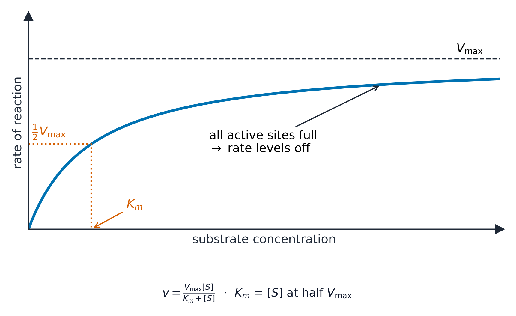
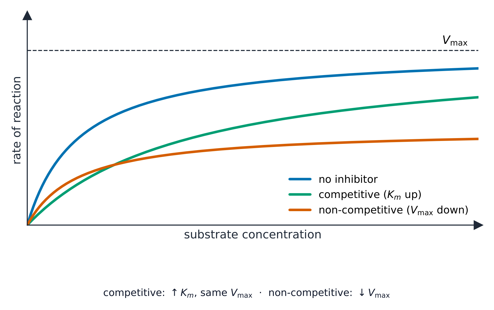


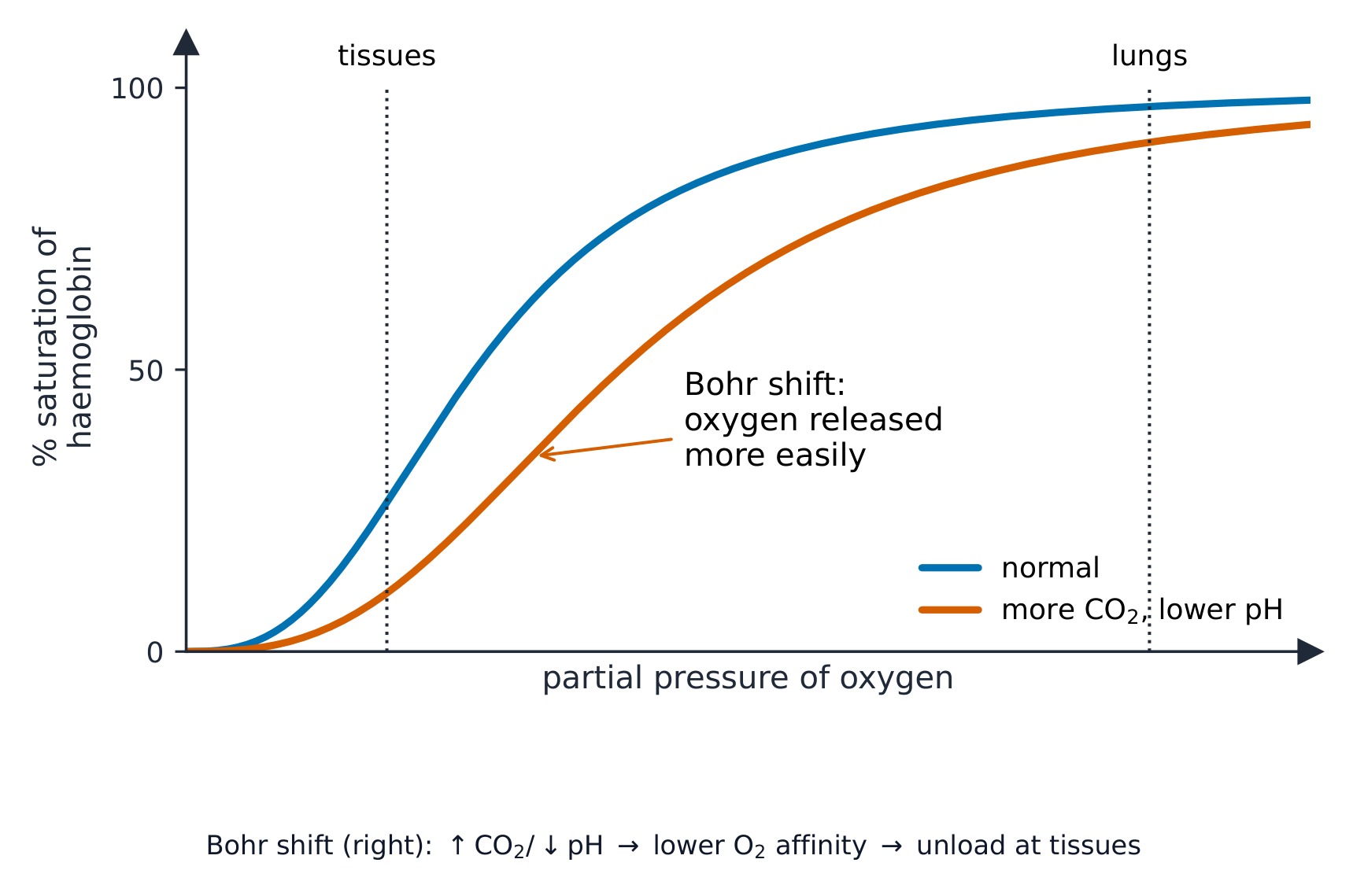


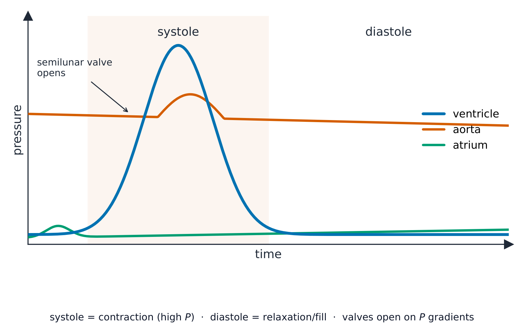


 *どの抗生物質が有効かをテストする方法。各紙円盤には異なる抗生物質が含まれる。透明な輪があることは、その抗生物質が周囲の細菌を殺したことを意味する。輪が大きいほど薬剤の有効性が高い。輪のない円盤は、細菌が耐性を示しているものである。