- state that the mammalian circulatory system is a closed double circulation consisting of a heart, blood and blood vessels including arteries, arterioles, capillaries, venules and veins
- describe the functions of the main blood vessels of the pulmonary and systemic circulations, limited to pulmonary artery, pulmonary vein, aorta and vena cava
- recognise arteries, veins and capillaries from microscope slides, photomicrographs and electron micrographs and make plan diagrams showing the structure of arteries and veins in transverse section (TS) and longitudinal section (LS)
- explain how the structure of muscular arteries, elastic arteries, veins and capillaries are each related to their functions
- recognise and draw red blood cells, monocytes, neutrophils and lymphocytes from microscope slides, photomicrographs and electron micrographs
- state that water is the main component of blood and tissue fluid and relate the properties of water to its role in transport in mammals, limited to solvent action and high specific heat capacity
- state the functions of tissue fluid and describe the formation of tissue fluid in a capillary network
哺乳动物体内的运输
A-Level 生物 · 第 8 主题
 9:17
9:17
The Circulatory System
Your heart beats about a hundred thousand times a day, and it never gets a day off. But here is the clever part. It is not one loop. It is two. Blood leaves…
英文讲解 · 内嵌中英文字幕
8.1
循环系统

大纲
来源:剑桥国际大纲
哺乳动物有一个闭合的双循环。"闭合"意味着血液一直待在血管(blood vessels)里面。"双"意味着每绕身体完整一圈,血液就通过心脏(heart)两次。这给出两个相连的环,所以我们把它叫双循环(double circulation),把整个东西叫一个循环系统(circulatory system):
- 肺循环(pulmonary circulation)把血液从心脏运到肺再运回。
- 体循环(systemic circulation)把血液从心脏运到身体的其余部分再运回。
血液按这个顺序行进:动脉(arteries)→ 小动脉(arterioles)→ 毛细血管(capillaries)→ 小静脉(venules)→ 静脉(veins)。
主要的血管是:
- 肺动脉(pulmonary artery)——把氧气(oxygen)少的血液从心脏运到肺。
- 肺静脉(pulmonary vein)——把富氧血液从肺运回心脏。
- 主动脉(aorta)——把富氧血液从心脏运到身体的大动脉。
- 腔静脉(vena cava)——把缺氧血液从身体送回心脏的大静脉。
How the vessels suit their jobs
| 血管 | 结构 | 功能 |
|---|---|---|
| 动脉 | 厚的肌肉(muscle)和弹性(elastic)纤维壁;窄的管腔(lumen,里面的空间) | 以高压把血液从心脏运走;弹性壁伸展和回弹以平滑流动;肌肉控制流动 |
| 毛细血管 | 壁只有一个细胞厚;非常窄 | 血液和细胞之间物质交换的短距离 |
| 静脉 | 薄壁;宽管腔;有瓣膜(valves) | 以低压把血液送回心脏;瓣膜阻止血液倒流 |

Blood cells
你应当识别:红细胞(red blood cells,它携带氧气),和三种白细胞——单核细胞(monocytes)、中性粒细胞(neutrophils)和淋巴细胞(lymphocytes)。

Water, plasma and tissue fluid
水是血液的主要部分。它是一个好的溶剂(solvent),所以它携带溶解的物质,而且它有一个高的比热容(specific heat capacity),所以血液的温度保持稳定。
在一个毛细血管的起点,高血压把液体(但不是细胞或大蛋白质)从血浆(plasma)中、穿过毛细血管壁推出去。细胞周围的这种液体是组织液(tissue fluid)。它给细胞供应氧气和葡萄糖并带走废物。它的大部分在远端返回毛细血管,那里压力较低。

探索
Double circulation
Follow one red blood cell around the loop. It passes through the heart twice each circuit — once to the lungs, once to the body.
词汇表
训练
| 英文 | 中文 | 拼音 |
|---|---|---|
| blood vessel/blʌd ˈvesl/ | 血管 | xuè guǎn |
| heart/hɑːt/ | 心脏 | xīn zàng |
| double circulation/ˈdʌbl ˌsɜːkjʊˈleɪʃn/ | 双循环 | shuāng xún huán |
| circulatory system/ˌsɜːkjʊˈleɪtəri ˈsɪstəm/ | 循环系统 | xún huán xì tǒng |
| pulmonary circulation/ˈpʌlmənəri ˌsɜːkjʊˈleɪʃn/ | 肺循环 | fèi xún huán |
| systemic circulation/sɪˈstemɪk ˌsɜːkjʊˈleɪʃn/ | 体循环 | tǐ xún huán |
| artery/ˈɑːtəri/ | 动脉 | dòng mài |
| arteriole/ɑːˈtɪəriəʊl/ | 小动脉 | xiǎo dòng mài |
| capillary/kəˈpɪləri/ | 毛细血管 | máo xì xuè guǎn |
| venule/ˈvenjuːl/ | 小静脉 | xiǎo jìng mài |
| vein/veɪn/ | 静脉 | jìng mài |
| pulmonary artery/ˈpʌlmənəri ˈɑːtəri/ | 肺动脉 | fèi dòng mài |
| pulmonary vein/ˈpʌlmənəri veɪn/ | 肺静脉 | fèi jìng mài |
| aorta/eɪˈɔːtə/ | 主动脉 | zhǔ dòng mài |
| vena cava/ˈviːnə ˈkɑːvə/ | 腔静脉 | qiāng jìng mài |
| muscle/ˈmʌsl/ | 肌肉 | jī ròu |
| elastic/ɪˈlæstɪk/ | 弹性 | tán xìng |
| lumen/ˈluːmen/ | 管腔 | guǎn qiāng |
| valve/vælv/ | 瓣膜 | bàn mó |
| red blood cell/red blʌd sel/ | 红细胞 | hóng xì bāo |
| monocyte/ˈmɒnəʊsaɪt/ | 单核细胞 | dān hé xì bāo |
| neutrophil/ˈnjuːtrəfɪl/ | 中性粒细胞 | zhōng xìng lì xì bāo |
| lymphocyte/ˈlɪmfəsaɪt/ | 淋巴细胞 | lín bā xì bāo |
| solvent/ˈsɒlvənt/ | 溶剂 | róng jì |
| specific heat capacity/spəˈsɪfɪk hiːt kəˈpæsɪti/ | 比热容 | bǐ rè róng |
| plasma/ˈplæzmə/ | 血浆 | xuè jiāng |
| tissue fluid/ˈtɪʃuː ˈfluːɪd/ | 组织液 | zǔ zhī yè |
8.2
氧气与二氧化碳的运输
大纲
- describe the role of red blood cells in transporting oxygen and carbon dioxide with reference to the roles of: • haemoglobin • carbonic anhydrase • the formation of haemoglobinic acid • the formation of carbaminohaemoglobin
- describe the chloride shift and explain the importance of the chloride shift
- describe the role of plasma in the transport of carbon dioxide
- describe and explain the oxygen dissociation curve of adult haemoglobin
- explain the importance of the oxygen dissociation curve at partial pressures of oxygen in the lungs and in respiring tissues
- describe the Bohr shift and explain the importance of the Bohr shift
来源:剑桥国际大纲
Carrying oxygen
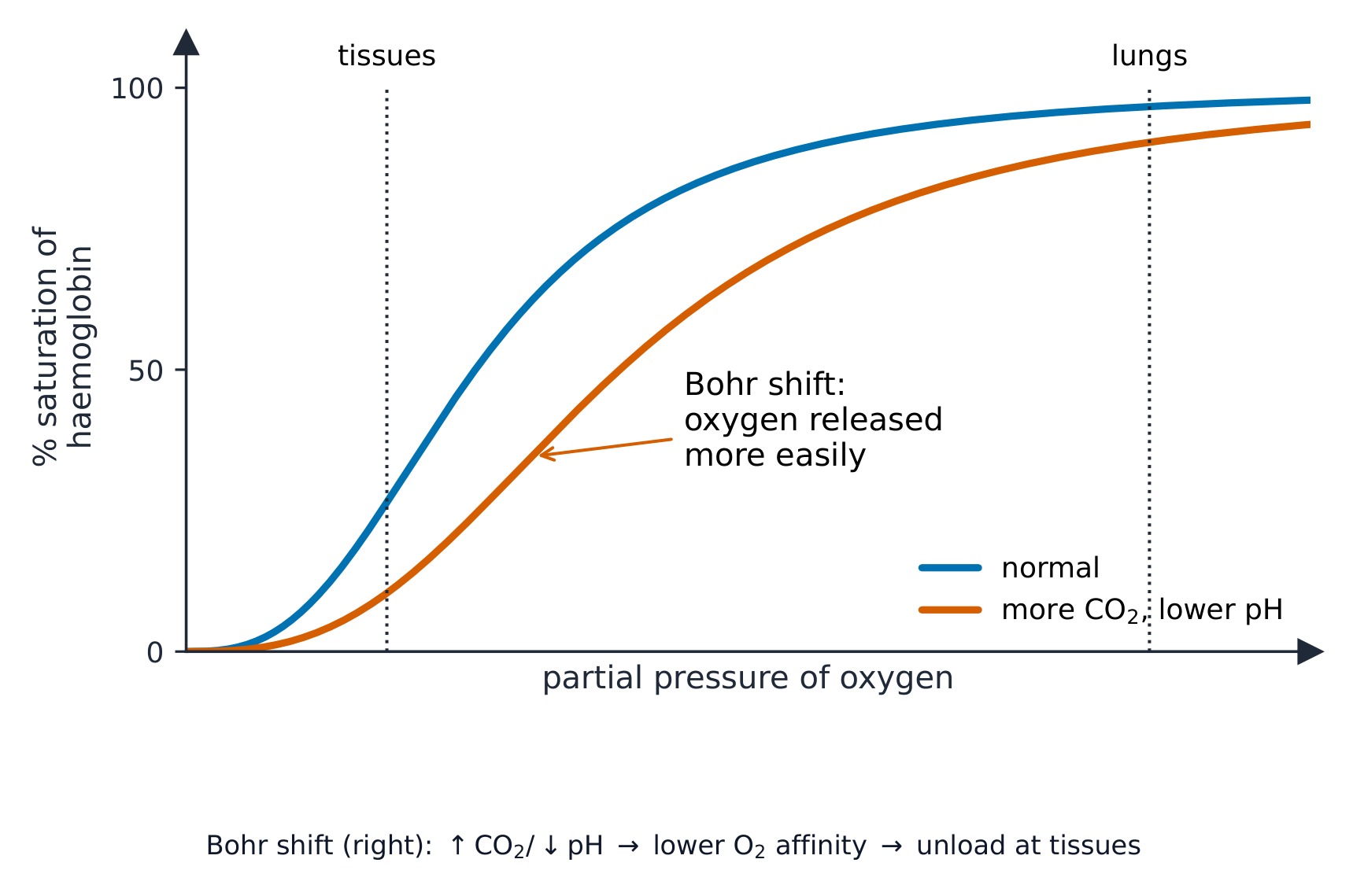
氧气由红细胞中的血红蛋白(haemoglobin)携带。我们用氧解离曲线(oxygen dissociation curve)显示血红蛋白多好地抓住氧气。这个 S 形的图把饱和度(saturation,血红蛋白装满氧气的程度)对氧气的分压(partial pressure)作图:
- 在氧气分压高的地方(在肺中),血红蛋白装载起来并变得几乎完全饱和。
- 在它低的地方(在进行呼吸作用的组织中),血红蛋白卸下它的氧气供细胞使用。
The Bohr shift
当组织非常活跃时,它们释放更多的二氧化碳(carbon dioxide),这降低 pH。这使血红蛋白更容易释放氧气,所以曲线向右移动。这个有益的变化是波尔位移(Bohr shift):氧气恰好在它最被需要的地方被交出。
Carrying carbon dioxide
一点点二氧化碳直接溶入血浆,但大部分在红细胞内部一个反应之后被携带:
- 酶(enzyme)碳酸酐酶(carbonic anhydrase)加速二氧化碳与水的反应以制造碳酸。
- 碳酸分裂成氢离子和碳酸氢根离子(hydrogencarbonate ions)。
- 碳酸氢根离子移出到血浆。这是二氧化碳被携带的主要方式。
- 为了保持电荷平衡,氯离子(chloride ions)移入红细胞。这种移动是氯转移(chloride shift)。
- 氢离子加入血红蛋白以构成血红蛋白酸(haemoglobinic acid);这吸收氢离子并保持 pH 稳定。
一些二氧化碳也直接加入血红蛋白以构成氨甲酰血红蛋白(carbaminohaemoglobin)。

例题。 在肺部,氧分压约为 $12\ \text{kPa}$,血红蛋白的饱和度约为 98%;在运动中的肌肉里,氧分压约为 $3\ \text{kPa}$,饱和度降到约 40%。有多少氧被卸下?为什么曲线呈 S 形?把饱和度相减:$98 - 40 =$ 58% 的血红蛋白结合氧在肌肉中被释放。S 形源于协同结合 - 第一个氧结合后改变了血红蛋白的形状,使后面的氧更容易结合,这正是曲线中段如此陡峭的原因。这种陡峭正是要点所在:在进行呼吸作用的组织里,分压小幅下降就会引起氧的大量释放。运动中肌肉产生的额外二氧化碳降低了 pH,使曲线右移,于是在相同分压下卸下的氧更多 - 这就是波尔位移在起作用。
探索
How blood carries oxygen
Haemoglobin picks up oxygen where there is lots of it (the lungs) and releases it where there is little (the tissues).
词汇表
训练
| 英文 | 中文 | 拼音 |
|---|---|---|
| oxygen/ˈɒksɪdʒn/ | 氧气 | yǎng qì |
| haemoglobin/ˌhiːməˈɡləʊbɪn/ | 血红蛋白 | xuè hóng dàn bái |
| oxygen dissociation curve/ˈɒksɪdʒn dɪˌsəʊsɪˈeɪʃn kɜːv/ | 氧解离曲线 | yǎng jiě lí qū xiàn |
| saturation/ˌsætʃəˈreɪʃn/ | 饱和度 | bǎo hé dù |
| partial pressure/ˈpɑːʃl ˈpreʃə/ | 分压 | fēn yā |
| Bohr shift/ˈbəʊə ʃɪft/ | 波尔位移 | bō ěr wèi yí |
| carbon dioxide/ˈkɑːbən daɪˈɒksaɪd/ | 二氧化碳 | èr yǎng huà tàn |
| enzyme/ˈenzaɪm/ | 酶 | méi |
| carbonic anhydrase/kɑːˈbɒnɪk ˈænhaɪdreɪz/ | 碳酸酐酶 | tàn suān gān méi |
| hydrogencarbonate ion/ˈhaɪdrəʊdʒeŋkɑːbɒneɪt ˈaɪɒn/ | 碳酸氢根离子 | tàn suān qīng gēn lí zi |
| chloride ion/ˈklɔːraɪd ˈaɪɒn/ | 氯离子 | lǜ lí zi |
| chloride shift/ˈklɔːraɪd ʃɪft/ | 氯转移 | lǜ zhuǎn yí |
| haemoglobinic acid/ˌhiːməˌɡləʊˈbɪnɪk ˈæsɪd/ | 血红蛋白酸 | xuè hóng dàn bái suān |
| carbaminohaemoglobin/ˌkɑːbəˌmɪnəʊˌhiːməˈɡləʊbɪn/ | 氨甲酰血红蛋白 | ān jiǎ xiān xuè hóng dàn bái |
8.3
心脏

大纲
- describe the external and internal structure of the mammalian heart
- explain the differences in the thickness of the walls of the: • atria and ventricles • left ventricle and right ventricle
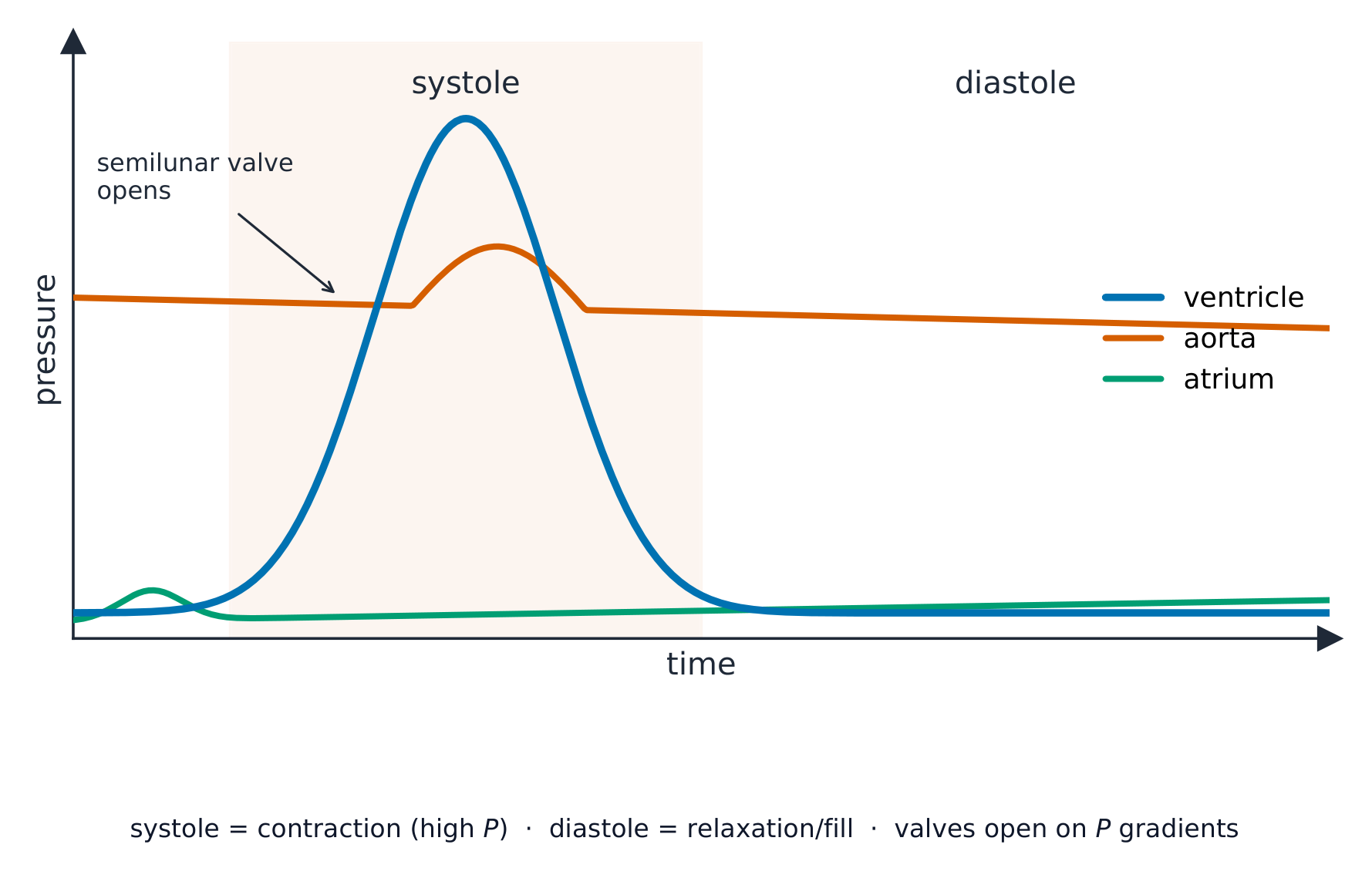
- describe the cardiac cycle, with reference to the relationship between blood pressure changes during systole and diastole and the opening and closing of valves
- explain the roles of the sinoatrial node, the atrioventricular node and the Purkyne tissue in the cardiac cycle (knowledge of nervous and hormonal control is not expected)
来源:剑桥国际大纲
Structure
心脏有四个腔。两个上腔是心房(atria,单数:atrium);它们有薄壁,因为它们只把血液往下推入下面的腔。两个下腔是心室(ventricles);它们有厚的肌肉壁,因为它们把血液泵出心脏。
左心室壁比右心室壁厚,因为左侧必须把血液泵绕全身,而右侧只泵到附近的肺。
The cardiac cycle
一次心跳是心动周期(cardiac cycle)。它有两部分:收缩期(systole,当心肌收缩时)和舒张期(diastole,当它放松并充盈时)。当一个腔收缩时,里面的压力上升;这种压力变化打开和关闭瓣膜,使血液只往一个方向流。
Controlling the heartbeat
心脏设定它自己的节律:
- 右心房中的窦房结(sinoatrial node)是起搏器。它发出一个电兴奋波,它铺开到心房并使它们收缩。
- 房室结(atrioventricular node)接收这个波、把它扣住片刻(所以心房先排空),然后传递它。
- 浦肯野组织(Purkyne tissue)把波往下带并穿过心室壁,所以心室从底部向上收缩并把血液推出。

探索
Explore the heart
Tap each part. The right side pumps blood to the lungs; the thicker-walled left side pumps it round the whole body.
探索
The cardiac cycle
Step through one heartbeat — atria contract, then ventricles contract, then everything relaxes and refills.
探索
The cardiac cycle
Step through one heartbeat. Pressure changes open and close the valves so blood always flows one way.
词汇表
训练
| 英文 | 中文 | 拼音 |
|---|---|---|
| atrium/ˈeɪtrɪəm/ | 心房 | xīn fáng |
| ventricle/ˈventrɪkl/ | 心室 | xīn shì |
| cardiac cycle/ˈkɑːdiæk ˈsaɪkl/ | 心动周期 | xīn dòng zhōu qī |
| systole/ˈsɪstəʊl/ | 收缩期 | shōu suō qī |
| diastole/ˈdaɪəstəʊl/ | 舒张期 | shū zhāng qī |
| sinoatrial node/sɪˈnəʊtrɪəl nəʊd/ | 窦房结 | dòu fáng jié |
| atrioventricular node/ˌeɪtrɪˌəʊvənˈtrɪkjʊlə nəʊd/ | 房室结 | fáng shì jié |
| Purkyne tissue/ˈpɜːkaɪn ˈtɪʃuː/ | 浦肯野组织 | pǔ kěn yě zǔ zhī |
8.3
考试技巧
- 追踪双循环并命名腔、瓣膜和血管;左心室壁更厚(以更高的压力泵得更远)。
- 读心动周期压力图:当跨一个瓣膜的压力交叉时它打开/关闭。
- 解释氧解离曲线(S 形)和波尔位移(更多 CO2 把它右移,向活跃的组织卸下更多 O2)。
- 大部分 CO2 作为碳酸氢根携带(通过氯转移)。
本主题的互动课程
逐步学习,并即时检测练习。