- understand that a piezo-electric crystal changes shape when a p.d. is applied across it and that the crystal generates an e.m.f. when its shape changes
- understand how ultrasound waves are generated and detected by a piezoelectric transducer
- understand how the reflection of pulses of ultrasound at boundaries between tissues can be used to obtain diagnostic information about internal structures
- define the specific acoustic impedance of a medium as $Z = \rho c$, where $c$ is the speed of sound in the medium
- use $I_{\text{R}} / I_0 = (Z_1 - Z_2)^2 / (Z_1 + Z_2)^2$ for the intensity reflection coefficient of a boundary between two media
- recall and use $I = I_0 e^{-\mu x}$ for the attenuation of ultrasound in matter
医学物理
A-Level 物理 · 第 24 主题
 17:14
17:14
Ultrasound & Medical Imaging
The very first photograph of your life was probably taken with sound. Before you were born, a doctor pressed a small probe to your mother's skin, and out of…
英文讲解 · 内嵌中英文字幕
24.1
超声波的产生与应用

大纲
来源:剑桥国际大纲
压电效应
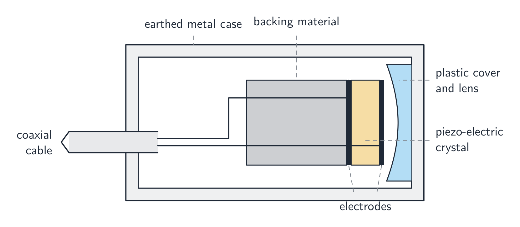
一个压电(piezo-electric)晶体在它两端加上电势差时会略微改变形状,反过来也是:当它的形状被改变时,它在自身两端产生一个电动势(electromotive force,e.m.f.)。石英和 PZT 是常见的例子。两个相联系的效应:
- 加一个电势差 → 晶体改变形状(用来产生振动)。
- 改变形状(一个波挤压它)→ 出现一个电动势(用来探测振动)。
压电换能器
一个换能器(transducer)用这个效应既产生又探测 超声波(ultrasound)。
- 一个交流电势差(几 MHz)使晶体以相同的频率振动,发出高于 $20\ \text{kHz}$($1$–$10\ \text{MHz}$ 用于医学成像)的纵波(longitudinal)。
- 同一晶体然后探测:返回的超声波使它振动并产生一个电动势。
产生超声波(三分)。 (1) 在晶体两端加一个交变电势差。(2) 晶体改变形状,以电势差的频率膨胀和收缩,从而振动。(3) 电势差选在晶体的共振频率,所以振动很大,晶体的表面推动组织,发出该频率的纵波。探测超声波(两分)。 返回的波的压强变化改变晶体的形状;形状改变产生电动势,以波的频率交变,被放大并记录。同一晶体兼作两用,在发射和接收之间切换。
所以一个换能器既是发射器又是探测器,在发送和收听之间切换。
脉冲回波成像
要看身体内部(脉冲回波(pulse-echo)成像):
- 换能器向身体发送一个短脉冲。
- 在每个组织边界,一部分脉冲被反射,一部分继续前进。
- 换能器探测每个反射的脉冲。
- 时间延迟给出深度:$d = c t / 2$(往返)。回波的振幅给出反射的强度。
例题。 一个超声脉冲在发出后 $60\ \mu\text{s}$ 返回换能器。声音在组织中的速率是 $1500\ \text{m s}^{-1}$。求反射边界的深度。
脉冲往返传播,所以 $d = \dfrac{ct}{2}$:
$$d = \frac{1500 \times 60 \times 10^{-6}}{2} = 0.045\ \text{m}\ (= 4.5\ \text{cm}).$$
- 在身体上扫过就建立起一幅二维图像。

在换能器和皮肤之间涂上耦合剂(coupling gel)以挤出空气;没有它几乎所有的超声波都会在皮肤-空气边界反射,从不进入身体。

"概述用超声波获取诊断信息的方法"(四分)。 (1) 换能器用凝胶耦合到皮肤上,向身体发送一个超声脉冲。(2) 在组织之间的每个边界上,一部分脉冲被反射;反射的脉冲返回换能器并被探测。(3) 发射与回波之间的时间延迟,结合声速,给出边界的深度($d = ct/2$)。(4) 回波的强度由边界处声阻抗的变化决定,表明这是什么样的边界。扫动换能器或使用阵列就建立起图像。
为什么用脉冲,为什么用兆赫。 换能器发出一个短脉冲然后倾听:每个边界的回波必须在下一个脉冲发出之前到达,否则不同脉冲的回波无法区分,而且晶体不能同时发射和接收。频率是一种折中:更高的频率意味着更短的波长、更精细的细节(分辨率(resolution)),但衰减更强,所以深部器官需要比浅部更低的频率。典型的医学扫描用 $1$–$15\ \text{MHz}$,在组织中波长不到一毫米。
声阻抗
一种介质的声阻抗(specific acoustic impedance)是
$$Z = \rho c,$$
其中 $\rho$ 是密度(density),$c$ 是声速。单位:$\text{kg m}^{-2}\ \text{s}^{-1}$。骨骼有大的 $Z$;空气有小的 $Z$;软组织介于两者之间。
例题。 求软组织的声阻抗。(密度 $1060\ \text{kg m}^{-3}$,声速 $1540\ \text{m s}^{-1}$。)
$$Z = \rho c = 1060 \times 1540 \approx 1.6 \times 10^{6}\ \text{kg m}^{-2}\ \text{s}^{-1}.$$
定义(两分)。 介质的声阻抗是它的密度与其中声速的乘积。 两个量都必须点名;"声音通过的难度"不得分。因为 $Z$ 结合了密度和速度,两种密度差异很大的介质仍可能有相近的 $Z$(水和软组织),决定反射多少声音的是 $Z$,而不是单独的密度。
边界处的反射
在阻抗为 $Z_{1}$ 和 $Z_{2}$ 的介质之间的边界处,强度反射系数(intensity reflection coefficient,被反射的比例)是
$$\frac{I_{\text{R}}}{I_{0}} = \left(\frac{Z_{1} - Z_{2}}{Z_{1} + Z_{2}}\right)^{2}.$$
- 阻抗差异很大:几乎全部被反射(皮肤/空气——因此用耦合剂)。
- 阻抗很相近:几乎没有被反射,所以看不到边界。
- 成像最佳:差异足够大到给出回波,但又没有大到什么都不能继续前进。

例题(为什么用凝胶)。 空气:密度 $1.29\ \text{kg m}^{-3}$,声速 $343\ \text{m s}^{-1}$。凝胶:$Z = 1.50 \times 10^{6}\ \text{kg m}^{-2}\ \text{s}^{-1}$。软组织:$Z = 1.63 \times 10^{6}\ \text{kg m}^{-2}\ \text{s}^{-1}$。求空气–组织边界和凝胶–组织边界的强度反射系数。
$Z_{\text{air}} = 1.29 \times 343 = 442\ \text{kg m}^{-2}\ \text{s}^{-1}$。空气–组织:$\left(\dfrac{442 - 1.63 \times 10^{6}}{442 + 1.63 \times 10^{6}}\right)^{2} = 0.9989$:$99.9\%$ 的强度被反射,只有 $0.1\%$ 进入身体。凝胶–组织:$\left(\dfrac{1.50 - 1.63}{1.50 + 1.63}\right)^{2} = 0.0017$:只有 $0.2\%$ 被反射。所以阻抗接近皮肤的凝胶取代了本来会几乎全部反射的空气层,几乎全部脉冲进入身体(回波也能沿同一路径回来)。
例题。 水:密度 $1000\ \text{kg m}^{-3}$,声速 $1480\ \text{m s}^{-1}$。玻璃:密度 $2500\ \text{kg m}^{-3}$,声速 $5600\ \text{m s}^{-1}$。在水–玻璃边界处强度的多大比例被反射?
$Z_{\text{water}} = 1.48 \times 10^{6}$,$Z_{\text{glass}} = 1.40 \times 10^{7}\ \text{kg m}^{-2}\ \text{s}^{-1}$。系数 $= \left(\dfrac{1.48 - 14.0}{1.48 + 14.0}\right)^{2} = 0.65$:三分之二被反射,三分之一透射。用 $10^{6}$ 为单位计算保持算术简洁,因为重要的是比值。组织中的钢植入物($Z_{\text{steel}} = 4.7 \times 10^{7}$)给出 $0.87$:几乎全部脉冲从它的表面反射,所以它后面的东西无法成像,但植入物本身显得非常亮。
衰减
当超声波穿过组织时,它的强度随距离下降:
$$I = I_{0} e^{-\mu x},$$
其中 $\mu$ 是衰减系数(attenuation coefficient,单位 $\text{m}^{-1}$)。同样的形式适用于 X 射线。

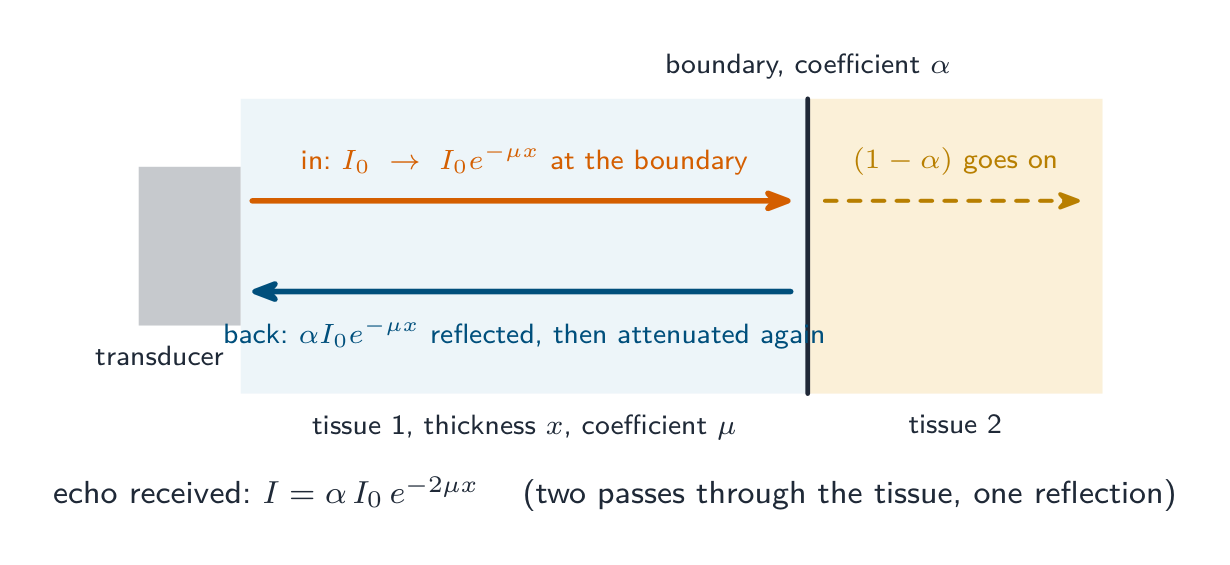
例题(回波的强度)。 强度为 $I_{0}$ 的脉冲穿过 $4.0\ \text{cm}$、衰减系数 $0.50\ \text{cm}^{-1}$ 的组织到达一个强度反射系数为 $0.010$ 的边界。求返回换能器的回波强度占 $I_{0}$ 的比例。
到边界:$e^{-\mu x} = e^{-0.50 \times 4.0} = 0.135$。反射:$\times 0.010$。再穿回同样的 $4.0\ \text{cm}$:再 $\times 0.135$。回波:$I/I_{0} = 0.135 \times 0.010 \times 0.135 = 1.8 \times 10^{-4}$,约 $0.02\%$。考官检查两点:组织被穿过两次,反射系数乘的是边界处的强度,不是 $I_{0}$。这就是深部边界的回波在显示前比浅部的放大更多的原因。
探索
Ultrasound scan route
Follow a pulse from transducer to echo image.
词汇表
训练
| 英文 | 中文 | 拼音 |
|---|---|---|
| ultrasound/ˌʊltrəˈsaʊnd/ | 超声波 | chāo shēng bō |
| piezo-electric/piˌeɪtsəʊ ɪˈlektrɪk/ | 压电 | yā diàn |
| electromotive force/ɪˌlektrəʊˈməʊtɪv fɔːs/ | 电动势 | diàn dòng shì |
| transducer/trænsˈdjuːsə/ | 换能器 | huàn néng qì |
| longitudinal/ˌlɒŋɡɪˈtjuːdɪnl/ | 纵波 | zòng bō |
| pulse-echo/pʌls ˈekəʊ/ | 脉冲回波 | mài chōng huí bō |
| coupling gel/ˈkʌplɪŋ dʒel/ | 耦合剂 | ǒu hé jì |
| resolution/ˌrezəˈluːʃn/ | 分辨率 | fēn biàn lǜ |
| specific acoustic impedance/spəˈsɪfɪk əˈkuːstɪk ɪmˈpiːdəns/ | 声阻抗 | shēng zǔ kàng |
| density/ˈdensɪti/ | 密度 | mì dù |
| intensity reflection coefficient/ɪnˈtensɪti rɪˈflekʃn ˌkəʊɪˈfɪʃənt/ | 强度反射系数 | qiáng dù fǎn shè xì shù |
| attenuation coefficient/əˌtenjuːˈeɪʃn ˌkəʊɪˈfɪʃənt/ | 衰减系数 | shuāi jiǎn xì shù |
24.2
X射线的产生与应用

大纲
- explain that X-rays are produced by electron bombardment of a metal target and calculate the minimum wavelength of X-rays produced from the accelerating p.d.
- understand the use of X-rays in imaging internal body structures, including an understanding of the term contrast in X-ray imaging
- recall and use $I = I_0 e^{-\mu x}$ for the attenuation of X-rays in matter
- understand that computed tomography (CT) scanning produces a 3D image of an internal structure by first combining multiple X-ray images taken in the same section from different angles to obtain a 2D image of the section, then repeating this process along an axis and combining 2D images of multiple sections
来源:剑桥国际大纲
产生
X 射线来自一个 X 射线管(X-ray tube):
- 一个加热的阴极(cathode)通过热电子发射(thermionic emission)发射电子。
- 一个高电势差(几十到几百 kV)使电子穿过真空(vacuum)加速到一个金属靶(target,即阳极(anode),常常是钨(tungsten))。
- 电子撞击靶并急剧减速。它们大部分动能(kinetic energy)变成热;一小部分作为 X 射线光子(photons)发射(轫致辐射(Bremsstrahlung),"刹车辐射")。一些电子把金属原子的内层电子打出来,重新填充时发射特征(characteristic)X 射线线。
"解释 X 射线是如何产生的"(四分)。 (1) 电子通过热电子发射从加热的灯丝(阴极)发出。(2) 它们在抽真空的管中经高电势差(几十 kV)加速射向金属靶(阳极)。(3) 撞击靶时它们急剧减速,并且 (4) 失去的动能以 X 射线光子的形式发出(其中大部分变成靶中的热,这就是阳极要冷却或旋转的原因)。
控制射线束。 强度(单位面积每秒的能量)由每秒撞击靶的电子数决定,所以由灯丝电流控制(更热的灯丝发出更多电子);硬度(hardness),即穿透能力,由光子能量决定,所以由加速电势差控制:更大的电势差给出能量更高、波长更短、穿透力更强的 X 射线。一块金属滤片除去最软的 X 射线,它们会在患者皮肤中被吸收而到不了探测器。
例题(靶的发热)。 在 $75\ \text{kV}$ 下运行的管中电子束电流为 $30\ \text{mA}$,电子能量的 $99\%$ 变成质量 $15\ \text{g}$ 的钨靶(比热容 $134\ \text{J kg}^{-1}\ \text{K}^{-1}$)中的热。若没有热量损失,求温度上升的速率。
输入功率 $= IV = 0.030 \times 75\,000 = 2250\ \text{W}$;发热功率 $= 0.99 \times 2250 = 2230\ \text{W}$。于是 $mc\,\Delta\theta/\Delta t = 2230$,所以 $\Delta\theta/\Delta t = 2230/(0.015 \times 134) = 1.1 \times 10^{3}\ \text{K s}^{-1}$。靶几秒内就会熔化,这就是曝光很短、阳极旋转以分散热量的原因。
最小波长
一个 X 射线光子能有的最大能量是一个被加速电子的全部动能,在单次事件中损失。对于加速电势差 $V$,动能是 $eV$,所以
$$h f_{\text{max}} = e V, \qquad \lambda_{\text{min}} = \frac{h c}{e V}.$$
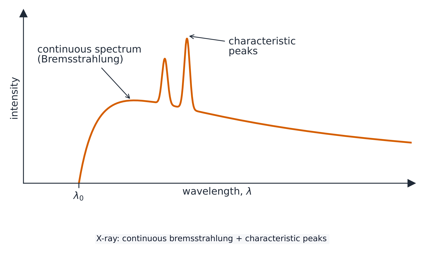
这是短波长截止。连续的轫致辐射谱在 $\lambda_{\text{min}}$ 以上拖尾,带由靶金属决定的尖锐特征峰。
例题(最小波长)。 电子经 $75\ \text{kV}$ 加速。求 X 射线的最大光子能量和最小波长。
最大能量 $= eV = 75\ \text{keV} = 75\,000 \times 1.60 \times 10^{-19} = 1.2 \times 10^{-14}\ \text{J}$(或 $0.075\ \text{MeV}$)。于是 $\lambda_{\text{min}} = hc/(eV) = (6.63 \times 10^{-34})(3.00 \times 10^{8})/(1.2 \times 10^{-14}) = 1.7 \times 10^{-11}\ \text{m}$。最大光子动量为 $p = E/c = 4.0 \times 10^{-23}\ \text{N s}$。电势差加倍,最小波长减半。
解释谱的形状。 谱是连续的,因为每个电子可以在一次或几次减速中失去其能量的任意比例,所以产生了直到最大值的每种能量的光子。它在 $\lambda_{\text{min}}$ 处有一个陡的截止,因为一个光子携带的能量不能超过一个电子所具有的 $eV$,而这只在一个电子在单次事件中失去全部能量时发生。峰是靶金属的特征:入射电子把靶原子的一个内层电子撞出,一个外层电子落入空位,发出能量等于两个能级之差的光子(第 22 讲)。
用 X 射线成像
X 射线穿过患者到达一个探测器。衰减(attenuate)更多的组织(骨骼,高 $Z$)投下更强的阴影,看起来更亮;衰减更少的组织(软组织、肺)看起来更暗。
对比度(contrast)是各组织之间衰减的差异。可以给予一种造影剂(contrast medium,例如钡餐)使软组织突显出来。

清晰度与对比度。 清晰度(sharpness)是图像中结构边缘的清楚程度:小的 X 射线源(靶上的小点)、不动的患者和靠近患者的探测器都能改善它。对比度是图像中相邻区域之间变黑程度(或亮度)的差异,由衰减的差异产生。两者相互独立:图像可以清晰但对比度差,或者相反。
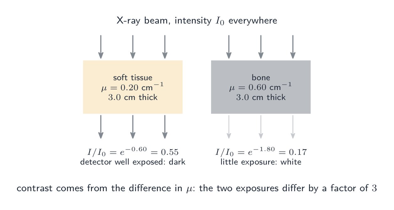
"解释为什么内部结构的 X 射线图像有好的对比度。" 骨和软组织的衰减系数差异很大,所以相等的厚度透射的强度非常不同,探测器在各自后面的曝光非常不同。当两种软组织的系数相近时(胃与其周围),就吞服或注射一种高衰减系数的造影剂(如钡)来勾勒其中一种。使用较低的电势差(较软的 X 射线)也能增大对比度,代价是更大的剂量。
例题。 X 射线在身体的一个区域穿过 $3.0\ \text{cm}$ 软组织($\mu = 0.20\ \text{cm}^{-1}$),在另一个区域穿过 $3.0\ \text{cm}$ 骨($\mu = 0.60\ \text{cm}^{-1}$)。比较透射强度。
软组织:$I/I_{0} = e^{-0.20 \times 3.0} = e^{-0.60} = 0.55$。骨:$e^{-0.60 \times 3.0} = e^{-1.80} = 0.17$。软组织区域得到的曝光是骨区域的三倍多,所以骨显白、组织显暗。曝光之比 $e^{(\mu_{\text{bone}} - \mu_{\text{tissue}})x}$ 随厚度增大:结构越厚对比度越好,但总衰减也越大。
衰减定律
$$I = I_{0} e^{-\mu x}.$$
能量更高的 X 射线穿透得更远(更小的 $\mu$);骨骼的 $\mu$ 比软组织大得多。要求给定比例所需的厚度,取对数:$x = \dfrac{1}{\mu} \ln\dfrac{I_{0}}{I}$。半值厚度(half-value thickness)$x_{1/2} = \ln 2 / \mu$ 使强度减半(像衰变中的半衰期)。
例题。 X 射线穿过衰减系数 $\mu = 40\ \text{m}^{-1}$ 的 $3.0\ \text{cm}$ 组织。求穿透过去的强度比例。
$$\frac{I}{I_{0}} = e^{-\mu x} = e^{-40 \times 0.030} = e^{-1.2} \approx 0.30.$$
例题(吸收的百分比)。 X 射线穿过 $2.8\ \text{cm}$、衰减系数 $1.4\ \text{cm}^{-1}$ 的介质。X 射线能量的百分之多少被吸收?
透射比例 $e^{-1.4 \times 2.8} = e^{-3.92} = 0.020$,所以 $98\%$ 被吸收。题目问的是被吸收的,不是透过的:读两遍。这里的半值厚度是 $\ln 2/\mu = 0.50\ \text{cm}$,所以 $2.8\ \text{cm}$ 是 $5.6$ 个半值厚度,$(1/2)^{5.6} = 0.02$ 验证了答案。
例题(两层)。 强度为 $I_{0}$ 的射线束穿过 $2.0\ \text{cm}$ 的材料 P($\mu = 0.35\ \text{cm}^{-1}$),然后穿过 $1.5\ \text{cm}$ 的材料 Q($\mu = 0.90\ \text{cm}^{-1}$)。求透射强度。
指数相乘:$I = I_{0}\, e^{-0.35 \times 2.0}\, e^{-0.90 \times 1.5} = I_{0}\, e^{-(0.70 + 1.35)} = I_{0}\, e^{-2.05} = 0.13\, I_{0}$。把指数相加($\mu_{1}x_{1} + \mu_{2}x_{2}$);决不把厚度或系数相加。各层的顺序对总透射强度没有影响。
计算机断层扫描(CT)
一次计算机断层扫描(computed tomography,CT)建立一幅三维图像:
- 管和探测器绕患者旋转,从不同角度对一个薄切片拍多幅图像。
- 一台计算机把这些合成为该切片的一个二维横截面。
- 患者被移动,对下一个切片成像。
- 各切片被堆叠成一幅三维图像。
CT 显示的远多于单张 X 射线,因为重叠的软组织被多角度重建分开。

"解释 CT 扫描如何产生三维图像"(五分)。 (1) X 射线管(和一圈探测器)绕患者旋转,使得 (2) 从不同角度拍摄一个切片(截面)的许多 X 射线图像。(3) 计算机把它们合成为该切片的二维图像。(4) 移动患者,对相继的切片重复该过程。(5) 各切片合并成一幅三维图像,可以旋转并从任何角度观看。**目的:**对身体的一个截面成像,避免单张 X 射线中结构的重叠,显示原本被遮挡的结构及其深度位置。代价是比单张 X 射线大得多的剂量。
探索
X-ray production route
Follow electrons from cathode to X-ray photons.
词汇表
训练
| 英文 | 中文 | 拼音 |
|---|---|---|
| X-ray tube/eks reɪ tjuːb/ | X射线管 | X shè xiàn guǎn |
| cathode/ˈkæθəʊd/ | 阴极 | yīn jí |
| thermionic emission/ˌθɜːmɪˈɒnɪk ɪˈmɪʃn/ | 热电子发射 | rè diàn zi fā shè |
| vacuum/ˈvækjuːm/ | 真空 | zhēn kōng |
| target/ˈtɑːɡɪt/ | 靶 | bǎ |
| anode/ˈænəʊd/ | 阳极 | yáng jí |
| tungsten/ˈtʌŋstn/ | 钨 | wū |
| kinetic energy/kɪˈnetɪk ˈenədʒi/ | 动能 | dòng néng |
| energy/ˈenədʒi/ | 能量 | néng liàng |
| photon/ˈfəʊtɒn/ | 光子 | guāng zi |
| Bremsstrahlung/ˈbremʃtrɑːlʊŋ/ | 轫致辐射 | rèn zhì fú shè |
| characteristic/ˌkærɪktəˈrɪstɪk/ | 特征 | tè zhēng |
| hardness/ˈhɑːdnəs/ | 硬度 | yìng dù |
| attenuate/əˈtenjuːeɪt/ | 衰减 | shuāi jiǎn |
| contrast/ˈkɒntræst/ | 对比度 | duì bǐ dù |
| contrast medium/ˈkɒntræst ˈmiːdɪəm/ | 造影剂 | zào yǐng jì |
| sharpness/ˈʃɑːpnəs/ | 清晰度 | qīng xī dù |
| half-value thickness/hɑːf ˈvæljuː ˈθɪknəs/ | 半值厚度 | bàn zhí hòu dù |
| computed tomography/kəmˈpjuːtɪd təˈmɒɡrəfi/ | 计算机断层扫描 | jì suàn jī duàn céng sǎo miáo |
24.3
PET扫描

大纲
- understand that a tracer is a substance containing radioactive nuclei that can be introduced into the body and is then absorbed by the tissue being studied
- recall that a tracer that decays by $\beta^+$ decay is used in positron emission tomography (PET scanning)
- understand that annihilation occurs when a particle interacts with its antiparticle and that mass–energy and momentum are conserved in the process
- explain that, in PET scanning, positrons emitted by the decay of the tracer annihilate when they interact with electrons in the tissue, producing a pair of gamma-ray photons travelling in opposite directions
- calculate the energy of the gamma-ray photons emitted during the annihilation of an electron-positron pair
- understand that the gamma-ray photons from an annihilation event travel outside the body and can be detected, and an image of the tracer concentration in the tissue can be created by processing the arrival times of the gamma-ray photons
来源:剑桥国际大纲
示踪剂
示踪剂(tracer)是一种放进身体、带放射性原子核的物质。它被所研究的组织更多地吸收(例如肿瘤由于其高代谢(metabolism)吸收更多带葡萄糖标记的示踪剂)。它的衰变从外部被探测。
在正电子发射断层扫描(positron emission tomography,PET)中,示踪剂是一个 $\beta^{+}$ 发射体——它放出一个正电子(positron)。一个常见的是葡萄糖类似物上的氟-18(FDG)。
"解释示踪剂是什么以及如何使用"(三分)。 示踪剂是一种含有放射性核(对 PET 是 $\beta^{+}$ 发射体,如氟-18)的物质,通常结合在葡萄糖这样的分子上,引入身体(注射或吞服),并被所研究的组织吸收;它发出的辐射离开身体并被探测,所以它的分布显示出该组织在哪里活跃。选择半衰期短的核素,使扫描期间活度高而之后患者的剂量小,但又不能短到还没到达组织就衰变完:氟-18($110$ 分钟)和氧-15($2$ 分钟,现场制备现场使用)是典型的。
湮灭
当一个粒子遇到它的反粒子(antiparticle)时它们湮灭(annihilate):它们的质量变成电磁能量。在 PET 中:
- 一个正电子在遇到一个电子(electron)之前传播几毫米。
- 它们湮灭。能量和动量(momentum)守恒。
- 由于总动量约为零,产生两个朝相反方向运动的光子,每个 $511\ \text{keV}$($= m_{e} c^{2}$)。
湮灭光子的能量
由能量守恒,总光子能量等于这一对的静止能量(energy):
$$2 h f = 2 m_{e} c^{2}, \qquad h f = m_{e} c^{2}.$$
每个光子有 $h f = m_{e} c^{2} \approx 8.2 \times 10^{-14}\ \text{J} \approx 0.51\ \text{MeV}$,$\lambda \approx 2.4 \times 10^{-12}\ \text{m}$。
解释湮灭(四分)。 (1) 示踪剂发出的正电子行进一小段距离后遇到组织中的一个电子。(2) 这一对湮灭:它们的质量转化为能量,(3) 以两个伽马射线光子发出,因为 (4) 动量必须守恒:这一对几乎没有动量,所以两个光子必须以相等的动量沿相反方向运动。每个光子携带一个粒子的静止能量 $0.51\ \text{MeV}$;单个光子不能满足动量守恒。
例题。 求一个正电子和一个电子在都缓慢运动时湮灭所释放的总能量,以及每个光子的波长。
$E = 2m_{\text{e}}c^{2} = 2 \times 9.11 \times 10^{-31} \times (3.00 \times 10^{8})^{2} = 1.64 \times 10^{-13}\ \text{J}$($1.02\ \text{MeV}$),平分:每个 $8.2 \times 10^{-14}\ \text{J}$。$\lambda = hc/E = (6.63 \times 10^{-34})(3.00 \times 10^{8})/(8.2 \times 10^{-14}) = 2.4 \times 10^{-12}\ \text{m}$,一条伽马射线。若电子和正电子各以 $4.9 \times 10^{7}\ \text{m s}^{-1}$ 反向运动,每个的动能为 $\tfrac{1}{2}mv^{2} = 1.1 \times 10^{-15}\ \text{J}$,约为其静止能量的 $1\%$,光子各携带 $8.3 \times 10^{-14}\ \text{J}$:静止能量占主导,这就是湮灭光子总是能量几乎相同的原因。
重建图像
两个光子朝相反方向离开身体,击中患者周围的探测器环(detector rings)。记录两个同时到达(一次"符合")确定湮灭发生所在的直线。来自多个角度的许多次符合让计算机建立一幅示踪剂的三维图——显示代谢活动高的组织。比较两个到达时间可以细化沿那条直线的位置(飞行时间 PET)。

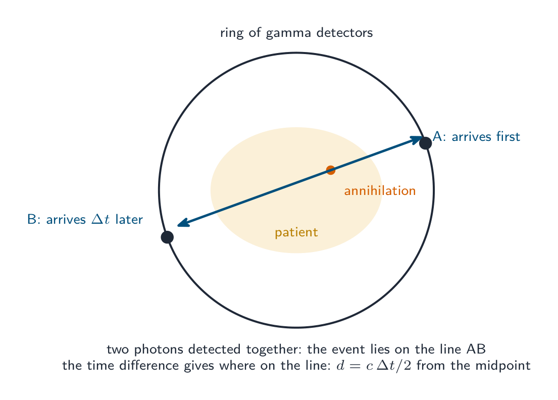
"解释如何用伽马光子形成图像"(四分)。 (1) 两个光子沿相反方向离开身体,被探测器环探测。(2) (几乎)同时到达的两个光子被认为来自同一次湮灭,因此它位于连接这两个探测器的直线上。(3) 到达时间的微小差异给出沿该直线的位置($d = c\,\Delta t/2$,从中点算起;$1\ \text{ns}$ 的差异对应 $15\ \text{cm}$,所以计时必须非常精确)。(4) 计算机收集许多这样的事件,绘出组织中示踪剂的浓度图,形成图像;浓度更高意味着更活跃的组织,如肿瘤。单独到达的光子(其伙伴在体内被吸收)被舍弃。
探索
PET scan route
Follow positron emission to a ring of detected photons.
词汇表
训练
| 英文 | 中文 | 拼音 |
|---|---|---|
| electron/ɪˈlektrɒn/ | 电子 | diàn zi |
| momentum/məʊˈmentəm/ | 动量 | dòng liàng |
| tracer/ˈtreɪsə/ | 示踪剂 | shì zōng jì |
| metabolism/məˈtæbəlɪzəm/ | 代谢 | dài xiè |
| positron emission tomography/ˈpɒzɪtrɒn ɪˈmɪʃn təˈmɒɡrəfi/ | 正电子发射断层扫描 | zhèng diàn zi fā shè duàn céng sǎo miáo |
| positron/ˈpɒzɪtrɒn/ | 正电子 | zhèng diàn zi |
| antiparticle/ˌæntɪˈpɑːtɪkl/ | 反粒子 | fǎn lì zi |
| annihilate/əˈnaɪəleɪt/ | 湮灭 | yān miè |
24.3
考官认可的定义
定义题按固定措辞评分。准确学会这些,只给一个答案。
| 术语 | 定义 |
|---|---|
| 压电效应 | 晶体在两端加电势差时改变形状,在形状改变时产生电动势 |
| 声阻抗 | 介质的密度与其中声速的乘积,$Z = \rho c$ |
| 强度反射系数 | 在边界处被反射的入射强度的比例,$(Z_{1} - Z_{2})^{2}/(Z_{1} + Z_{2})^{2}$ |
| 衰减系数 | $I = I_{0}e^{-\mu x}$ 中的常量 $\mu$;它越大,强度随厚度下降越快 |
| (X 射线的)硬度 | 射线束的穿透能力,由光子能量(加速电势差)决定 |
| 对比度 | 图像中相邻区域之间变黑程度的差异 |
| 清晰度 | 图像中结构边缘的清楚程度 |
| CT 扫描 | 从许多角度拍摄的一个截面的 X 射线图像由计算机合成为该切片的二维图像;相继的切片给出三维图像 |
| 示踪剂 | 引入身体并被所研究组织吸收的含放射性核的物质 |
| 湮灭 | 粒子与其反粒子相互作用,质量转化为能量(光子),质能和动量守恒 |
24.3
考试技巧
- 超声波:脉冲进、回波回;深度 $= ct/2$(去和回);$Z = \rho c$;反射系数由两个阻抗求出;凝胶使阻抗与皮肤匹配。衰减是指数式的,回波穿过组织两次。
- X 射线:产生是加热的灯丝、高电势差、在靶处突然减速;强度由灯丝电流控制,硬度由电势差控制;$\lambda_{\text{min}} = hc/(eV)$;$I = I_{0}e^{-\mu x}$,多层时指数相加;对比度是 $\mu$ 的差异。
- CT:许多角度、一个切片、计算机、相继的切片、三维。五点都要说。
- PET:$\beta^{+}$ 示踪剂,与电子湮灭,两个 $0.51\ \text{MeV}$ 的光子沿相反方向(动量),符合给出直线,计时给出位置。
- 逐字记住三个"解释"答案:晶体如何产生和探测、X 射线如何产生、光子如何定位示踪剂。
- 每个阻抗($\text{kg m}^{-2}\ \text{s}^{-1}$)和系数($\text{cm}^{-1}$ 或 $\text{m}^{-1}$)都要写单位,只有当 $\mu$ 以 $\text{m}^{-1}$ 计时才把 $\text{cm}$ 换成 $\text{m}$。
常见错误
- 在反射边界的深度中忘记因子 2,或在回波强度中忘记第二次穿过组织。
- 把声阻抗定义为"对声音的阻力"而不是密度乘声速。
- 把凝胶–皮肤的反射系数说成"零";它很小,而这正是要点。
- 说电势差控制 X 射线束的强度,或说灯丝电流控制硬度;正好相反。
- 解释最小波长时不说一个电子把全部动能给了单个光子。
- 在应用 $e^{-\mu x}$ 之前把不同材料的厚度相加;应把乘积 $\mu x$ 相加。
- 把 CT 描述成"从几个角度拍的 X 射线"而没有计算机重建、切片或相继的切片。
- 湮灭给出一个光子,或两个同向的光子;动量守恒要求两个反向的光子。
- 混淆示踪剂半衰期的道理:短是为了剂量小,但要长到足以到达组织并完成扫描。
本主题的互动课程
逐步学习,并即时检测练习。