Production and use of ultrasound
Introduced| English | Meaning |
|---|---|
| transducer/trænsˈdjuːsə/ | A device that converts a signal or energy from one form into another. |
| ultrasound/ˌʊltrəˈsaʊnd/ | Sound with a frequency above the upper limit of normal human hearing, conventionally above 20 kilohertz. |
| piezo-electric/piˌeɪtsəʊ ɪˈlektrɪk/ | Describing a material that develops electric charge when deformed and can deform when an electric field is applied. |
| electromotive force/ɪˌlektrəʊˈməʊtɪv fɔːs/ | The energy supplied by a source per unit charge passing through it. |
| longitudinal/ˌlɒŋɡɪˈtjuːdɪnl/ | Describing a wave in which particle oscillations are parallel to the direction of energy transfer. |
| resolution/ˌrezəˈluːʃn/ | The ability to distinguish fine detail or closely separated features. |
| specific acoustic impedance/spəˈsɪfɪk əˈkuːstɪk ɪmˈpiːdəns/ | For a travelling plane sound wave, the ratio of acoustic pressure to particle velocity; in a uniform medium it equals density times sound speed. |
| density/ˈdensɪti/ | Mass per unit volume. |
| intensity reflection coefficient/ɪnˈtensɪti rɪˈflekʃn ˌkəʊɪˈfɪʃənt/ | The ratio of reflected wave intensity to incident wave intensity at a boundary. |
Without the jelly you would see nothing at all
- Before a scan the sonographer smears cold gel on the skin. It looks like a comfort measure. It is not.
- Without it there is a thin layer of air between the transducer and the skin, and 99.9% of the pulse reflects straight back off it.
- With the gel, only about $0.2\%$ reflects and almost the whole pulse enters the body.
- One number, computed from $Z = \rho c$, decides that. This lesson is how ultrasound is made, how it is used, and what that number means.
The piezo-electric effect
- A piezo-electric 压电 crystal such as quartz or PZT does two linked things, and both are needed.
- Put a p.d. across it and it changes shape. That is how vibrations are made.
- Change its shape, by squeezing it, and an electromotive force 电动势 appears across it. That is how vibrations are detected.
- The two effects are exact opposites of each other, which is why one crystal can do both jobs.

Push it and it speaks, speak to it and it pushes back
A piezo-electric crystal:
Difficulty 1/5 · 100 coins + a small bonus on first completion today
Both effects let one crystal send ultrasound (apply p.d.) and detect it (read the e.m.f.).
The transducer
- A transducer 换能器 uses that effect to both make and detect ultrasound 超声波, meaning longitudinal 纵波 waves above $20\ \text{kHz}$.
- Generating, for three marks: an alternating p.d. is applied across the crystal; the crystal expands and contracts at the frequency of the p.d.; the p.d. is set at the crystal's resonant frequency so the vibration is large, and the faces push on the tissue.
- Detecting, for two marks: the returning wave's pressure variations change the shape of the crystal, and a changing shape generates an e.m.f., which is amplified and recorded.
- The same crystal does both, switched between transmitting and listening.
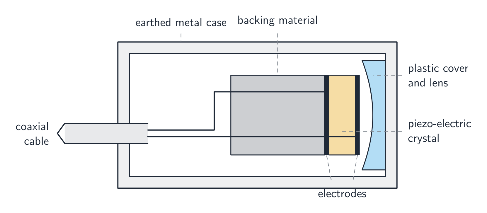
One crystal, two jobs
Put the generation of ultrasound by a transducer in order.
Difficulty 3/5 · 200 coins + a small bonus on first completion today
Detection is the reverse: the returning wave changes the crystal's shape, and a changing shape generates an e.m.f.
Pulse-echo imaging
- The four-mark outline runs: a pulse is sent in through coupling gel; at each boundary part reflects and returns; the time delay gives the depth; the intensity of the echo says what kind of boundary it was.
- Depth comes from the there-and-back trip:
- Sweeping the transducer, or using an array, builds a two-dimensional image.
- The factor of $2$ is the single commonest arithmetic slip in this topic.

One spike out, several back
Worked example: how deep is the boundary
- A pulse returns $60\ \mu\text{s}$ after it was sent, and the speed of sound in the tissue is $1500\ \text{m/s}$. Find the depth of the reflecting boundary.
- $d = \dfrac{ct}{2} = \dfrac{1500 \times 60\times10^{-6}}{2} = 0.045\ \text{m} = 4.5\ \text{cm}$.
- Halve it, because the pulse travelled there and back. Forgetting to gives $9.0\ \text{cm}$, which is a whole answer wrong for a reason worth remembering.
An echo returns after $2.0 \times 10^{-4}\ \text{s}$; the speed of sound is $1500\ \dfrac{\text{m}}{\text{s}}$. How deep is the boundary?
Difficulty 2/5 · 140 coins + a small bonus on first completion today
$d = \dfrac{ct}{2} = \dfrac{1500 \times 2.0 \times 10^{-4}}{2} = 0.15\ \text{m}$ (the pulse travels there and back).
An ultrasound pulse returns 60 microseconds after transmission, with a speed of sound of 1500 m/s. How deep is the boundary, in cm?
Difficulty 2/5 · 140 coins + a small bonus on first completion today
d = ct/2 = 4.5 cm. Halve it: the pulse travelled there AND back. Forgetting the 2 gives 9.0 cm.
Why pulses, and why megahertz
- The transducer sends a short pulse and then listens. Every echo must arrive before the next pulse leaves, or echoes from different pulses could not be told apart, and the crystal cannot transmit and receive at the same moment.
- The frequency is a compromise. A higher frequency means a shorter wavelength and finer detail, better resolution 分辨率.
- But a higher frequency is also attenuated more strongly, so a deep organ needs a lower frequency than a shallow one.
- Medical scanning uses $1$ to $15\ \text{MHz}$, giving wavelengths of a fraction of a millimetre in tissue.
A higher ultrasound frequency has which effects? Select all that apply.
Difficulty 2/5 · 140 coins + a small bonus on first completion today
Reflection is set by the impedance mismatch, not the frequency. The frequency choice is a resolution-against-penetration compromise, so a deep organ is scanned at a lower frequency.
Specific acoustic impedance
- The specific acoustic impedance 声阻抗 of a medium is the product of its density and the speed of sound in it.
- Both quantities must be named for the two marks. "How hard sound finds it to pass" scores nothing.
- Density 密度 alone does not decide it: water and soft tissue have very different compositions and nearly the same $Z$, and it is $Z$, not density, that fixes how much reflects.
- For soft tissue: $Z = 1060 \times 1540 = 1.6\times10^{6}\ \text{kg/(m}^2\text{ s)}$.
Acoustic impedance Z = density × ____.
Difficulty 1/5 · 100 coins + a small bonus on first completion today
$Z = \rho c$ — density times the speed of sound in the medium.
Match each boundary to what happens to the pulse.
Difficulty 2/5 · 140 coins + a small bonus on first completion today
Imaging needs the middle case. The gel exists to convert the first case into the third at the skin.
How much reflects at a boundary
- At a boundary between impedances $Z_1$ and $Z_2$, the intensity reflection coefficient 强度反射系数 is
- Very different impedances reflect almost everything, which is the skin-air problem.
- Very similar impedances reflect almost nothing, so the boundary is invisible.
- The useful case for imaging is in between: different enough to give an echo, similar enough that the pulse carries on to the next boundary.

The mismatch is the echo
Ultrasound scan route
Follow a pulse from transducer to echo image.
Two media with very similar acoustic impedance give almost no echo.
Difficulty 1/5 · 100 coins + a small bonus on first completion today
The reflected fraction depends on the impedance difference; similar impedances reflect very little, so the boundary is hard to see.
Worked example: why the gel is not optional
- Air has $\rho = 1.29\ \text{kg/m}^3$ and $c = 343\ \text{m/s}$. Gel has $Z = 1.50\times10^{6}$ and soft tissue $Z = 1.63\times10^{6}\ \text{kg/(m}^2\text{ s)}$. Compare the reflection at an air-tissue and a gel-tissue boundary.
- $Z_{\text{air}} = 1.29 \times 343 = 442\ \text{kg/(m}^2\text{ s)}$, which is smaller than tissue's by a factor of nearly $4000$.
- Air-tissue: $\left(\dfrac{442 - 1.63\times10^{6}}{442 + 1.63\times10^{6}}\right)^2 = 0.9989$, so $99.9\%$ reflects and only $0.1\%$ enters the body.
- Gel-tissue: $\left(\dfrac{1.50 - 1.63}{1.50 + 1.63}\right)^2 = 0.0017$, so only $0.2\%$ reflects.
- The gel replaces the air with something whose impedance nearly matches skin, so almost the whole pulse gets in, and the echoes get back out the same way.
The coupling gel between the probe and the skin:
Difficulty 1/5 · 100 coins + a small bonus on first completion today
Without it, the huge skin–air impedance difference would reflect almost all the ultrasound at the surface.
Gel has Z = 1.50e6 and soft tissue Z = 1.63e6 kg/(m^2 s). What percentage of the intensity reflects at the gel-tissue boundary?
Difficulty 2/5 · 140 coins + a small bonus on first completion today
((1.50-1.63)/(1.50+1.63))^2 = 0.0017, that is 0.17%. At an air-tissue boundary the same formula gives 99.9%, which is why the gel is not optional.
Attenuation
- Ultrasound weakens as it travels, by the same exponential law that governs X-rays and radioactive decay:
- $\mu$ is the attenuation coefficient of the medium, and a higher frequency has a larger $\mu$, which is the trade-off behind the choice of frequency.
- To find a thickness, take logs: $x = \dfrac{1}{\mu}\ln\dfrac{I_0}{I}$.
- Watch the units: $\mu$ in $\text{cm}^{-1}$ needs $x$ in cm. Mixing them is the standard error and gives an answer wrong by a factor of $100$ in the exponent.
Marks that slip away
- Halve the distance in the pulse-echo calculation. The pulse goes there and back.
- $Z = \rho c$ needs both density and speed named in the definition.
- The reflection formula uses the difference over the sum, squared. Forgetting to square it changes the answer completely.
- The gel matches impedances, it does not "help the sound travel". Say what it replaces and why.
- Higher frequency gives better resolution but more attenuation. A question about a deep organ is asking for that trade-off.
You've got it
- a piezo-electric crystal changes shape under a p.d. and generates an e.m.f. when its shape changes, so one transducer both sends and detects
- pulse-echo imaging gives depth from $d = ct/2$ and tissue type from the echo's intensity
- specific acoustic impedance is $Z = \rho c$, and $I_R/I_0 = \left(\frac{Z_1-Z_2}{Z_1+Z_2}\right)^2$, which is why an air gap reflects $99.9\%$ and the gel is essential
- ultrasound is attenuated as $I = I_0 e^{-\mu x}$, and a higher frequency trades better resolution for worse penetration