- understand that a piezo-electric crystal changes shape when a p.d. is applied across it and that the crystal generates an e.m.f. when its shape changes
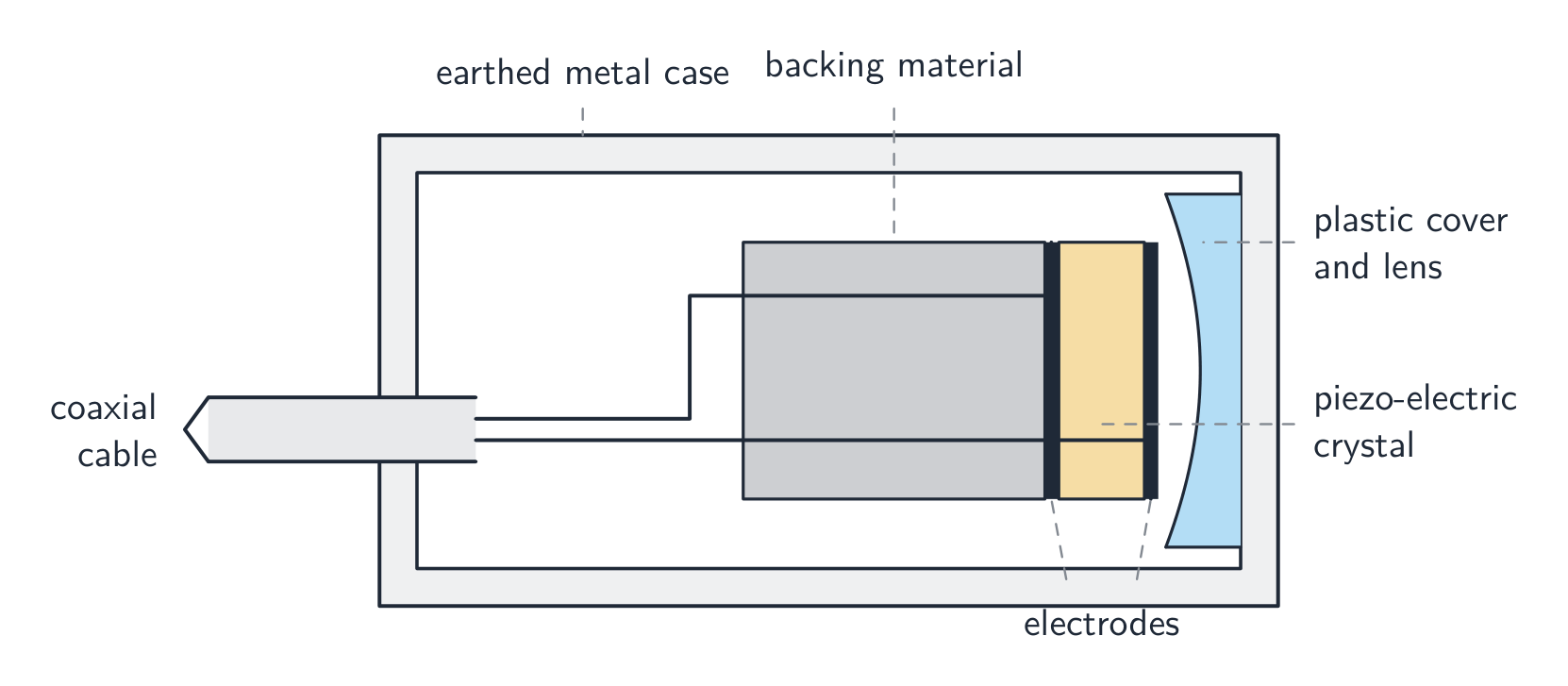
- understand how ultrasound waves are generated and detected by a piezoelectric transducer
- understand how the reflection of pulses of ultrasound at boundaries between tissues can be used to obtain diagnostic information about internal structures
- define the specific acoustic impedance of a medium as $Z = \rho c$, where $c$ is the speed of sound in the medium
- use $I_{\text{R}} / I_0 = (Z_1 - Z_2)^2 / (Z_1 + Z_2)^2$ for the intensity reflection coefficient of a boundary between two media
- recall and use $I = I_0 e^{-\mu x}$ for the attenuation of ultrasound in matter
Medical physics
A-Level Physics · Topic 24
 17:14
17:14
Ultrasound & Medical Imaging
The very first photograph of your life was probably taken with sound. Before you were born, a doctor pressed a small probe to your mother's skin, and out of…
English narration · English + 中文 subtitles burned in
24.1
Ultrasound

Syllabus
Source: Cambridge International syllabus
Piezo-electric effect
A piezo-electric 压电 crystal changes shape a little when a p.d. is put across it, and the reverse: it makes an electromotive force 电动势 (e.m.f.) across itself when its shape is changed. Quartz and PZT are common examples. Two linked effects:
- apply a p.d. → the crystal changes shape (used to make vibrations).
- change the shape (a wave squeezes it) → an e.m.f. appears (used to detect vibrations).
Piezo-electric transducer
A transducer 换能器 uses this effect to both make and detect ultrasound 超声波.
- an alternating p.d. (a few MHz) makes the crystal vibrate at the same frequency, sending out longitudinal 纵波 waves above $20\ \text{kHz}$ ($1$–$10\ \text{MHz}$ for medical imaging).
- the same crystal then detects: returning ultrasound makes it vibrate and produce an e.m.f.
Generating ultrasound (three marks). (1) An alternating p.d. is applied across the crystal. (2) The crystal changes shape, expanding and contracting at the frequency of the p.d., so it vibrates. (3) The p.d. is chosen at the crystal's resonant frequency, so the vibration is large and the crystal's faces push on the tissue, sending out a longitudinal wave of that frequency. Detecting ultrasound (two marks). The returning wave's pressure variations change the shape of the crystal; a changing shape generates an e.m.f. across it, alternating at the frequency of the wave, which is amplified and recorded. The same crystal is used for both, switched between transmitting and receiving.
So one transducer is both emitter and detector, switching between sending and listening.
Pulse-echo imaging
To see inside the body (pulse-echo 脉冲回波 imaging):
- the transducer sends a short pulse into the body.
- at each tissue boundary, part of the pulse is reflected and part goes on.
- the transducer detects each reflected pulse.
- the time delay gives the depth: $d = c t / 2$ (there and back). The echo's amplitude gives the strength of the reflection.
Worked example. An ultrasound pulse returns to the transducer $60\ \mu\text{s}$ after it was sent. The speed of sound in the tissue is $1500\ \text{m s}^{-1}$. Find the depth of the reflecting boundary.
The pulse travels there and back, so $d = \dfrac{ct}{2}$:
$$d = \frac{1500 \times 60 \times 10^{-6}}{2} = 0.045\ \text{m}\ (= 4.5\ \text{cm}).$$
- sweeping across the body builds a 2-D image.

A coupling gel 耦合剂 is put between the transducer and the skin to push out the air; without it almost all the ultrasound would reflect at the skin–air boundary and never enter the body.

"Outline the use of ultrasound to obtain diagnostic information" (four marks). (1) A pulse of ultrasound is sent into the body by the transducer, which is coupled to the skin with gel. (2) At each boundary between tissues part of the pulse is reflected; the reflected pulse returns to the transducer and is detected. (3) The time delay between emission and echo, with the speed of sound, gives the depth of the boundary ($d = ct/2$). (4) The intensity of the echo, set by the change in acoustic impedance at the boundary, shows what kind of boundary it is. Sweeping the transducer, or using an array, builds up an image.
Why pulses, and why megahertz. The transducer sends a short pulse and then listens: the echo from every boundary must arrive before the next pulse leaves, otherwise echoes from different pulses could not be told apart, and the crystal cannot transmit and receive at the same moment. The frequency is a compromise: a higher frequency means a shorter wavelength and so finer detail (resolution 分辨率), but it is attenuated more strongly, so a deep organ needs a lower frequency than a shallow one. Typical medical scanning uses $1$–$15\ \text{MHz}$, wavelengths of a fraction of a millimetre in tissue.
Specific acoustic impedance
The specific acoustic impedance 声阻抗 of a medium is
$$Z = \rho c,$$
where $\rho$ is the density 密度 and $c$ the speed of sound. Unit: $\text{kg m}^{-2}\ \text{s}^{-1}$. Bone has large $Z$; air has small $Z$; soft tissue is in between.
Worked example. Find the specific acoustic impedance of soft tissue. (Density $1060\ \text{kg m}^{-3}$, speed of sound $1540\ \text{m s}^{-1}$.)
$$Z = \rho c = 1060 \times 1540 \approx 1.6 \times 10^{6}\ \text{kg m}^{-2}\ \text{s}^{-1}.$$
The definition (two marks). The specific acoustic impedance of a medium is the product of its density and the speed of sound in it. Both quantities must be named; "how hard sound finds it to pass" scores nothing. Because $Z$ combines density and speed, two media with very different densities can still have similar $Z$ (water and soft tissue), and it is $Z$, not density alone, that decides how much sound reflects.
Reflection at a boundary
At a boundary between media of impedance $Z_{1}$ and $Z_{2}$, the intensity reflection coefficient 强度反射系数 (fraction reflected) is
$$\frac{I_{\text{R}}}{I_{0}} = \left(\frac{Z_{1} - Z_{2}}{Z_{1} + Z_{2}}\right)^{2}.$$
- very different impedances: almost all is reflected (skin/air — hence the gel).
- very similar impedances: almost nothing is reflected, so the boundary cannot be seen.
- best for imaging: different enough to give an echo, but not so different that nothing passes on.

Worked example (why gel). Air: density $1.29\ \text{kg m}^{-3}$, speed of sound $343\ \text{m s}^{-1}$. Gel: $Z = 1.50 \times 10^{6}\ \text{kg m}^{-2}\ \text{s}^{-1}$. Soft tissue: $Z = 1.63 \times 10^{6}\ \text{kg m}^{-2}\ \text{s}^{-1}$. Find the intensity reflection coefficient at an air–tissue boundary and at a gel–tissue boundary.
$Z_{\text{air}} = 1.29 \times 343 = 442\ \text{kg m}^{-2}\ \text{s}^{-1}$. Air–tissue: $\left(\dfrac{442 - 1.63 \times 10^{6}}{442 + 1.63 \times 10^{6}}\right)^{2} = 0.9989$: $99.9\%$ of the intensity reflects and only $0.1\%$ enters the body. Gel–tissue: $\left(\dfrac{1.50 - 1.63}{1.50 + 1.63}\right)^{2} = 0.0017$: only $0.2\%$ reflects. So the gel, whose impedance is close to that of skin, replaces the air layer that would otherwise reflect nearly everything, and almost all of the pulse enters the body (and the echoes can get back out again by the same route).
Worked example. Water: density $1000\ \text{kg m}^{-3}$, speed $1480\ \text{m s}^{-1}$. Glass: density $2500\ \text{kg m}^{-3}$, speed $5600\ \text{m s}^{-1}$. What fraction of the intensity is reflected at a water–glass boundary?
$Z_{\text{water}} = 1.48 \times 10^{6}$, $Z_{\text{glass}} = 1.40 \times 10^{7}\ \text{kg m}^{-2}\ \text{s}^{-1}$. Coefficient $= \left(\dfrac{1.48 - 14.0}{1.48 + 14.0}\right)^{2} = 0.65$: two-thirds reflected, one-third transmitted. Working in units of $10^{6}$ keeps the arithmetic clean, since the ratio is what matters. A steel implant in tissue ($Z_{\text{steel}} = 4.7 \times 10^{7}$) gives $0.87$: nearly all the pulse reflects from its surface, so nothing behind it can be imaged, but the implant itself shows up very brightly.
Attenuation
As ultrasound goes through tissue, its intensity falls with distance:
$$I = I_{0} e^{-\mu x},$$
where $\mu$ is the attenuation coefficient 衰减系数 (unit $\text{m}^{-1}$). The same form applies to X-rays.

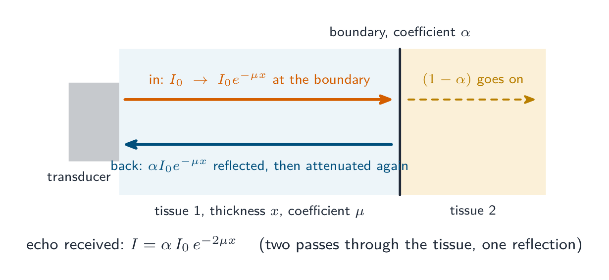
Worked example (an echo's intensity). A pulse of intensity $I_{0}$ passes through $4.0\ \text{cm}$ of tissue with attenuation coefficient $0.50\ \text{cm}^{-1}$ to a boundary whose intensity reflection coefficient is $0.010$. Find the intensity of the echo returning to the transducer, as a fraction of $I_{0}$.
To the boundary: $e^{-\mu x} = e^{-0.50 \times 4.0} = 0.135$. Reflected: $\times 0.010$. Back through the same $4.0\ \text{cm}$: $\times 0.135$ again. Echo: $I/I_{0} = 0.135 \times 0.010 \times 0.135 = 1.8 \times 10^{-4}$, about $0.02\%$. Two things the examiner checks: the tissue is crossed twice, and the reflection coefficient multiplies the intensity at the boundary, not $I_{0}$. This is why echoes from deep boundaries are amplified more than shallow ones before display.
Explore
Ultrasound scan route
Follow a pulse from transducer to echo image.
Vocabulary
Train
| English |
|---|
| ultrasound/ˌʊltrəˈsaʊnd/ |
| piezo-electric/piˌeɪtsəʊ ɪˈlektrɪk/ |
| electromotive force/ɪˌlektrəʊˈməʊtɪv fɔːs/ |
| transducer/trænsˈdjuːsə/ |
| longitudinal/ˌlɒŋɡɪˈtjuːdɪnl/ |
| pulse-echo/pʌls ˈekəʊ/ |
| coupling gel/ˈkʌplɪŋ dʒel/ |
| resolution/ˌrezəˈluːʃn/ |
| specific acoustic impedance/spəˈsɪfɪk əˈkuːstɪk ɪmˈpiːdəns/ |
| density/ˈdensɪti/ |
| intensity reflection coefficient/ɪnˈtensɪti rɪˈflekʃn ˌkəʊɪˈfɪʃənt/ |
| attenuation coefficient/əˌtenjuːˈeɪʃn ˌkəʊɪˈfɪʃənt/ |
24.2
X-rays

Syllabus
- explain that X-rays are produced by electron bombardment of a metal target and calculate the minimum wavelength of X-rays produced from the accelerating p.d.
- understand the use of X-rays in imaging internal body structures, including an understanding of the term contrast in X-ray imaging
- recall and use $I = I_0 e^{-\mu x}$ for the attenuation of X-rays in matter
- understand that computed tomography (CT) scanning produces a 3D image of an internal structure by first combining multiple X-ray images taken in the same section from different angles to obtain a 2D image of the section, then repeating this process along an axis and combining 2D images of multiple sections
Source: Cambridge International syllabus
Production
X-rays come from an X-ray tube X射线管:
- a heated cathode 阴极 emits electrons by thermionic emission 热电子发射.
- a high p.d. (tens to hundreds of kV) accelerates the electrons across a vacuum 真空 to a metal target 靶 (the anode 阳极, often tungsten 钨).
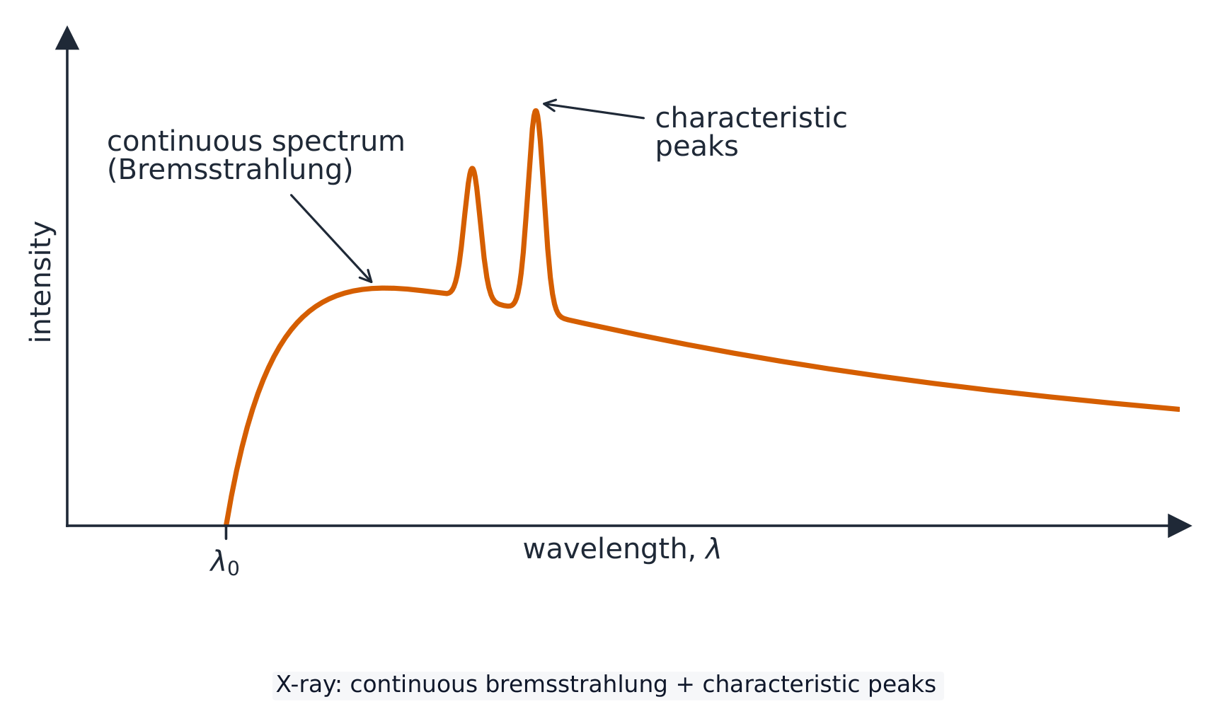
- the electrons hit the target and slow sharply. Most of their kinetic energy 动能 becomes heat; a small part is emitted as X-ray photons 光子 (Bremsstrahlung 轫致辐射, "braking radiation"). Some electrons knock out inner electrons of the metal atoms, and the refilling emits characteristic 特征 X-ray lines.
"Explain how X-rays are produced" (four marks). (1) Electrons are emitted from a heated filament (cathode) by thermionic emission. (2) They are accelerated through a high potential difference (tens of kV) across an evacuated tube towards a metal target (the anode). (3) They decelerate rapidly when they strike the target, and (4) the kinetic energy lost is emitted as X-ray photons (with most of it becoming heat in the target, which is why the anode is cooled or rotated).
Controlling the beam. The intensity (energy per unit area per second) is set by the number of electrons hitting the target per second, so it is controlled by the filament current (a hotter filament emits more electrons); the hardness 硬度, meaning the penetrating power, is set by the photon energies, so it is controlled by the accelerating p.d.: a larger p.d. gives higher-energy, shorter-wavelength, more penetrating X-rays. A metal filter removes the softest X-rays, which would be absorbed in the patient's skin without reaching the detector.
Worked example (heating of the target). In a tube run at $75\ \text{kV}$ the electron beam current is $30\ \text{mA}$ and $99\%$ of the electrons' energy becomes heat in a tungsten target of mass $15\ \text{g}$ (specific heat capacity $134\ \text{J kg}^{-1}\ \text{K}^{-1}$). Find the rate of temperature rise if no heat were lost.
Power delivered $= IV = 0.030 \times 75\,000 = 2250\ \text{W}$; heating power $= 0.99 \times 2250 = 2230\ \text{W}$. Then $mc\,\Delta\theta/\Delta t = 2230$, so $\Delta\theta/\Delta t = 2230/(0.015 \times 134) = 1.1 \times 10^{3}\ \text{K s}^{-1}$. A target would melt within seconds, which is why exposures are short and anodes rotate to spread the heat.
Minimum wavelength
The most energy one X-ray photon can have is the full kinetic energy of one accelerated electron, lost in a single event. For accelerating p.d. $V$, the KE is $eV$, so
$$h f_{\text{max}} = e V, \qquad \lambda_{\text{min}} = \frac{h c}{e V}.$$
This is the short-wavelength cut-off. The continuous Bremsstrahlung spectrum tails off above $\lambda_{\text{min}}$, with sharp characteristic peaks set by the target metal.
Worked example (minimum wavelength). Electrons are accelerated through $75\ \text{kV}$. Find the maximum photon energy and the minimum wavelength of the X-rays.
Maximum energy $= eV = 75\ \text{keV} = 75\,000 \times 1.60 \times 10^{-19} = 1.2 \times 10^{-14}\ \text{J}$ (or $0.075\ \text{MeV}$). Then $\lambda_{\text{min}} = hc/(eV) = (6.63 \times 10^{-34})(3.00 \times 10^{8})/(1.2 \times 10^{-14}) = 1.7 \times 10^{-11}\ \text{m}$. The maximum photon momentum is $p = E/c = 4.0 \times 10^{-23}\ \text{N s}$. Doubling the p.d. halves the minimum wavelength.
Explaining the shape of the spectrum. The spectrum is continuous because each electron may lose any fraction of its energy in one or several decelerations, so photons of every energy up to the maximum are produced. It has a sharp cut-off at $\lambda_{\text{min}}$ because a photon cannot carry more energy than one electron has, $eV$, and that happens only when an electron loses all its energy in a single event. The peaks are characteristic of the target metal: an incoming electron knocks an inner electron out of a target atom, and an outer electron falls into the vacancy, emitting a photon whose energy is the difference between the two levels (Topic 22).
Imaging with X-rays
X-rays pass through the patient onto a detector. Tissues that attenuate 衰减 more (bone, high $Z$) cast a stronger shadow and look lighter; tissues that attenuate less (soft tissue, lung) look darker.
The contrast 对比度 is the difference in attenuation between tissues. A contrast medium 造影剂 (e.g. a barium meal) can be given to make soft tissues stand out.

Sharpness and contrast. Sharpness 清晰度 is how well defined the edges of structures are in the image: a small X-ray source (a small spot on the target), a stationary patient and a detector close to the patient all improve it. Contrast is the difference in degree of blackening (or brightness) between neighbouring regions of the image, produced by a difference in attenuation. The two are independent: an image can be sharp but have poor contrast, or the reverse.
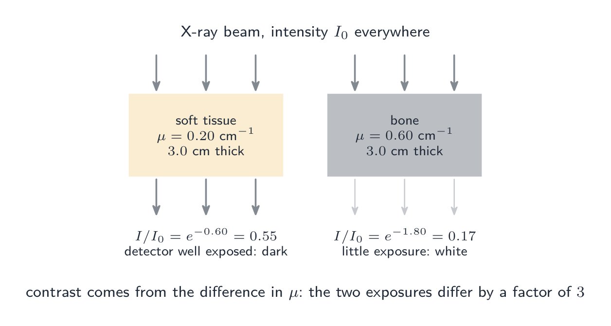
"Explain why X-ray images of internal structures have good contrast." Bone and soft tissue have very different attenuation coefficients, so equal thicknesses transmit very different intensities and the detector is exposed very differently behind each. Where two soft tissues have similar coefficients (the stomach and its surroundings), a contrast medium with a high attenuation coefficient, such as barium, is swallowed or injected to outline one of them. Using a lower p.d. (softer X-rays) also increases contrast, at the cost of a larger dose.
Worked example. X-rays pass through $3.0\ \text{cm}$ of soft tissue ($\mu = 0.20\ \text{cm}^{-1}$) in one region of the body and $3.0\ \text{cm}$ of bone ($\mu = 0.60\ \text{cm}^{-1}$) in another. Compare the transmitted intensities.
Soft tissue: $I/I_{0} = e^{-0.20 \times 3.0} = e^{-0.60} = 0.55$. Bone: $e^{-0.60 \times 3.0} = e^{-1.80} = 0.17$. The soft-tissue region receives more than three times the exposure of the bone region, so the bone appears white and the tissue dark. The ratio of exposures, $e^{(\mu_{\text{bone}} - \mu_{\text{tissue}})x}$, grows with thickness: contrast improves for thicker structures, but so does the total attenuation.
Attenuation law
$$I = I_{0} e^{-\mu x}.$$
Higher-energy X-rays penetrate further (smaller $\mu$); bone has a much larger $\mu$ than soft tissue. To find the thickness for a given fraction, take logs: $x = \dfrac{1}{\mu} \ln\dfrac{I_{0}}{I}$. The half-value thickness 半值厚度 $x_{1/2} = \ln 2 / \mu$ halves the intensity (like half-life in decay).
Worked example. X-rays pass through $3.0\ \text{cm}$ of tissue with attenuation coefficient $\mu = 40\ \text{m}^{-1}$. Find the fraction of the intensity that gets through.
$$\frac{I}{I_{0}} = e^{-\mu x} = e^{-40 \times 0.030} = e^{-1.2} \approx 0.30.$$
Worked example (percentage absorbed). X-rays pass through $2.8\ \text{cm}$ of a medium with attenuation coefficient $1.4\ \text{cm}^{-1}$. What percentage of the X-ray energy is absorbed?
Transmitted fraction $e^{-1.4 \times 2.8} = e^{-3.92} = 0.020$, so $98\%$ is absorbed. The question asks for what is absorbed, not what gets through: read it twice. The half-value thickness here is $\ln 2/\mu = 0.50\ \text{cm}$, so $2.8\ \text{cm}$ is $5.6$ half-value thicknesses and $(1/2)^{5.6} = 0.02$ checks the answer.
Worked example (two layers). A beam of intensity $I_{0}$ passes through $2.0\ \text{cm}$ of material P ($\mu = 0.35\ \text{cm}^{-1}$) and then $1.5\ \text{cm}$ of material Q ($\mu = 0.90\ \text{cm}^{-1}$). Find the transmitted intensity.
The exponentials multiply: $I = I_{0}\, e^{-0.35 \times 2.0}\, e^{-0.90 \times 1.5} = I_{0}\, e^{-(0.70 + 1.35)} = I_{0}\, e^{-2.05} = 0.13\, I_{0}$. Add the exponents ($\mu_{1}x_{1} + \mu_{2}x_{2}$); never add the thicknesses or the coefficients. The order of the layers makes no difference to the total transmitted intensity.
Computed tomography (CT)
A computed tomography 计算机断层扫描 (CT) scan builds a 3-D image:
- the tube and detectors rotate around the patient, taking many images of one thin slice from different angles.
- a computer combines these into a 2-D cross-section of the slice.
- the patient is moved along, and the next slice is imaged.
- the slices are stacked into a 3-D image.
CT shows far more than a single X-ray, because overlapping soft tissues are separated by the many-angle reconstruction.

"Explain how CT scanning produces a three-dimensional image" (five marks). (1) The X-ray tube (and a ring of detectors) rotates around the patient, so that (2) many X-ray images of one slice (section) are taken from different angles. (3) A computer combines them to produce a two-dimensional image of that slice. (4) The patient is moved and the process is repeated for successive slices. (5) The slices are combined to build a 3-D image, which can be rotated and viewed from any angle. Purpose: to image a section of the body without the overlapping of structures that a single X-ray suffers, revealing structures that would be hidden and their positions in depth. The price is a much larger dose than a single X-ray.
Explore
X-ray production route
Follow electrons from cathode to X-ray photons.
Vocabulary
Train
| English |
|---|
| X-ray tube/eks reɪ tjuːb/ |
| cathode/ˈkæθəʊd/ |
| thermionic emission/ˌθɜːmɪˈɒnɪk ɪˈmɪʃn/ |
| vacuum/ˈvækjuːm/ |
| target/ˈtɑːɡɪt/ |
| anode/ˈænəʊd/ |
| tungsten/ˈtʌŋstn/ |
| kinetic energy/kɪˈnetɪk ˈenədʒi/ |
| photon/ˈfəʊtɒn/ |
| Bremsstrahlung/ˈbremʃtrɑːlʊŋ/ |
| characteristic/ˌkærɪktəˈrɪstɪk/ |
| hardness/ˈhɑːdnəs/ |
| attenuate/əˈtenjuːeɪt/ |
| contrast/ˈkɒntræst/ |
| contrast medium/ˈkɒntræst ˈmiːdɪəm/ |
| sharpness/ˈʃɑːpnəs/ |
| half-value thickness/hɑːf ˈvæljuː ˈθɪknəs/ |
| computed tomography/kəmˈpjuːtɪd təˈmɒɡrəfi/ |
24.3
PET scanning

Syllabus
- understand that a tracer is a substance containing radioactive nuclei that can be introduced into the body and is then absorbed by the tissue being studied
- recall that a tracer that decays by $\beta^+$ decay is used in positron emission tomography (PET scanning)
- understand that annihilation occurs when a particle interacts with its antiparticle and that mass–energy and momentum are conserved in the process
- explain that, in PET scanning, positrons emitted by the decay of the tracer annihilate when they interact with electrons in the tissue, producing a pair of gamma-ray photons travelling in opposite directions
- calculate the energy of the gamma-ray photons emitted during the annihilation of an electron-positron pair
- understand that the gamma-ray photons from an annihilation event travel outside the body and can be detected, and an image of the tracer concentration in the tissue can be created by processing the arrival times of the gamma-ray photons
Source: Cambridge International syllabus
Tracer
A tracer 示踪剂 is a substance with radioactive nuclei put into the body. It is taken up more by the tissue being studied (e.g. a tumour takes up more glucose-tagged tracer due to its high metabolism 代谢). Its decay is detected from outside.
In positron emission tomography 正电子发射断层扫描 (PET), the tracer is a $\beta^{+}$ emitter — it gives out a positron 正电子. A common one is fluorine-18 on a glucose analogue (FDG).
"Explain what is meant by a tracer and how it is used" (three marks). A tracer is a substance containing radioactive nuclei (for PET, a $\beta^{+}$ emitter such as fluorine-18) that is introduced into the body (injected or swallowed), usually bound to a molecule such as glucose, and is absorbed by the tissue being studied; the radiation it emits leaves the body and is detected, so its distribution shows where that tissue is active. The nuclide is chosen with a short half-life, so the activity is high during the scan and the patient's dose afterwards is small, but not so short that it decays before it reaches the tissue: fluorine-18 ($110$ minutes) and oxygen-15 ($2$ minutes, made and used on the spot) are typical.
Annihilation
When a particle meets its antiparticle 反粒子 they annihilate 湮灭: their mass turns into electromagnetic energy. In PET:
- a positron travels a few mm before meeting an electron 电子.
- they annihilate. Energy and momentum 动量 are conserved.
- since the total momentum is about zero, two photons are produced going in opposite directions, each $511\ \text{keV}$ ($= m_{e} c^{2}$).
Energy of the annihilation photons
By energy conservation, the total photon energy equals the pair's rest energy 能量:
$$2 h f = 2 m_{e} c^{2}, \qquad h f = m_{e} c^{2}.$$
Each photon has $h f = m_{e} c^{2} \approx 8.2 \times 10^{-14}\ \text{J} \approx 0.51\ \text{MeV}$, with $\lambda \approx 2.4 \times 10^{-12}\ \text{m}$.
Explaining the annihilation (four marks). (1) The positron emitted by the tracer travels a short distance and meets an electron in the tissue. (2) The pair annihilates: their mass is converted to energy, (3) emitted as two gamma-ray photons, since (4) momentum must be conserved: the pair had almost no momentum, so the two photons must travel in opposite directions with equal momenta. Each photon carries the rest energy of one particle, $0.51\ \text{MeV}$; a single photon could not conserve momentum.
Worked example. Find the total energy released when a positron and an electron, each moving slowly, annihilate, and the wavelength of each photon.
$E = 2m_{\text{e}}c^{2} = 2 \times 9.11 \times 10^{-31} \times (3.00 \times 10^{8})^{2} = 1.64 \times 10^{-13}\ \text{J}$ ($1.02\ \text{MeV}$), shared equally: $8.2 \times 10^{-14}\ \text{J}$ each. $\lambda = hc/E = (6.63 \times 10^{-34})(3.00 \times 10^{8})/(8.2 \times 10^{-14}) = 2.4 \times 10^{-12}\ \text{m}$, a gamma ray. If instead the electron and positron each move at $4.9 \times 10^{7}\ \text{m s}^{-1}$ in opposite directions, each has kinetic energy $\tfrac{1}{2}mv^{2} = 1.1 \times 10^{-15}\ \text{J}$, about $1\%$ of its rest energy, and the photons carry $8.3 \times 10^{-14}\ \text{J}$ each: the rest energy dominates, which is why the annihilation photons always have very nearly the same energy.
Reconstructing the image
The two photons leave the body in opposite directions and hit detector rings around the patient. Recording the two simultaneous arrivals (a "coincidence") fixes the line the annihilation happened on. Many coincidences from many angles let the computer build a 3-D map of the tracer — showing tissues with high metabolic activity. Comparing the two arrival times can refine the position along that line (time-of-flight PET).

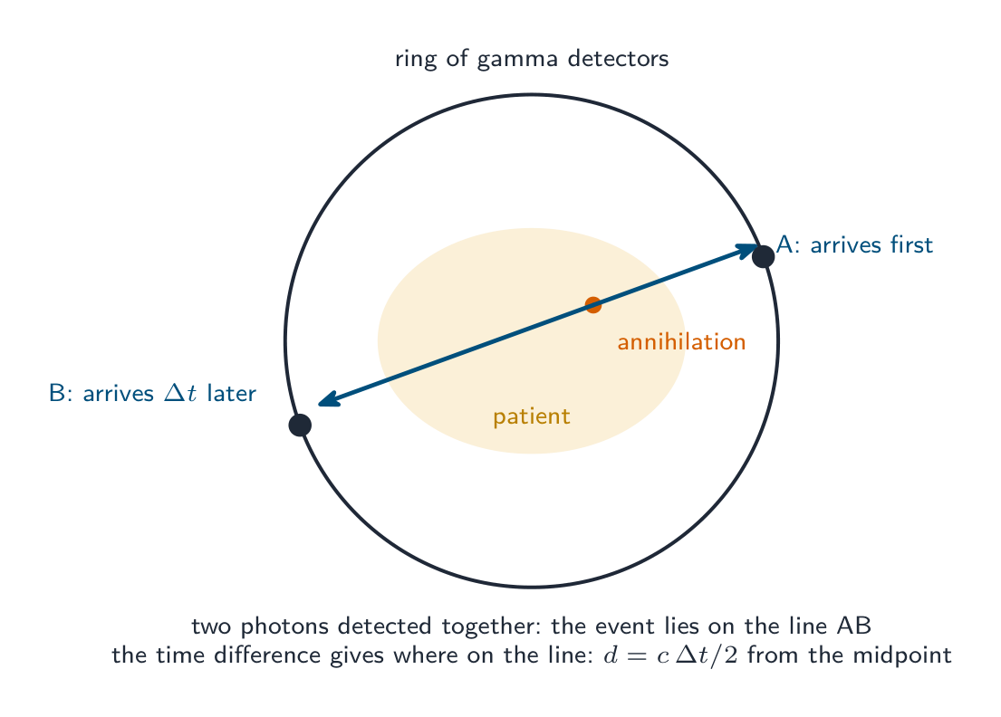
"Explain how the gamma photons are used to form an image" (four marks). (1) The two photons leave the body in opposite directions and are detected by the ring of detectors. (2) Two photons arriving at (almost) the same time are taken to come from one annihilation, which therefore lies on the line joining the two detectors. (3) The small difference in arrival times gives the position along that line ($d = c\,\Delta t/2$ from the midpoint; a $1\ \text{ns}$ difference is $15\ \text{cm}$, so the timing must be very precise). (4) A computer collects many such events and maps the concentration of tracer in the tissue, producing an image; a higher concentration means more active tissue, such as a tumour. Photons that arrive singly (their partner absorbed in the body) are rejected.
Explore
PET scan route
Follow positron emission to a ring of detected photons.
Vocabulary
Train
| English |
|---|
| electron/ɪˈlektrɒn/ |
| energy/ˈenədʒi/ |
| momentum/məʊˈmentəm/ |
| tracer/ˈtreɪsə/ |
| metabolism/məˈtæbəlɪzəm/ |
| positron emission tomography/ˈpɒzɪtrɒn ɪˈmɪʃn təˈmɒɡrəfi/ |
| positron/ˈpɒzɪtrɒn/ |
| antiparticle/ˌæntɪˈpɑːtɪkl/ |
| annihilate/əˈnaɪəleɪt/ |
24.3
Definitions the examiner accepts
A definition question is marked against fixed wording. Learn these exactly, and give one answer only.
| Term | Definition |
|---|---|
| piezo-electric effect | a crystal changes shape when a p.d. is applied across it, and generates an e.m.f. when its shape is changed |
| specific acoustic impedance | the product of the density of a medium and the speed of sound in it, $Z = \rho c$ |
| intensity reflection coefficient | the fraction of the incident intensity reflected at a boundary, $(Z_{1} - Z_{2})^{2}/(Z_{1} + Z_{2})^{2}$ |
| attenuation coefficient | the constant $\mu$ in $I = I_{0}e^{-\mu x}$; the larger it is, the faster the intensity falls with thickness |
| hardness (of X-rays) | the penetrating power of the beam, set by the photon energies (the accelerating p.d.) |
| contrast | the difference in degree of blackening between neighbouring regions of an image |
| sharpness | how well defined the edges of structures are in an image |
| CT scanning | X-ray images of one section taken from many angles are combined by computer into a 2-D image of the slice; successive slices give a 3-D image |
| tracer | a substance containing radioactive nuclei that is introduced into the body and absorbed by the tissue under study |
| annihilation | a particle and its antiparticle interact and their mass is converted to energy (photons), with mass–energy and momentum conserved |
24.3
Exam tips
- Ultrasound: pulse in, echo back; depth $= ct/2$ (there and back); $Z = \rho c$; reflection coefficient from the two impedances; gel matches impedance to the skin. Attenuation is exponential and an echo crosses the tissue twice.
- X-rays: production is heated filament, high p.d., sudden deceleration at the target; intensity by filament current, hardness by p.d.; $\lambda_{\text{min}} = hc/(eV)$; $I = I_{0}e^{-\mu x}$ with exponents added for layers; contrast is a difference in $\mu$.
- CT: many angles, one slice, computer, successive slices, 3-D. Say all five.
- PET: $\beta^{+}$ tracer, annihilation with an electron, two $0.51\ \text{MeV}$ photons in opposite directions (momentum), coincidence gives the line, timing gives the position.
- Know the three "explain" answers word for word: how the crystal generates and detects, how X-rays are produced, how the photons locate the tracer.
- Write units with every impedance ($\text{kg m}^{-2}\ \text{s}^{-1}$) and coefficient ($\text{cm}^{-1}$ or $\text{m}^{-1}$), and convert $\text{cm}$ to $\text{m}$ only if $\mu$ is in $\text{m}^{-1}$.
Common mistakes
- Forgetting the factor of 2 in the depth of a reflecting boundary, or the second pass through the tissue in an echo's intensity.
- Defining acoustic impedance as "the resistance to sound" instead of density times speed of sound.
- Quoting the reflection coefficient for gel–skin as "zero"; it is small, and that is the point.
- Saying the p.d. controls the intensity of the X-ray beam, or that the filament current controls the hardness; it is the other way round.
- Explaining the minimum wavelength without saying that one electron gives all its kinetic energy to a single photon.
- Adding thicknesses of different materials before applying $e^{-\mu x}$; add the products $\mu x$ instead.
- Describing CT as "an X-ray from several angles" without the computer reconstruction, the slice, or the successive slices.
- Giving one photon for annihilation, or two photons in the same direction; momentum conservation demands two in opposite directions.
- Confusing the tracer's half-life reasoning: short so that the dose is small, but long enough to reach the tissue and be scanned.
Interactive lessons on this topic
Work through it step by step, with instant-check exercises.