Skip to content · ⁨ข้ามไปยังเนื้อหา⁩

Transport in mammals · ⁨การขนส่งในสัตว์เลี้ยงลูกด้วยนม⁩

A-Level Biology · ⁨Biology A-Level⁩ · Topic 8 · ⁨หัวข้อ 8⁩

Video lesson for this topic · ⁨บทเรียนวิดีโอสำหรับหัวข้อนี้⁩ Open the video page · ⁨เปิดหน้าวิดีโอ⁩
9:17

ระบบไหลเวียนเลือด

หัวใจของคุณเต้นประมาณแสนครั้งต่อวันและไม่เคยหยุดพัก แต่ส่วนที่ฉลาดคือ มันไม่ใช่วงจรเดียว มันคือสอง วงเลือดออกจาก…

English narration · English + 中文 subtitles burned in · ⁨การบรรยายภาษาอังกฤษ · คำบรรยายภาษาอังกฤษ + 中文 ลอยตัวบนภาพ⁩

8.1

The circulatory system · ⁨ระบบไหลเวียนเลือด⁩

Syllabus · ⁨หลักสูตร⁩
English
  1. state that the mammalian circulatory system is a closed double circulation consisting of a heart, blood and blood vessels including arteries, arterioles, capillaries, venules and veins
  2. describe the functions of the main blood vessels of the pulmonary and systemic circulations, limited to pulmonary artery, pulmonary vein, aorta and vena cava
  3. recognise arteries, veins and capillaries from microscope slides, photomicrographs and electron micrographs and make plan diagrams showing the structure of arteries and veins in transverse section (TS) and longitudinal section (LS)
  4. explain how the structure of muscular arteries, elastic arteries, veins and capillaries are each related to their functions
  5. recognise and draw red blood cells, monocytes, neutrophils and lymphocytes from microscope slides, photomicrographs and electron micrographs
  6. state that water is the main component of blood and tissue fluid and relate the properties of water to its role in transport in mammals, limited to solvent action and high specific heat capacity
  7. state the functions of tissue fluid and describe the formation of tissue fluid in a capillary network
ไทย
  1. ระบุว่าระบบไหลเวียนเลือดของสัตว์เลี้ยงลูกด้วยนมเป็นระบบไหลเวียนแบบ ปิด (closed double circulation) ประกอบด้วยหัวใจ เลือด และหลอดเลือด รวมถึง หลอดเลือดแดง (arteries), หลอดเลือดฝอยเล็ก (arterioles), หลอดเลือดฝอย (capillaries), หลอดเลือดฝอยใหญ่ (venules) และ หลอดเลือดดำ (veins)
  2. อธิบายหน้าที่ของหลอดเลือดหลักของการไหลเวียนเลือด ปอด (pulmonary) และการไหลเวียนเลือด ทั่วร่างกาย (systemic) โดยจำกัดอยู่ที่ หลอดเลือดแดงปอด (pulmonary artery), หลอดเลือดดำปอด (pulmonary vein), หลอดเลือดเอออร์ตา (aorta) และ หลอดเลือดดำใหญ่ (vena cava)
  3. จำแนกหลอดเลือดแดง หลอดเลือดดำ และหลอดเลือดฝอยได้จากสไลด์กล้องจุลทรรศน์ ภาพถ่ายภายใต้กล้องจุลทรรศน์ และภาพจากกล้องจุลทรรศน์อิเล็กตรอน และสร้างแผนภาพแสดงโครงสร้างของหลอดเลือดแดงและหลอดเลือดดำในส่วนขวาง (TS) และส่วนยาว (LS)
  4. อธิบายว่าโครงสร้างของหลอดเลือดแดงกล้ามเนื้อ หลอดเลือดยืดหยุ่น หลอดเลือดดำ และหลอดเลือดฝอย มีความสัมพันธ์กับหน้าที่ของแต่ละชนิดอย่างไร
  5. จำแนกและวาด เม็ดเลือดแดง, โมโนไซต์ (monocytes), นิวโทรฟิล (neutrophils) และ ลิมโฟไซต์ (lymphocytes) จากสไลด์กล้องจุลทรรศน์ ภาพถ่ายภายใต้กล้องจุลทรรศน์ และภาพจากกล้องจุลทรรศน์อิเล็กตรอน
  6. ระบุว่าน้ำเป็นองค์ประกอบหลักของเลือดและของเหลวในเนื้อเยื่อ และเชื่อมโยงคุณสมบัติของน้ำเข้ากับบทบาทในการขนส่งในสัตว์เลี้ยงลูกด้วยนม โดยจำกัดเฉพาะ作用การละลาย (solvent action) และความจุความร้อนจำเพาะสูง (high specific heat capacity)
  7. ระบุหน้าที่ของ ของเหลวในเนื้อเยื่อ (tissue fluid) และอธิบายการสร้างของเหลวในเนื้อเยื่อใน เครือข่ายหลอดเลือดฝอย (capillary network)

Source: Cambridge International syllabus · ⁨แหล่งที่มา: หลักสูตร Cambridge International⁩

English

Mammals have a closed double circulation. "Closed" means the blood stays inside blood vessels 血管 the whole time. "Double" means the blood passes through the heart 心脏 twice for each full trip around the body. This gives two linked loops, so we call it a double circulation 双循环 and the whole thing a circulatory system 循环系统:

  • the pulmonary circulation 肺循环 carries blood from the heart to the lungs and back.
  • the systemic circulation 体循环 carries blood from the heart to the rest of the body and back.

Blood travels in this order: arteries 动脉 → arterioles 小动脉 → capillaries 毛细血管 → venules 小静脉 → veins 静脉.

The main vessels are:

  • the pulmonary artery 肺动脉 — carries blood low in oxygen 氧气 from the heart to the lungs.
  • the pulmonary vein 肺静脉 — carries oxygen-rich blood from the lungs back to the heart.
  • the aorta 主动脉 — the big artery that carries oxygen-rich blood from the heart to the body.
  • the vena cava 腔静脉 — the big vein that returns oxygen-poor blood from the body to the heart.

How the vessels suit their jobs

Vessel Structure Function
artery thick wall of muscle 肌肉 and elastic 弹性 fibres; narrow lumen 管腔 (the space inside) carries blood at high pressure away from the heart; elastic walls stretch and recoil to smooth the flow; muscle controls the flow
capillary wall just one cell thick; very narrow short distance for exchange of substances between blood and cells
vein thin wall; wide lumen; has valves 瓣膜 returns blood at low pressure to the heart; valves stop blood flowing backwards

Blood cells

You should recognise: red blood cells 红细胞 (which carry oxygen), and three white blood cells — monocytes 单核细胞, neutrophils 中性粒细胞 and lymphocytes 淋巴细胞.

Water, plasma and tissue fluid

Water is the main part of blood. It is a good solvent 溶剂, so it carries dissolved substances, and it has a high specific heat capacity 比热容, so the blood's temperature stays steady.

At the start of a capillary, the high blood pressure pushes liquid (but not the cells or large proteins) out of the plasma 血浆 and through the capillary wall. This liquid around the cells is tissue fluid 组织液. It supplies the cells with oxygen and glucose and carries waste away. Most of it returns to the capillary at the far end, where the pressure is lower.

ไทย

สัตว์เลี้ยงลูกด้วยนมมี ระบบไหลเวียนเลือดแบบปิดสองรอบ คำว่า "ปิด" หมายความว่าเลือดอยู่ใน หลอดเลือด ตลอดเวลา คำว่า "สองรอบ" หมายความว่าเลือดผ่าน หัวใจ สองครั้งสำหรับการหมุนเวียนเต็มรอบหนึ่ง สิ่งนี้สร้างวงจรสองวงที่เชื่อมโยงกัน เราจึงเรียกว่าเป็น ระบบไหลเวียนเลือดแบบสองรอบ และทั้งหมดนี้เรียกว่า ระบบไหลเวียนเลือด:

  • ระบบไหลเวียนเลือดปอด ขนส่งเลือดจากหัวใจไปยังปอดและกลับมา
  • ระบบไหลเวียนเลือดทั่วไป ขนส่งเลือดจากหัวใจไปยังส่วนอื่นๆ ของร่างกายและกลับมา

เลือดเดินทางตามลำดับนี้: หลอดเลือดแดง → หลอดเลือดฝอยเล็ก → หลอดเลือดฝอย → หลอดเลือดดำเล็ก → หลอดเลือดดำ

แผนภาพวงกลมของการไหลเวียนเลือดสองรอบ: หัวใจด้านขวาสูบเลือดขาดออกซิเจนไปยังปอดและกลับมาที่หัวใจด้านซ้าย ซึ่งสูบเลือดอุดมออกซิเจนไปยังร่างกายและกลับมาที่หัวใจด้านขวา
การไหลเวียนเลือดสองรอบ: วงจรปอดไปที่ปอด, วงจรทั่วไปไปที่ร่างกาย; เลือดผ่านหัวใจสองครั้ง

หลอดเลือดหลักมีดังนี้:

  • หลอดเลือดแดงปอด — ขนส่งเลือดที่มี ออกซิเจน ต่ำจากหัวใจไปยังปอด
  • หลอดเลือดดำปอด — ขนส่งเลือดที่มีออกซิเจนสูงจากปอดกลับมาที่หัวใจ
  • หลอดเลือดแองโธรา — หลอดเลือดแดงใหญ่ที่ขนส่งเลือดที่มีออกซิเจนสูงจากหัวใจไปยังร่างกาย
  • หลอดเลือดวีนาคาวา — หลอดเลือดดำใหญ่ที่นำเลือดที่มีออกซิเจนต่ำจากร่างกายกลับมาที่หัวใจ

ความเหมาะสมของหลอดเลือดต่อหน้าที่

หลอดเลือด โครงสร้าง หน้าที่
หลอดเลือดแดง ผนังหนาประกอบด้วย กล้ามเนื้อ และ เส้นใยยืดหยุ่น; ช่องกลาง (พื้นที่ภายใน)แคบ ขนส่งเลือดที่แรงดันสูงออกจากหัวใจ; ผนังยืดหยุ่นสามารถขยายและหดตัวเพื่อปรับการไหล; กล้ามเนื้อมีบทบาทควบคุมการไหล
หลอดเลือดฝอย ผนังบางเพียงชั้นเซลล์เดียว; แคบมาก ระยะทางสั้นสำหรับการแลกเปลี่ยนสารระหว่างเลือดกับเซลล์
หลอดเลือดดำ ผนังบาง; ช่องกลางกว้าง; มี วาล์ว นำเลือดที่แรงดันต่ำกลับมาที่หัวใจ; วาล์วป้องกันไม่ให้เลือดไหลย้อนกลับ
ภาพตัดขวางของหลอดเลือดแดงที่มีผนังหนาและช่องกลางแคบ, หลอดเลือดดำที่มีผนังบาง ช่องกลางกว้าง และมีวาล์ว, และหลอดเลือดฝอยขนาดเล็กบางเพียงชั้นเซลล์
หลอดเลือดแดงมีผนังหนาและช่องกลางแคบ; หลอดเลือดดำมีผนังบาง ช่องกลางกว้างและมีวาล์ว; หลอดเลือดฝอยบางเพียงชั้นเซลล์

เซลล์เม็ดเลือด

คุณควรจำได้: เม็ดเลือดแดง (ซึ่งขนส่งออกซิเจน), และเม็ดเลือดขาวสามชนิด — โมโนไซต์, นิวโทรฟิล และ ไลม์โฟซิต

ภาพpluginเลือดที่ถูกย้อมสีภายใต้กล้องจุลทรรศน์แสง: เม็ดเลือดแดงขนาดเล็กทรงกลมจำนวนมากพร้อมศูนย์สีอ่อน, ไลม์โฟซิตขนาดใหญ่หนึ่งตัวที่มีนิวเคลียสสีม่วงเข้มอยู่ใกล้ศูนย์กลาง, และนิวโทรฟิลสองตัวที่มีนิวเคลียสแบ่งlobe อยู่ด้านล่าง
ภาพpluginเลือดที่ถูกย้อมสี: เม็ดเลือดแดงขนาดเล็กจำนวนมาก, บวกกับไลม์โฟซิต (ตรงกลาง) และนิวโทรฟิล (นิวเคลียสแบ่งlobe) — เม็ดเลือดขาวมีขนาดใหญ่กว่าและมีนิวเคลียส

น้ำ พลาสมา และของเหลวในเนื้อเยื่อ

水是血液的主要成分。它是一种良好的溶剂,因此能携带溶解的物质,并且具有高的比热容,所以血液的温度保持稳定。

ที่ปลายต้นของหลอดเลือดฝอย แรงดันเลือดที่สูงจะดันของเหลว (แต่ไม่ใช่เซลล์หรือโปรตีนขนาดใหญ่) ออกมาจาก พลาสมา ผ่านผนังหลอดเลือดฝอย ของเหลวรอบๆ เซลล์นี้คือ ของเหลวในเนื้อเยื่อ มันให้สารอาหารแก่เซลล์และนำของเสียออก ส่วนใหญ่จะกลับเข้าสู่หลอดเลือดฝอยที่ปลาย另一端 ซึ่งแรงดันต่ำกว่า

หลอดเลือดฝอยที่มีของเหลวถูกดันออกที่ฝั่งหลอดเลือดแดงที่มีแรงดันสูงเพื่อแช่เซลล์ในร่างกาย, และส่วนใหญ่กลับคืนที่ฝั่งหลอดเลือดดำที่มีแรงดันต่ำกว่า
แรงดันสูงที่ฝั่งหลอดเลือดแดงดันของเหลวออกเพื่อสร้างของเหลวในเนื้อเยื่อ; ส่วนใหญ่จะกลับคืนที่ฝั่งหลอดเลือดดำ
Explore · ⁨สำรวจ⁩

Double circulation · ⁨การไหลเวียนเลือดแบบสองรอบ⁩

Follow one red blood cell around the loop. It passes through the heart twice each circuit — once to the lungs, once to the body. · ⁨ติดตามการไหลของเม็ดเลือดแดงหนึ่งเม็ดตลอดวงจร มันผ่านหัวใจสองครั้งในแต่ละรอบ — ครั้งแรกไปปอด ครั้งที่สองไปส่วนอื่นๆ ของร่างกาย⁩

Vocabulary · ⁨คำศัพท์⁩ Train · ⁨ฝึกฝน⁩
English ไทย
blood vessel/blʌd ˈvesl/ หลอดเลือด
double circulation/ˈdʌbl ˌsɜːkjʊˈleɪʃn/ การไหลเวียนเลือดแบบสองรอบ
circulatory system/ˌsɜːkjʊˈleɪtəri ˈsɪstəm/ ระบบไหลเวียนเลือด
pulmonary circulation/ˈpʌlmənəri ˌsɜːkjʊˈleɪʃn/ pulmonary circulation
systemic circulation/sɪˈstemɪk ˌsɜːkjʊˈleɪʃn/ systemic circulation
artery/ˈɑːtəri/ หลอดเลือดแดง
arteriole/ɑːˈtɪəriəʊl/ หลอดเลือดแดงเล็ก (arteriole)
capillary/kəˈpɪləri/ หลอดเลือดฝอย
venule/ˈvenjuːl/ หลอดเลือดดำเล็ก (venule)
vein/veɪn/ หลอดเลือดดำ
pulmonary artery/ˈpʌlmənəri ˈɑːtəri/ หลอดเลือดปอด
tissue fluid/ˈtɪʃuː ˈfluːɪd/ tissue fluid
haemoglobin/ˌhiːməˈɡləʊbɪn/ ฮีโมโกลบิน
oxygen dissociation curve/ˈɒksɪdʒn dɪˌsəʊsɪˈeɪʃn kɜːv/ กราฟการแยกตัวของออกซิเจน
saturation/ˌsætʃəˈreɪʃn/ saturation
partial pressure/ˈpɑːʃl ˈpreʃə/ ความดันย่อย
8.2

Transport of oxygen and carbon dioxide · ⁨การขนส่งออกซิเจนและคาร์บอนไดออกไซด์⁩

Syllabus · ⁨หลักสูตร⁩
English
  1. describe the role of red blood cells in transporting oxygen and carbon dioxide with reference to the roles of: • haemoglobin • carbonic anhydrase • the formation of haemoglobinic acid • the formation of carbaminohaemoglobin
  2. describe the chloride shift and explain the importance of the chloride shift
  3. describe the role of plasma in the transport of carbon dioxide
  4. describe and explain the oxygen dissociation curve of adult haemoglobin
  5. explain the importance of the oxygen dissociation curve at partial pressures of oxygen in the lungs and in respiring tissues
  6. describe the Bohr shift and explain the importance of the Bohr shift
ไทย
  1. อธิบายบทบาทของเม็ดเลือดแดงในการขนส่ง ออกซิเจน และ คาร์บอนไดออกไซด์ โดยอ้างอิงถึงบทบาทของ: • ฮีโมโกลบิน (haemoglobin) • คาร์บอนิคแอนไฮเดรส (carbonic anhydrase) • การเกิด กรดฮีโมโกลบิน (haemoglobinic acid) • การเกิด คาร์บามิโนฮีโมโกลบิน (carbaminohaemoglobin)
  2. อธิบายปรากฏการณ์ คลอไรด์ชิฟต์ (chloride shift) และอธิบายความสำคัญของคลอไรด์ชิฟต์
  3. อธิบายบทบาทของพลาสมาในการขนส่งคาร์บอนไดออกไซด์
  4. อธิบายและอธิบาย กราฟแยกออกซิเจน (oxygen dissociation curve) ของฮีโมโกลบินผู้ใหญ่
  5. อธิบายความสำคัญของกราฟแยกออกซิเจนที่ความดันย่อยของออกซิเจนในปอดและในเนื้อเยื่อที่กำลังหายใจ
  6. อธิบายปรากฏการณ์ โบร์ชิฟต์ (Bohr shift) และอธิบายความสำคัญของโบร์ชิฟต์

Source: Cambridge International syllabus · ⁨แหล่งที่มา: หลักสูตร Cambridge International⁩

English

Carrying oxygen

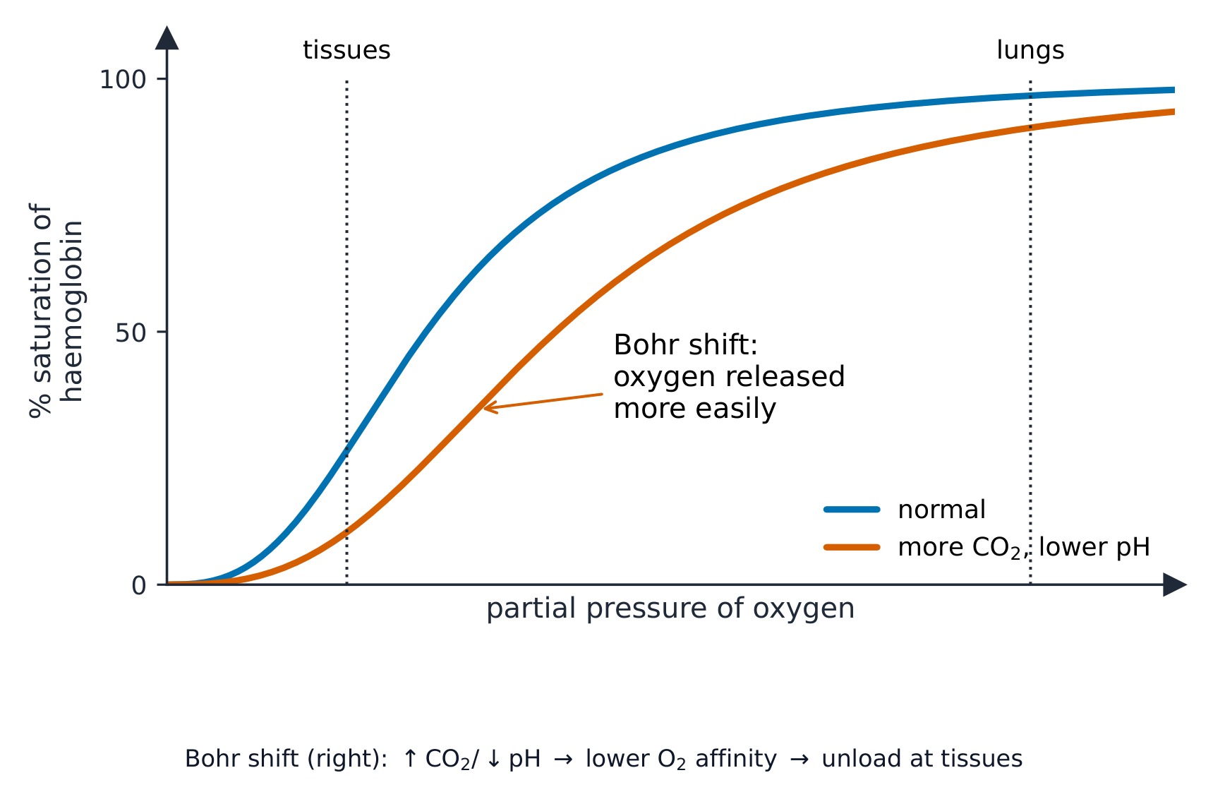
Oxygen is carried by haemoglobin 血红蛋白 in the red blood cells. We show how well haemoglobin holds oxygen with the oxygen dissociation curve 氧解离曲线. This S-shaped graph plots the saturation 饱和度 (how full of oxygen the haemoglobin is) against the partial pressure 分压 of oxygen:

  • where the partial pressure of oxygen is high (in the lungs), haemoglobin loads up and becomes almost fully saturated.
  • where it is low (in respiring tissues), haemoglobin unloads its oxygen for the cells to use.

The Bohr shift

When tissues are very active, they release more carbon dioxide 二氧化碳, which lowers the pH. This makes haemoglobin release oxygen more easily, so the curve moves to the right. This helpful change is the Bohr shift 波尔位移: oxygen is given up exactly where it is most needed.

Carrying carbon dioxide

A little carbon dioxide dissolves straight into the plasma, but most is carried after a reaction inside the red blood cells:

  1. the enzyme 酶 carbonic anhydrase 碳酸酐酶 speeds up the reaction of carbon dioxide with water to make carbonic acid.
  2. the carbonic acid splits into hydrogen ions and hydrogencarbonate ions 碳酸氢根离子.
  3. the hydrogencarbonate ions move out into the plasma. This is the main way carbon dioxide is carried.
  4. to keep the charge balanced, chloride ions 氯离子 move into the red blood cells. This movement is the chloride shift 氯转移.
  5. the hydrogen ions join haemoglobin to form haemoglobinic acid 血红蛋白酸; this mops up the hydrogen ions and keeps the pH steady.

Some carbon dioxide also joins haemoglobin directly to form carbaminohaemoglobin 氨甲酰血红蛋白.

Worked example. At the lungs the partial pressure of oxygen is about $12\ \text{kPa}$ and haemoglobin is about 98% saturated; in an exercising muscle it is about $3\ \text{kPa}$ and saturation falls to about 40%. How much oxygen is unloaded, and why is the curve S-shaped? Subtract the saturations: $98 - 40 =$ 58% of the haemoglobin's oxygen is released in the muscle. The S shape comes from cooperative binding - the first oxygen to bind changes haemoglobin's shape so the next ones bind more easily, which is why the middle of the curve is so steep. That steepness is the point: a small fall in partial pressure in a respiring tissue causes a large release of oxygen. The extra carbon dioxide in an exercising muscle lowers the pH and shifts the curve right, so even more is unloaded at the same partial pressure - that is the Bohr shift doing its job.

ไทย

การขนส่งออกซิเจน

ออกซิเจนถูกขนส่งโดย ฮีโมโกลบิน ในเซลล์เม็ดเลือดแดง เราแสดงถึงประสิทธิภาพที่ฮีโมโกลบินจับออกซิเจนได้ด้วย กราฟการแยกตัวของออกซิเจน กราฟรูปตัว S นี้แสดงระดับ ความอิ่มตัว (ปริมาณออกซิเจนที่ฮีโมโกลบินมีอยู่) เทียบกับ ความดันย่อยของออกซิเจน:

  • เมื่อความดันย่อยของออกซิเจน สูง (ในปอด) ฮีโมโกลบินจะจับออกซิเจนเข้าไว้และมีความอิ่มตัวเกือบเต็ม
  • เมื่อความดันย่อย ต่ำ (ในเนื้อเยื่อที่กำลังมีการหายใจ) ฮีโมโกลบินจะปล่อยออกซิเจนออกมาเพื่อให้เซลล์นำไปใช้

การเลื่อนไปทางขวาของบอร์ (Bohr shift)

เมื่อเนื้อเยื่อทำงานหนักมาก จะปล่อย คาร์บอนไดออกไซด์ ออกมามากขึ้น ซึ่งจะทำให้ค่า pH ลดลง สิ่งนี้ทำให้ฮีโมโกลบินปล่อยออกซิเจนได้ง่ายขึ้น ทำให้กราฟเลื่อนไปทางขวา การเปลี่ยนแปลงที่เป็นประโยชน์นี้เรียกว่า การเลื่อนไปทางขวาของบอร์: ออกซิเจนจะถูกปล่อยออกมาที่ตำแหน่งที่ต้องการมากที่สุดพอดี

กราฟการแยกตัวของออกซิเจนรูปตัว S ที่แสดงความสัมพันธ์ระหว่างความอิ่มตัวเป็นเปอร์เซ็นต์กับความดันย่อยของออกซิเจน พร้อมกราฟที่สองเลื่อนไปทางขวาเพื่อแสดงผลจากคาร์บอนไดออกไซด์ที่สูงขึ้นและค่า pH ที่ต่ำกว่า
ฮีโมโกลบินจับออกซิเจนในปอดและปล่อยในเนื้อเยื่อ; การเลื่อนไปทางขวาของบอร์ทำให้กราฟเคลื่อนที่ไปทางขวาเพื่อปล่อยออกซิเจนมากขึ้น

การขนส่งคาร์บอนไดออกไซด์

คาร์บอนไดออกไซด์บางส่วนละลายเข้าไปในพลาสมาโดยตรง แต่ส่วนใหญ่จะถูกขนส่งหลังจากเกิดปฏิกิริยาภายในเซลล์เม็ดเลือดแดง:

  1. เอนไซม์ คาร์บอนิกแอนไฮเดรซ ช่วยเร่งปฏิกิริยาระหว่างคาร์บอนไดออกไซด์กับน้ำเพื่อสร้างกรดคาร์บอนิค
  2. กรดคาร์บอนิคแตกตัวเป็นไอออนไฮโดรเจนและ ไอออนไฮโดรเจนคาร์บอเนต
  3. ไอออนไฮโดรเจนคาร์บอเนตเคลื่อนที่ออกไปยังพลาสมา นี่คือวิธีหลักในการขนส่งคาร์บอนไดออกไซด์
  4. เพื่อรักษาสมดุลของประจุ ไอออนคลอไรด์ จะเคลื่อนที่เข้าสู่เซลล์เม็ดเลือดแดง การเคลื่อนที่นี้เรียกว่า การเลื่อนคลอไรด์ (chloride shift)
  5. ไอออนไฮโดรเจนจะรวมกับฮีโมโกลบินเพื่อสร้าง กรดฮีโมโกลบิน; สิ่งนี้จะดูดซับไอออนไฮโดรเจนและรักษาค่า pH ให้คงที่

คาร์บอนไดออกไซด์บางส่วนยังรวมกับฮีโมโกลบินโดยตรงเพื่อสร้าง คาร์บามิโนฮีโมโกลบิน

ภายในเซลล์เม็ดเลือดแดง: คาร์บอนไดออกไซด์และน้ำกลายเป็นกรดคาร์บอนิค (โดยคาร์บอนิกแอนไฮเดรซ), ซึ่งแตกตัวเป็นไอออนไฮโดรเจนและไฮโดรเจนคาร์บอเนต; ไฮโดรเจนคาร์บอเนตออกจากเซลล์ในขณะที่คลอไรด์เข้า, และไอออนไฮโดรเจนจับกับฮีโมโกลบิน
CO₂ ส่วนใหญ่เดินทางในฐานะไอออนไฮโดรเจนคาร์บอเนต; การเลื่อนคลอไรด์รักษาสมดุลประจุและฮีโมโกลบินดูดซับ H⁺

ตัวอย่างคำนวณ. ที่ปอด ความดันย่อยของออกซิเจนอยู่ที่ประมาณ $12\ \text{kPa}$ และฮีโมโกลบินมีความอิ่มตัวประมาณ 98%; ในกล้ามเนื้อที่กำลังออกกำลังกายอยู่ที่ประมาณ $3\ \text{kPa}$ และความอิ่มตัวลดลงเหลือประมาณ 40% มีออกซิเจนถูกปล่อยออกมาเท่าไหร่ และทำไมกราฟจึงมีรูปตัว S? หักค่าความอิ่มตัว: $98 - 40 =$ 58% ของออกซิเจนในฮีโมโกลบินถูกปล่อยในกล้ามเนื้อ รูปร่างตัว S เกิดจาก การจับแบบร่วมมือ: ออกซิเจนตัวแรกที่จับเปลี่ยนรูปร่างของฮีโมโกลบินทำให้ตัวถัดๆ ไปจับได้ง่ายขึ้น นี่คือเหตุผลว่าทำไมส่วนกลางของกราฟจึงชันมาก ความชันนี้เป็นจุดสำคัญ: การลดลงเล็กน้อยของความดันย่อยในเนื้อเยื่อที่กำลังหายใจ ทำให้การปล่อยออกซิเจน มาก คาร์บอนไดออกไซด์เพิ่มในกล้ามเนื้อที่กำลังออกกำลังกาย会降低ค่า pH และเลื่อนกราฟ ไปทางขวา ทำให้มีการปล่อยออกซิเจนมากขึ้นที่ความดันย่อยเท่าเดิม นั่นคือหน้าที่ของการเลื่อนไปทางขวาของบอร์

Explore · ⁨สำรวจ⁩

How blood carries oxygen · ⁨วิธีการที่เลือดพาออกซิเจน⁩

Haemoglobin picks up oxygen where there is lots of it (the lungs) and releases it where there is little (the tissues). · ⁨ฮีโมโกลบินจับออกซิเจนเมื่อมีมาก (ในปอด) และปล่อยออกซิเจนเมื่อมีน้อย (ในเนื้อเยื่อ)⁩

Vocabulary · ⁨คำศัพท์⁩ Train · ⁨ฝึกฝน⁩
English ไทย
oxygen/ˈɒksɪdʒn/ ออกซิเจน
pulmonary vein/ˈpʌlmənəri veɪn/ หลอดเลือดดำปอด
Bohr shift/ˈbəʊə ʃɪft/ การเคลื่อนที่ของบอร์
carbon dioxide/ˈkɑːbən daɪˈɒksaɪd/ คาร์บอนไดออกไซด์
enzyme/ˈenzaɪm/ เอนไซม์
carbonic anhydrase/kɑːˈbɒnɪk ˈænhaɪdreɪz/ carbonic anhydrase
hydrogencarbonate ion/ˈhaɪdrəʊdʒeŋkɑːbɒneɪt ˈaɪɒn/ ไอออนไฮโดรเจนคาร์บอเนต
chloride ion/ˈklɔːraɪd ˈaɪɒn/ ไอออนคลอไรด์
chloride shift/ˈklɔːraɪd ʃɪft/ chloride shift
haemoglobinic acid/ˌhiːməˌɡləʊˈbɪnɪk ˈæsɪd/ haemoglobinic acid
carbaminohaemoglobin/ˌkɑːbəˌmɪnəʊˌhiːməˈɡləʊbɪn/ คาร์บามิโนฮีโมโกลบิน
8.3

The heart · ⁨หัวใจ⁩

Syllabus · ⁨หลักสูตร⁩
English
  1. describe the external and internal structure of the mammalian heart
  2. explain the differences in the thickness of the walls of the: • atria and ventricles • left ventricle and right ventricle
  3. describe the cardiac cycle, with reference to the relationship between blood pressure changes during systole and diastole and the opening and closing of valves
  4. explain the roles of the sinoatrial node, the atrioventricular node and the Purkyne tissue in the cardiac cycle (knowledge of nervous and hormonal control is not expected)
ไทย
  1. อธิบายโครงสร้างภายนอกและภายในของ หัวใจของสัตว์เลี้ยงลูกด้วยนม (mammalian heart)
  2. อธิบายความแตกต่างของความหนาของผนังของ: • ห้องบน (atria) และ ห้องล่าง (ventricles) • ห้องล่างซ้าย (left ventricle) และ ห้องล่างขวา (right ventricle)
  3. อธิบาย รอบการเต้นของหัวใจ (cardiac cycle) โดยอ้างอิงถึงความสัมพันธ์ระหว่างการเปลี่ยนแปลงความดันเลือดระหว่าง ** Systole** และ ** Diastole** และการเปิดและปิดของ วาล์ว (valves)
  4. อธิบายบทบาทของ โหนดไซโนแอทเรียล (sinoatrial node), โหนดแอทริโอเวนทริคูลาร์ (atrioventricular node) และ เนื้อเยื่อเปอร์กิน (Purkyne tissue) ในรอบการเต้นของหัวใจ (ไม่จำเป็นต้องรู้เรื่องการควบคุมด้วยระบบประสาทและฮอร์โมน)

Source: Cambridge International syllabus · ⁨แหล่งที่มา: หลักสูตร Cambridge International⁩

English

Structure

The heart has four chambers. The two upper chambers are the atria 心房 (singular: atrium); they have thin walls because they only push blood down into the chambers below. The two lower chambers are the ventricles 心室; they have thick muscular walls because they pump blood out of the heart.

The left ventricle wall is thicker than the right ventricle wall, because the left side must pump blood all the way round the body, while the right side only pumps to the nearby lungs.

The cardiac cycle

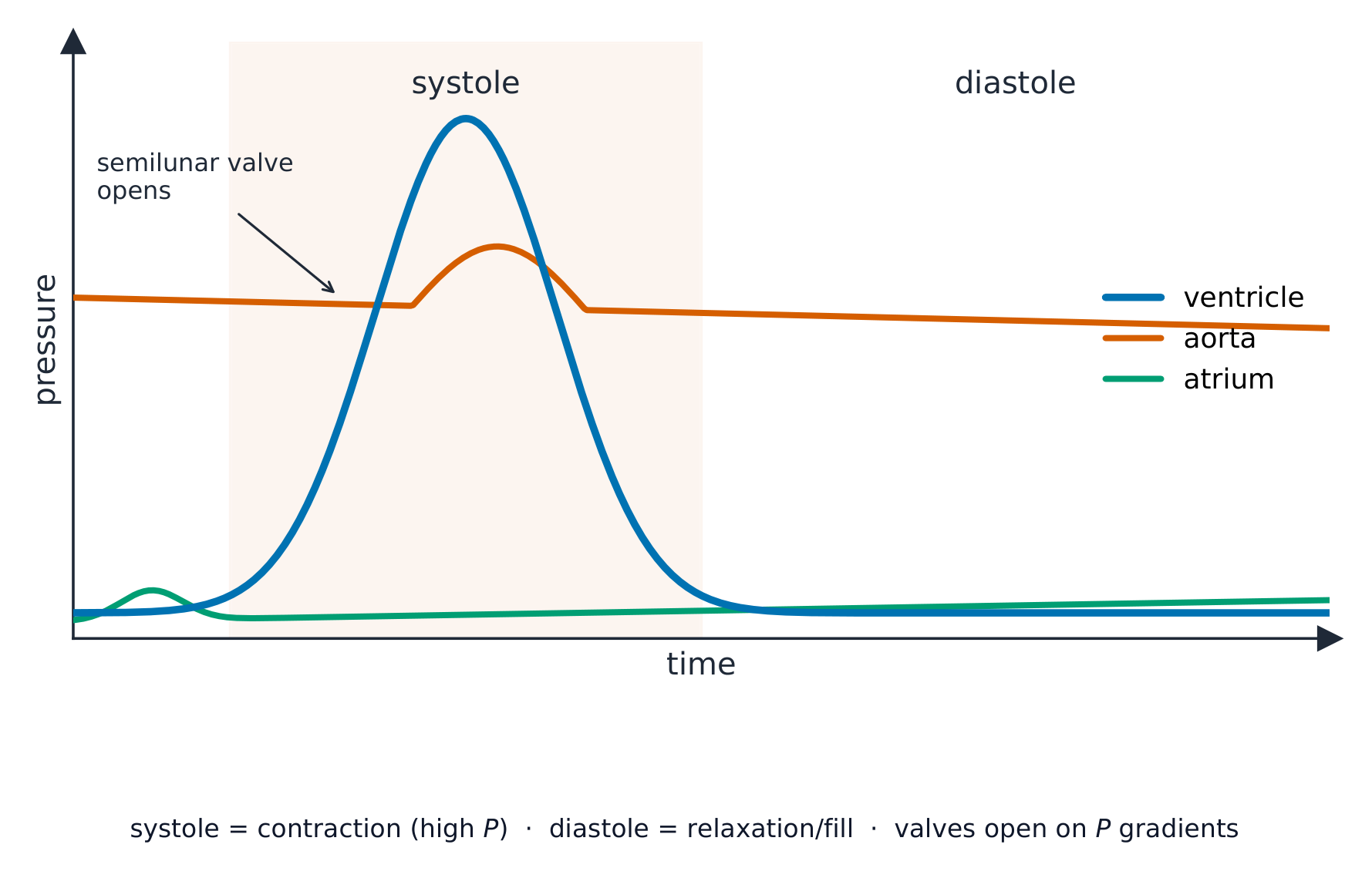
One heartbeat is the cardiac cycle 心动周期. It has two parts: systole 收缩期 (when heart muscle contracts) and diastole 舒张期 (when it relaxes and fills). When a chamber contracts, the pressure inside rises; this pressure change opens and closes the valves so that blood flows one way only.

Controlling the heartbeat

The heart sets its own rhythm:

  • the sinoatrial node 窦房结 in the right atrium is the pacemaker. It sends out a wave of electrical excitation that spreads across the atria and makes them contract.
  • the atrioventricular node 房室结 picks up the wave, holds it back for a moment (so the atria empty first), then passes it on.
  • the Purkyne tissue 浦肯野组织 carries the wave down and through the ventricle walls, so the ventricles contract from the bottom upwards and push blood out.
ไทย

โครงสร้าง

หัวใจมีสี่ห้อง ห้องบนสองห้องคือ ** atria ** (เอกพจน์: atrium);它们有薄的壁因为它们只把血液推向下方的腔室。两下腔是 ** ventricles **;它们有厚的肌肉壁因为它们在将血液泵出心脏。

左心室壁比右心室壁厚,因为左侧必须将血液泵至全身,而右侧仅泵至附近的肺部。

A labelled cut-away diagram of the human heart showing the four chambers, the valves, and the main vessels with arrows for the direction of blood flow
ห้องหัวใจทั้งสี่ chambers, วาล์ว และหลอดเลือด; ผนังของ ventricle ซ้ายมีความหนาที่สุด

รอบการเต้นของหัวใจ (cardiac cycle)

หนึ่งรอบการเต้นของหัวใจคือ cardiac cycle ซึ่งประกอบด้วยสองส่วน: systole (เมื่อกล้ามเนื้อหัวใจหดตัว) และ diastole (เมื่อผ่อนคลายและเติมเลือดเข้า) เมื่อห้องใดห้องหนึ่งหดตัว ความดันภายในจะเพิ่มขึ้น การเปลี่ยนแปลงความดันนี้จะทำให้วาล์วเปิดและปิด เพื่อให้เลือดไหลได้ทางเดียวเท่านั้น

กราฟแสดงความสัมพันธ์ระหว่างความดันกับเวลาสำหรับการเต้นของหัวใจหนึ่งรอบ โดยเส้นโค้งแสดงค่าความดันของ ventricle, aorta และ atrium; ความดันของ ventricle จะพุ่งสูงขึ้นในช่วง systole และ semilunar valve จะเปิดเมื่อความดันนั้นสูงกว่า aorta
ความดันของ ventricle พุ่งสูงขึ้นใน systole และผลักเลือดเข้าสู่ aorta; วาล์วจะเปิดหรือปิด.annotations ความดันสองเส้นกราฟตัดกัน

การควบคุมการเต้นของหัวใจ

หัวใจกำหนดจังหวะของตัวเอง:

  • sinoatrial node ใน right atrium เป็น pacemaker它将ส่งคลื่นการกระตุ้นไฟฟ้ากระจายผ่าน atria ทำให้ atria หดตัว
  • atrioventricular node รับคลื่นนี้ไว้ชั่วคราว (เพื่อให้ atria เติมเลือดออกหมดก่อน) แล้วส่งต่อไป
  • Purkyne tissue นำคลื่นลงผ่านผนัง ventricle ทำให้ ventricles หดตัวจากด้านล่างขึ้นด้านบนและผลักเลือดออก
ภาพร่างหัวใจแสดงคลื่นการกระตุ้นเริ่มต้นที่ SAN ใน right atrium กระจายไปยัง AVN จากนั้นลงและขึ้นตามผนัง ventricle ผ่าน Purkyne tissue
SAN กำหนดจังหวะ; คลื่นส่งไปยัง AVN จากนั้น Purkyne tissue ทำให้ ventricles หดตัวจากล่างขึ้นบน
Explore · ⁨สำรวจ⁩

Explore the heart · ⁨สำรวจหัวใจ⁩

Tap each part. The right side pumps blood to the lungs; the thicker-walled left side pumps it round the whole body. · ⁨แตะแต่ละส่วน ด้านขวาสูบเลือดไปยังปอด; ด้านซ้ายที่มีผนังหนากว่าสูบไปทั่วร่างกาย⁩

Explore · ⁨สำรวจ⁩

The cardiac cycle

Step through one heartbeat — atria contract, then ventricles contract, then everything relaxes and refills. · ⁨พิจารณารอบการเต้นของหัวใจหนึ่งรอบ — atria กระตึงตัว, แล้ว ventricles กระตึงตัว, แล้วทุกอย่างผ่อนคลายและเติมเต็ม⁩

Explore · ⁨สำรวจ⁩

The cardiac cycle

Step through one heartbeat. Pressure changes open and close the valves so blood always flows one way. · ⁨逐步浏览一次心跳。压力变化开闭瓣膜,确保血液始终单向流动。⁩

Vocabulary · ⁨คำศัพท์⁩ Train · ⁨ฝึกฝน⁩
English ไทย
heart/hɑːt/ หัวใจ
aorta/eɪˈɔːtə/ หลอดเลือดแดงเอออร์ตา
vena cava/ˈviːnə ˈkɑːvə/ หลอดเลือดดำใหญ่ (vena cava)
muscle/ˈmʌsl/ กล้ามเนื้อ
elastic/ɪˈlæstɪk/ ยืดหยุ่น
lumen/ˈluːmen/ lumen
valve/vælv/ วาล์ว
red blood cell/red blʌd sel/ red blood cell
monocyte/ˈmɒnəʊsaɪt/ โมโนไซต์
neutrophil/ˈnjuːtrəfɪl/ นิวโทรฟิล
lymphocyte/ˈlɪmfəsaɪt/ ไลม์โฟไซต์
solvent/ˈsɒlvənt/ ตัวทำละลาย
specific heat capacity/spəˈsɪfɪk hiːt kəˈpæsɪti/ ความจุความร้อนจำเพาะ
plasma/ˈplæzmə/ พลาสมา
atrium/ˈeɪtrɪəm/ ห้องหัวใจบน
ventricle/ˈventrɪkl/ ห้องหัวใจ
cardiac cycle/ˈkɑːdiæk ˈsaɪkl/ รอบหัวใจ
systole/ˈsɪstəʊl/ การบีบตัว
diastole/ˈdaɪəstəʊl/ ช่วงการคลายตัวของหัวใจ
sinoatrial node/sɪˈnəʊtrɪəl nəʊd/ นೋดซिनูเอตริอัล
atrioventricular node/ˌeɪtrɪˌəʊvənˈtrɪkjʊlə nəʊd/ นోดอะทริโอเวนตริคูลาร์
Purkyne tissue/ˈpɜːkaɪn ˈtɪʃuː/ เนื้อเยื่อเพอร์कीเน
8.3

Exam tips · ⁨ข้อแนะนำสำหรับการสอบ⁩

English
  • Trace the double circulation and name the chambers, valves and vessels; the left ventricle wall is thicker (pumps further at higher pressure).
  • Read the cardiac-cycle pressure graph: a valve opens/closes when the pressures across it cross over.
  • Explain the oxygen dissociation curve (S-shaped) and the Bohr shift (more CO2 shifts it right, unloading more O2 to active tissue).
  • Most CO2 is carried as hydrogencarbonate (via the chloride shift).
ไทย
  • ติดตามการไหลเวียนเลือดแบบคู่ (double circulation) และระบุชื่อ chambers, วาล์ว และหลอดเลือด; ผนัง left ventricle หนากว่า (สูบฉีดไกลกว่าด้วยความดันสูง)
  • อ่านกราฟความดันของ cardiac-cycle: วาล์วจะเปิด/ปิดเมื่อความดันทั้งสองข้างตัดกัน
  • อธิบาย เส้นโค้งการแยกตัวของออกซิเจน (รูปตัว S) และ Bohr shift (เมื่อมี CO2 มากขึ้น จะทำให้เส้นโค้งเคลื่อนที่ไปทางขวา ช่วยปล่อย O2 ไปยังเนื้อเยื่อที่มีกิจกรรมสูงมากขึ้น)
  • ส่วนใหญ่ CO2 จะถูกขนย้ายในรูปของ ไฮโดรเจนคาร์บอเนต (ผ่านกระบวนการ chloride shift)

Interactive lessons on this topic · ⁨บทเรียนเชิงโต้ตอบสำหรับหัวข้อนี้⁩

Work through it step by step, with instant-check exercises. · ⁨ทำทีละขั้นตอน พร้อมแบบฝึกหัดตรวจสอบผลทันที⁩

Past Papers · ⁨ข้อสอบย้อนหลัง⁩

More topics in A-Level Biology · ⁨Biology A-Level⁩ · ⁨หัวข้อเพิ่มเติมใน A-Level Biology · ⁨Biology A-Level⁩⁩

Log in or create account · ⁨เข้าสู่ระบบหรือสร้างบัญชี⁩

IGCSE, A-Level & AP