Skip to content · ⁨الانتقال إلى المحتوى⁩

Transport in mammals · ⁨النقل في الثدييات⁩

A-Level Biology · ⁨A-Level علم الأحياء⁩ · Topic 8 · ⁨الموضوع 8⁩

Video lesson for this topic · ⁨درس فيديو لهذا الموضوع⁩ Open the video page · ⁨افتح صفحة الفيديو⁩
9:17

الجهاز الدوري

ينبض قلبك حوالي مائة ألف مرة يومياً، ولا يعرف الراحة أبداً. لكن الجزء الذكي هو أنه ليس حلقة واحدة، بل حلقتان. يغادر الدم...

English narration · English + 中文 subtitles burned in · ⁨سرد باللغة الإنجليزية · ترجمة مدمجة بالإنجليزية + الصينية⁩

8.1

The circulatory system · ⁨الجهاز الدوري⁩

Syllabus · ⁨المنهج⁩
English
  1. state that the mammalian circulatory system is a closed double circulation consisting of a heart, blood and blood vessels including arteries, arterioles, capillaries, venules and veins
  2. describe the functions of the main blood vessels of the pulmonary and systemic circulations, limited to pulmonary artery, pulmonary vein, aorta and vena cava
  3. recognise arteries, veins and capillaries from microscope slides, photomicrographs and electron micrographs and make plan diagrams showing the structure of arteries and veins in transverse section (TS) and longitudinal section (LS)
  4. explain how the structure of muscular arteries, elastic arteries, veins and capillaries are each related to their functions
  5. recognise and draw red blood cells, monocytes, neutrophils and lymphocytes from microscope slides, photomicrographs and electron micrographs
  6. state that water is the main component of blood and tissue fluid and relate the properties of water to its role in transport in mammals, limited to solvent action and high specific heat capacity
  7. state the functions of tissue fluid and describe the formation of tissue fluid in a capillary network
العربية
  1. بيّن أن الجهاز الدوري للثدييات هو دورة مغلقة مزدوجة تتكون من قلب، دم، وأوعية دموية تشمل الشرايين، والشرايين الصغيرة، والشعيرات الدموية، والأوردة الصغيرة، والأوردة
  2. صف وظائف الأوعية الدموية الرئيسية في الدورة الرئوية والدورة الجسمية، مقتصراً على الشريان الرئوي، والوريد الرئوي، والشريان الأبهر، والوريد الأجوف
  3. تعرّف على الشرايين والأوردة والشعيرات الدموية من شرائح مجهرية، وصور مجهرية، وصور مجهرية إلكترونية، وارسم مخططات تخطيطية تظهر بنية الشرايين والأوردة في المقطع العرضي (TS) والمقطع الطولي (LS)
  4. اشرح كيف ترتبط بنية الشرايين العضلية، والشرايين المرنة، والأوردة، والشعيرات الدموية بوظائف كل منها
  5. تعرّف وارسم كريات الدم الحمراء، والخلايا الوحيدة، والخلايا المتعادلة، والخلايا الليمفاوية من شرائح مجهرية، وصور مجهرية، وصور مجهرية إلكترونية
  6. بيّن أن الماء هو المكون الرئيسي للدم والسائل النسيجي، وارتبط بخصائص الماء ودوره في النقل في الثدييات، مقتصراً على عمل المذيب والسعة الحرارية النوعية العالية
  7. بيّن وظائف السائل النسيجي وصف تكوين السائل النسيجي في **شبكة الشعيرات الدموية

Source: Cambridge International syllabus · ⁨المصدر: منهج كامبريدج الدولي⁩

English

Mammals have a closed double circulation. "Closed" means the blood stays inside blood vessels 血管 the whole time. "Double" means the blood passes through the heart 心脏 twice for each full trip around the body. This gives two linked loops, so we call it a double circulation 双循环 and the whole thing a circulatory system 循环系统:

  • the pulmonary circulation 肺循环 carries blood from the heart to the lungs and back.
  • the systemic circulation 体循环 carries blood from the heart to the rest of the body and back.

Blood travels in this order: arteries 动脉 → arterioles 小动脉 → capillaries 毛细血管 → venules 小静脉 → veins 静脉.

The main vessels are:

  • the pulmonary artery 肺动脉 — carries blood low in oxygen 氧气 from the heart to the lungs.
  • the pulmonary vein 肺静脉 — carries oxygen-rich blood from the lungs back to the heart.
  • the aorta 主动脉 — the big artery that carries oxygen-rich blood from the heart to the body.
  • the vena cava 腔静脉 — the big vein that returns oxygen-poor blood from the body to the heart.

How the vessels suit their jobs

Vessel Structure Function
artery thick wall of muscle 肌肉 and elastic 弹性 fibres; narrow lumen 管腔 (the space inside) carries blood at high pressure away from the heart; elastic walls stretch and recoil to smooth the flow; muscle controls the flow
capillary wall just one cell thick; very narrow short distance for exchange of substances between blood and cells
vein thin wall; wide lumen; has valves 瓣膜 returns blood at low pressure to the heart; valves stop blood flowing backwards

Blood cells

You should recognise: red blood cells 红细胞 (which carry oxygen), and three white blood cells — monocytes 单核细胞, neutrophils 中性粒细胞 and lymphocytes 淋巴细胞.

Water, plasma and tissue fluid

Water is the main part of blood. It is a good solvent 溶剂, so it carries dissolved substances, and it has a high specific heat capacity 比热容, so the blood's temperature stays steady.

At the start of a capillary, the high blood pressure pushes liquid (but not the cells or large proteins) out of the plasma 血浆 and through the capillary wall. This liquid around the cells is tissue fluid 组织液. It supplies the cells with oxygen and glucose and carries waste away. Most of it returns to the capillary at the far end, where the pressure is lower.

العربية

تمتلك الثدييات دورة دموية مزدوجة مغلقة. "مغلقة" تعني أن الدم يبقى داخل الأوعية الدموية طوال الوقت. "مزدوجة" تعني أن الدم يمر عبر القلب مرتين لكل دورة كاملة حول الجسم. هذا يوفر حلقتين مترابطتين، لذا نسميه دورة دموية مزدوجة والنظام ككل جهازا دوريا:

  • الدورة الرئوية: تنقل الدم من القلب إلى الرئتين وعادته.
  • الدورة الجسدية: تنقل الدم من القلب إلى بقية الجسم وعادته.

يسافر الدم بهذا الترتيب: شرايين → شرايين صغيرة → أوعية شعرية → وريدات → أوردة.

مخطط حلقي للدورة المزدوجة: يضخ القلب الأيمن الدم غير المؤكسج إلى الرئتين وعادته للقلب الأيسر، الذي يضخ الدم المؤكسج إلى الجسم وعادته للقلب الأيمن
الدورة المزدوجة: الحلقة الرئوية تذهب إلى الرئتين، والحلقة الجسدية للجسم؛ يمر الدم عبر القلب مرتين

الأوعية الرئيسية هي:

  • الشريان الرئوي — ينقل دماً فقيراً بـالأكسجين من القلب إلى الرئتين.
  • الوريد الرئوي — ينقل دماً غنياً بالأكسجين من الرئتين عائدة إلى القلب.
  • الأورطي — الشريان الكبير الذي ينقل دماً غنياً بالأكسجين من القلب إلى الجسم.
  • الوريد الأجوف — الوريد الكبير الذي يعيد الدم الفقير بالأكسجين من الجسم إلى القلب.

كيف تتناسب الأوعية مع وظائفها

الوعاء البنية الوظيفة
شريان جدار سميك من عضلات وألياف مرنة؛ لمن ضيق (المكان الداخلي) ينقل الدم بضغط عالٍ بعيداً عن القلب؛ تمتد الجدران المرنة وترتد لتنعيم التدفق؛ تتحكم العضلات في التدفق
وعاء شعري جدار سمايته خلية واحدة فقط؛ ضيق جداً مسافة قصيرة لتبادل المواد بين الدم والخلايا
وريد جدار رقيق؛ لمن واسع؛ يحتوي على صمامات يعيد الدم بضغط منخفض إلى القلب؛ تمنع الصمامات رجوع الدم
قطع عرضية لشريان بجدار سميك ومن ضيق، ووريد بجدار رقيق ومن واسع وصمام، ووعاء شعري صغير سمايته خلية واحدة
الشريان له جدار سميك ومن ضيق؛ الوريد جدار رقيق ومن واسع وصمامات؛ الوعاء الشعري سمايته خلية واحدة

خلايا الدم

يجب أن تتعرف على: خلايا الدم الحمراء (التي تحمل الأكسجين)، وثلاثة أنواع من خلايا الدم البيضاء — الخلايا الوحيدة، الخلايا المتعادلة والخلايا الليمفاوية.

شريحة دم ملونة تُرى تحت مجهر ضوئي: الكثير من خلايا الدم الحمراء الصغيرة الدائرية ذات مراكز باهتة، خلية ليمفاوية كبيرة واحدة بنواة بنفسجية داكنة بالقرب من المركز، وخلايا متعادلتان بنوى مفصصة أسفلهما
شريحة دم ملونة: العديد من الخلايا الحمراء الصغيرة، بالإضافة إلى خلية ليمفاوية (في المنتصف) وخلايا متعادلة (نواة مفصصة) — خلايا الدم البيضاء أكبر ولها نواة

الماء، البلازما وسائل الأنسجة

الماء هو الجزء الرئيسي من الدم. إنه مذيب جيد، لذا يحمل مواد مذابة، ولديه حرارة نوعية عالية، لذا تبقى درجة حرارة الدم ثابتة.

في بداية الوعاء الشعري، يدفع ضغط الدم المرتفع السائل (ولكن ليس الخلايا أو البروتينات الكبيرة) خارج البلازما وعبر جدار الوعاء الشعري. هذا السائل المحيط بالخلايا هو سائل الأنسجة. يزود الخلايا بالأكسجين والجلوكوز ويحمل الفضلات بعيداً. معظمه يعود إلى الوعاء الشعري في الطرف البعيد، حيث يكون الضغط أقل.

وعاء شعري يُدفع منه سائل عند الطرف الشرياني عالي الضغط ليغمر خلايا الجسم، ومعظمه يعود عند الطرف الوريدي منخفض الضغط
الضغط العالي عند الطرف الشرياني يدفع السائل للخارج لتكوين سائل الأنسجة؛ معظمه يعود عند الطرف الوريدي
Explore · ⁨استكشف⁩

Double circulation · ⁨الدورة المزدوجة⁩

Follow one red blood cell around the loop. It passes through the heart twice each circuit — once to the lungs, once to the body. · ⁨تابع خلايا الدم الحمراء في دورة كاملة. تمر عبر القلب مرتين في كل دورة — مرة إلى الرئتين، ومرة إلى الجسم.⁩

Vocabulary · ⁨مفردات⁩ Train · ⁨تدريب⁩
English العربية
blood vessel/blʌd ˈvesl/ وعاء دموي
double circulation/ˈdʌbl ˌsɜːkjʊˈleɪʃn/ الدورة المزدوجة
circulatory system/ˌsɜːkjʊˈleɪtəri ˈsɪstəm/ الجهاز الدوري
pulmonary circulation/ˈpʌlmənəri ˌsɜːkjʊˈleɪʃn/ pulmonary circulation
systemic circulation/sɪˈstemɪk ˌsɜːkjʊˈleɪʃn/ systemic circulation
artery/ˈɑːtəri/ الشريان
arteriole/ɑːˈtɪəriəʊl/ شريان صغير
capillary/kəˈpɪləri/ الشعيرة الدموية
venule/ˈvenjuːl/ وريد صغير
vein/veɪn/ الوريد
pulmonary artery/ˈpʌlmənəri ˈɑːtəri/ شريان رئوي
tissue fluid/ˈtɪʃuː ˈfluːɪd/ tissue fluid
haemoglobin/ˌhiːməˈɡləʊbɪn/ الهيموغلوبين
oxygen dissociation curve/ˈɒksɪdʒn dɪˌsəʊsɪˈeɪʃn kɜːv/ منحنى انفصال الأكسجين
saturation/ˌsætʃəˈreɪʃn/ تشبع اللون
partial pressure/ˈpɑːʃl ˈpreʃə/ الضغط الجزئي
Bohr shift/ˈbəʊə ʃɪft/ انزياح بور
carbon dioxide/ˈkɑːbən daɪˈɒksaɪd/ ثاني أكسيد الكربون
enzyme/ˈenzaɪm/ إنزيم
carbonic anhydrase/kɑːˈbɒnɪk ˈænhaɪdreɪz/ كربونات آنهيدراز
hydrogencarbonate ion/ˈhaɪdrəʊdʒeŋkɑːbɒneɪt ˈaɪɒn/ أيون البيكربونات
chloride ion/ˈklɔːraɪd ˈaɪɒn/ أيون الكلوريد
chloride shift/ˈklɔːraɪd ʃɪft/ انزياح الكلوريد
haemoglobinic acid/ˌhiːməˌɡləʊˈbɪnɪk ˈæsɪd/ حمض الهيموغلوبيني
carbaminohaemoglobin/ˌkɑːbəˌmɪnəʊˌhiːməˈɡləʊbɪn/ كاربامينوهيموغلوبين
atrium/ˈeɪtrɪəm/ أذين
ventricle/ˈventrɪkl/ البطين
8.2

Transport of oxygen and carbon dioxide · ⁨نقل الأكسجين وثاني أكسيد الكربون⁩

Syllabus · ⁨المنهج⁩
English
  1. describe the role of red blood cells in transporting oxygen and carbon dioxide with reference to the roles of: • haemoglobin • carbonic anhydrase • the formation of haemoglobinic acid • the formation of carbaminohaemoglobin
  2. describe the chloride shift and explain the importance of the chloride shift
  3. describe the role of plasma in the transport of carbon dioxide
  4. describe and explain the oxygen dissociation curve of adult haemoglobin
  5. explain the importance of the oxygen dissociation curve at partial pressures of oxygen in the lungs and in respiring tissues
  6. describe the Bohr shift and explain the importance of the Bohr shift
العربية
  1. صف دور كريات الدم الحمراء في نقل الأكسجين وثاني أكسيد الكربون مع الإشارة إلى أدوار: • الهيموجلوبين • إنزيم الأنهيدرة الكربونية • تكوين حمض الهيموغلوبيني • تكوين كاربامينوهيموجلوبين
  2. صف انزياح الكلوريد واشرح أهمية انزياح الكلوريد
  3. صف دور البلازما في نقل ثاني أكسيد الكربون
  4. صف واشرح منحنى تفكك الأكسجين للهيموجلوبين البالغ
  5. اشرح أهمية منحنى تفكك الأكسجين عند الضغوط الجزئية للأكسجين في الرئتين وأنسجة التنفس
  6. صف إزاحة بور واشرح أهمية إزاحة بور

Source: Cambridge International syllabus · ⁨المصدر: منهج كامبريدج الدولي⁩

English

Carrying oxygen

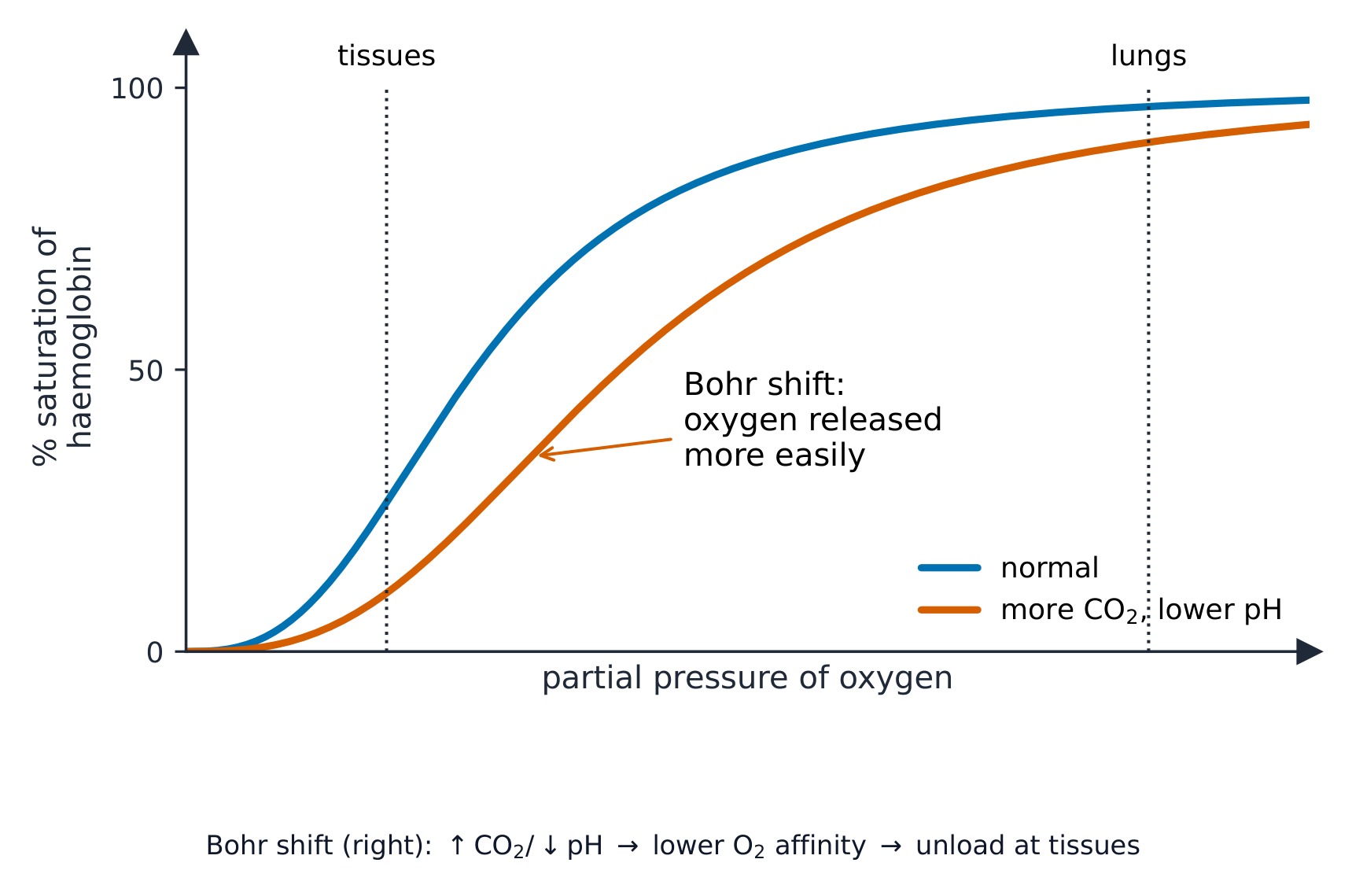
Oxygen is carried by haemoglobin 血红蛋白 in the red blood cells. We show how well haemoglobin holds oxygen with the oxygen dissociation curve 氧解离曲线. This S-shaped graph plots the saturation 饱和度 (how full of oxygen the haemoglobin is) against the partial pressure 分压 of oxygen:

  • where the partial pressure of oxygen is high (in the lungs), haemoglobin loads up and becomes almost fully saturated.
  • where it is low (in respiring tissues), haemoglobin unloads its oxygen for the cells to use.

The Bohr shift

When tissues are very active, they release more carbon dioxide 二氧化碳, which lowers the pH. This makes haemoglobin release oxygen more easily, so the curve moves to the right. This helpful change is the Bohr shift 波尔位移: oxygen is given up exactly where it is most needed.

Carrying carbon dioxide

A little carbon dioxide dissolves straight into the plasma, but most is carried after a reaction inside the red blood cells:

  1. the enzyme 酶 carbonic anhydrase 碳酸酐酶 speeds up the reaction of carbon dioxide with water to make carbonic acid.
  2. the carbonic acid splits into hydrogen ions and hydrogencarbonate ions 碳酸氢根离子.
  3. the hydrogencarbonate ions move out into the plasma. This is the main way carbon dioxide is carried.
  4. to keep the charge balanced, chloride ions 氯离子 move into the red blood cells. This movement is the chloride shift 氯转移.
  5. the hydrogen ions join haemoglobin to form haemoglobinic acid 血红蛋白酸; this mops up the hydrogen ions and keeps the pH steady.

Some carbon dioxide also joins haemoglobin directly to form carbaminohaemoglobin 氨甲酰血红蛋白.

Worked example. At the lungs the partial pressure of oxygen is about $12\ \text{kPa}$ and haemoglobin is about 98% saturated; in an exercising muscle it is about $3\ \text{kPa}$ and saturation falls to about 40%. How much oxygen is unloaded, and why is the curve S-shaped? Subtract the saturations: $98 - 40 =$ 58% of the haemoglobin's oxygen is released in the muscle. The S shape comes from cooperative binding - the first oxygen to bind changes haemoglobin's shape so the next ones bind more easily, which is why the middle of the curve is so steep. That steepness is the point: a small fall in partial pressure in a respiring tissue causes a large release of oxygen. The extra carbon dioxide in an exercising muscle lowers the pH and shifts the curve right, so even more is unloaded at the same partial pressure - that is the Bohr shift doing its job.

العربية

حمل الأكسجين

يتم نقل الأكسجين بواسطة الهيموغلوبين في خلايا الدم الحمراء. نوضح مدى كفاءة ارتباط الهيموغلوبين بالأكسجين من خلال منحنى تفكك الأكسجين. هذا المنحنى على شكل حرف S يرسم نسبة التشبع (مدى امتلاء الهيموغلوبين بالأكسجين) مقابل الضغط الجزئي للأكسجين:

  • حيث يكون الضغط الجزئي للأكسجين مرتفعاً (في الرئتين)، يمتلئ الهيموغلوبين ويصبح مشبعاً تقريباً بالكامل.
  • حيث يكون منخفضاً (في الأنسجة التي تقوم بالتنفس)، يفقد الهيموغلوبين أكسجينا لتستخدمه الخلايا.

انزياح بور

عندما تكون الأنسجة نشطة جداً، تطلق كميات أكبر من ثاني أكسيد الكربون، مما يخفض درجة الحموضة (pH). هذا يجعل الهيموغلوبين يحرر الأكسجين بسهولة أكبر، لذلك يتحرك المنحنى نحو اليمين. هذه التغير المفيد هو انزياح بور: يتم إطلاق الأكسجين تماماً عند المكان الذي يكون فيه أكثر حاجة إليه.

منحنى تفكك الأكسجين على شكل حرف S يرسم نسبة التشبع المئوية مقابل الضغط الجزئي للأكسجين، مع منحنى ثانٍ منزاحاً نحو اليمين لزيادة ثاني أكسيد الكربون وانخفاض درجة الحموضة
يحمل الهيموغلوبين الأكسجين في الرئتين ويحرره في الأنسجة؛ ينزاح انزياح بور المنحنى لليمين ليتم إطلاق المزيد

نقل ثاني أكسيد الكربون

يتحلل القليل من ثاني أكسيد الكربون مباشرة في البلازما، لكن معظمه يُنقل بعد تفاعل داخل خلايا الدم الحمراء:

  1. إنزيم أنهيدراز الكربونات يسرع تفاعل ثاني أكسيد الكربون مع الماء لتكوين حمض الكربونيك.
  2. يتفكك حمض الكربونيك إلى أيونات هيدروجين وأيونات بيكربونات.
  3. تتحرك أيونات البيكربونات إلى خارج الخلية إلى البلازما. وهذا هو الطريقة الرئيسية لنقل ثاني أكسيد الكربون.
  4. للحفاظ على توازن الشحنة، تدخل أيونات الكلوريد إلى خلايا الدم الحمراء. هذا الحركة تسمى انزياح الكلوريد.
  5. ترتبط أيونات الهيدروجين بالهيموغلوبين لتكوين حمض الهيموغلوبيني؛ هذا يمتص أيونات الهيدروجين ويحافظ على ثبات درجة الحموضة.

يرتبط بعض ثاني أكسيد الكربون أيضاً بالهيموغلوبين مباشرة لتكوين كاربامينوهيموغلوبين.

داخل خلية دم حمراء: يتحول ثاني أكسيد الكربون والماء إلى حمض كربوني (بواسطة أنهيدراز الكربونات)، والذي يتفكك إلى أيونات هيدروجين وبيكربونات؛ تغادر البيكربونات بينما يدخل الكلوريد، وترتبط أيونات الهيدروجين بالهيموغلوبين
يعبر معظم CO₂ على شكل أيونات بيكربونات؛ يحافظ انزياح الكلوريد على توازن الشحنة ويمتص الهيموغلوبين H⁺

مثال محلول. عند الرئتين يكون الضغط الجزئي للأكسجين حوالي $12\ \text{kPa}$ ويكون تشبع الهيموغلوبين حوالي 98%؛ في عضلة تمرين يكون حوالي $3\ \text{kPa}$ وينخفض التشبع إلى حوالي 40%. كم مقدار الأكسجين المحرر، ولماذا المنحنى على شكل S؟ اطرح التشبعات: $98 - 40 =$ 58% من أكسجين الهيموغلوبين يتم إطلاقه في العضلة. يأتي الشكل S من الارتباط التعاوني - أول جزيء أكسجين يرتبط يغير شكل الهيموغلوبين بحيث ترتبط الجزيئات التالية بشكل أسهل، ولهذا السبب يكون وسط المنحنى حاداً جداً. هذه الحدة هي النقطة الأساسية: انخفاض صغير في الضغط الجزئي في نسيج تنفسي يسبب إطلاقاً كبيراً للأكسجين. زيادة ثاني أكسيد الكربون في العضلة النشطة تخفض درجة الحموضة وتنزح المنحنى ليميناً، لذا يتم إطلاق المزيد عند نفس الضغط الجزئي - هذا هو انزياح بور يؤدي دوره.

Explore · ⁨استكشف⁩

How blood carries oxygen · ⁨كيف ينقل الدم الأكسجين⁩

Haemoglobin picks up oxygen where there is lots of it (the lungs) and releases it where there is little (the tissues). · ⁨يأخذ الهيموجلوبين الأكسجين حيث يوجد بكثرة (الرئتين) ويحرره حيث يكون قليلاً (الأنسجة).⁩

Vocabulary · ⁨مفردات⁩ Train · ⁨تدريب⁩
English العربية
oxygen/ˈɒksɪdʒn/ الأكسجين
pulmonary vein/ˈpʌlmənəri veɪn/ وريد رئوي
aorta/eɪˈɔːtə/ الشريان الأبهر
vena cava/ˈviːnə ˈkɑːvə/ الوريد الأجوف
muscle/ˈmʌsl/ عضلة
elastic/ɪˈlæstɪk/ مرن
lumen/ˈluːmen/ lumen
valve/vælv/ صمام
red blood cell/red blʌd sel/ خلية دم حمراء
monocyte/ˈmɒnəʊsaɪt/ خلية وحيدة
neutrophil/ˈnjuːtrəfɪl/ خلايا متعادلة
lymphocyte/ˈlɪmfəsaɪt/ خلايا لمفاوية
solvent/ˈsɒlvənt/ المذيب
specific heat capacity/spəˈsɪfɪk hiːt kəˈpæsɪti/ السعة الحرارية النوعية
plasma/ˈplæzmə/ البلازما
8.3

The heart · ⁨القلب⁩

Syllabus · ⁨المنهج⁩
English
  1. describe the external and internal structure of the mammalian heart
  2. explain the differences in the thickness of the walls of the: • atria and ventricles • left ventricle and right ventricle
  3. describe the cardiac cycle, with reference to the relationship between blood pressure changes during systole and diastole and the opening and closing of valves
  4. explain the roles of the sinoatrial node, the atrioventricular node and the Purkyne tissue in the cardiac cycle (knowledge of nervous and hormonal control is not expected)
العربية
  1. صف البنية الخارجية والداخلية لـ قلب الثدييات
  2. اشرح الفروق في سماكة جدران: • الأذينين والبطينين • البطين الأيسر والبطين الأيمن
  3. صف الدورة القلبية، مع الإشارة إلى العلاقة بين تغيرات ضغط الدم أثناء الانقباض والانبساط وفتح وإغلاق الصمامات
  4. اشرح أدوار العقدة الجيبية الأذينية، والعقدة الأذينية البطينية، وأنسجة بركني في الدورة القلبية (لا يُتوقع معرفة التحكم العصبي والهرموني)

Source: Cambridge International syllabus · ⁨المصدر: منهج كامبريدج الدولي⁩

English

Structure

The heart has four chambers. The two upper chambers are the atria 心房 (singular: atrium); they have thin walls because they only push blood down into the chambers below. The two lower chambers are the ventricles 心室; they have thick muscular walls because they pump blood out of the heart.

The left ventricle wall is thicker than the right ventricle wall, because the left side must pump blood all the way round the body, while the right side only pumps to the nearby lungs.

The cardiac cycle

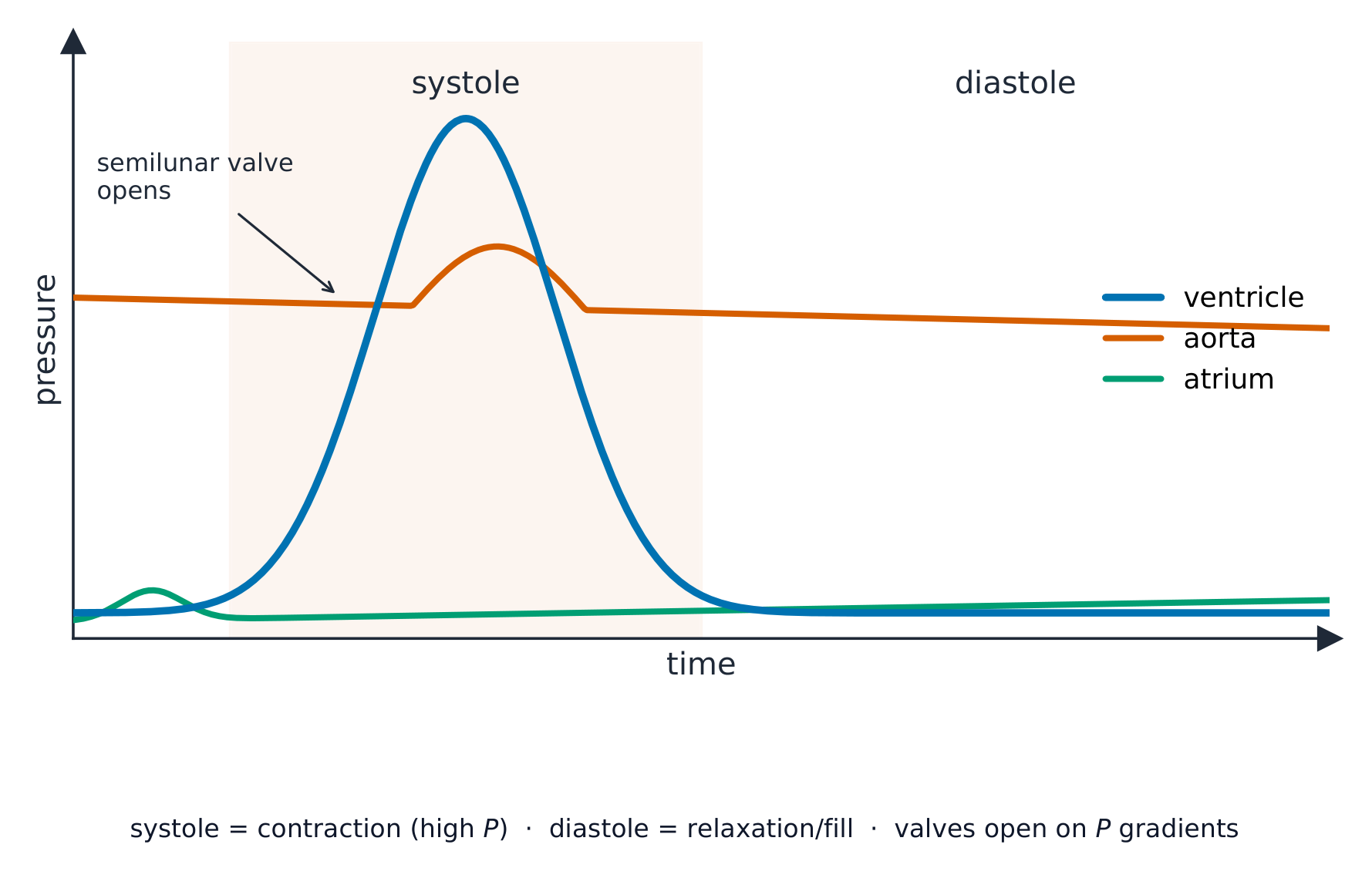
One heartbeat is the cardiac cycle 心动周期. It has two parts: systole 收缩期 (when heart muscle contracts) and diastole 舒张期 (when it relaxes and fills). When a chamber contracts, the pressure inside rises; this pressure change opens and closes the valves so that blood flows one way only.

Controlling the heartbeat

The heart sets its own rhythm:

  • the sinoatrial node 窦房结 in the right atrium is the pacemaker. It sends out a wave of electrical excitation that spreads across the atria and makes them contract.
  • the atrioventricular node 房室结 picks up the wave, holds it back for a moment (so the atria empty first), then passes it on.
  • the Purkyne tissue 浦肯野组织 carries the wave down and through the ventricle walls, so the ventricles contract from the bottom upwards and push blood out.
العربية

البنية

يحتوي القلب على أربع حجرات. الحجرتان العلويتان هما الأذينان (المفرد: أذين)؛ وجدرانها رقيقة لأنها تدفع الدم فقط للأسفل إلى الحجرات السفلية. الحجرتان السفليتان هما البطينان؛ وجدرانهما العضلية سميكة لأنها تضخ الدم خارج القلب.

جدار البطين الأيسر أكثر سماكة من جدار البطين الأيمن، لأن الجانب الأيسر يجب أن يضخ الدم عبر الجسم بأكمله، بينما الجانب الأيمن يضخ فقط إلى الرئتين القريبة.

مخطط مقطعي ملخص للقلب البشري يظهر الحجرات الأربع، الصمامات، والأوعية الرئيسية مع أسهم لاتجاه تدفق الدم
الحجرات الأربع، الصمامات والأوعية الرئيسية؛ جدار البطين الأيسر هو الأكثر سماكة

الدورة القلبية

نبضة واحدة للقلب هي الدورة القلبية. تتكون من جزأين: الانقباض (عندما تنقبض عضلة القلب) والانبساط (عندما ترتخي وتمتلئ). عندما تنقبض حجرة، يرتفع الضغط بداخلها؛ هذا التغير في الضغط يفتح ويغلق الصمامات بحيث يتدفق الدم في اتجاه واحد فقط.

رسم بياني للضغط مقابل الزمن لنبضة واحدة، مع منحنيات للبطين والأورطي والأذين؛ يرتفع ضغط البطين خلال الانقباض ويفتح الصمام النصفي حيث يتجاوز ضغط الأورطي
يرتفع ضغط البطين في مرحلة الانقباض ويدفع الدم إلى الأورطي؛ يفتح أو يغلق الصمام كلما تقاطعت منحني ضغطين

التحكم في ضربات القلب

يحدد القلب إيقاعه الخاص:

  • العقدة الجيبية الأذينية في الأذين الأيمن هي ناظم النبض. ترسل موجة من الإثارة الكهربائية تنتشر عبر الأذينين وتجعلهما ينقبضان.
  • العقدة الأذينية البطينية تستقبل الموجة، وتؤخرها للحظة (حتى يفرغ الأذينان أولاً)، ثم تنقلها.
  • نسيج بوركيه ينقل الموجة لأسفل عبر جدران البطين، sehingga تنقبض البطينات من الأسفل إلى الأعلى وتدفع الدم للخارج.
رسم تخطيطي للقلب يوضح موجة الإثارة التي تبدأ من العقدة الجيبية الأذينية في الأذين الأيمن، وتنتشر إلى العقدة الأذينية البطينية، ثم تنزل وتصعد عبر جدران البطين عن طريق نسيج بوركيه
تحدد العقدة الجيبية الأذينية الإيقاع؛ تنتقل الموجة إلى العقدة الأذينية البطينية، ثم يجعل نسيج بوركيه البطينين ينقبضان من الأسفل للأعلى
Explore · ⁨استكشف⁩

Explore the heart · ⁨استكشف القلب⁩

Tap each part. The right side pumps blood to the lungs; the thicker-walled left side pumps it round the whole body. · ⁨اضغط على كل جزء. الجانب الأيمن يضخ الدم إلى الرئتين؛ الجانب الأيسر ذو الجدار الأكثر سماكة يضخه حول الجسم بأكمله.⁩

Explore · ⁨استكشف⁩

The cardiac cycle · ⁨الدورة القلبية⁩

Step through one heartbeat — atria contract, then ventricles contract, then everything relaxes and refills. · ⁨مرر عبر نبضة قلبية واحدة — تنقبض الأذينة، ثم تنقبض البطينات، ثم يستريح الجميع ويمتلئ مرة أخرى.⁩

Explore · ⁨استكشف⁩

The cardiac cycle · ⁨الدورة القلبية⁩

Step through one heartbeat. Pressure changes open and close the valves so blood always flows one way. · ⁨مرر عبر نبضة قلبية واحدة. تغير الضغط يفتح ويغلق الصمامات لضمان تدفق الدم باتجاه واحد دائماً.⁩

Vocabulary · ⁨مفردات⁩ Train · ⁨تدريب⁩
English العربية
heart/hɑːt/ قلب
cardiac cycle/ˈkɑːdiæk ˈsaɪkl/ الدورة القلبية
systole/ˈsɪstəʊl/ الانقباض
diastole/ˈdaɪəstəʊl/ انبساط
sinoatrial node/sɪˈnəʊtrɪəl nəʊd/ العقدة الجيبية الأذينية
atrioventricular node/ˌeɪtrɪˌəʊvənˈtrɪkjʊlə nəʊd/ العقدة الأذينية البطينية
Purkyne tissue/ˈpɜːkaɪn ˈtɪʃuː/ نسيج بيركنجي
8.3

Exam tips · ⁨نصائح للامتحان⁩

English
  • Trace the double circulation and name the chambers, valves and vessels; the left ventricle wall is thicker (pumps further at higher pressure).
  • Read the cardiac-cycle pressure graph: a valve opens/closes when the pressures across it cross over.
  • Explain the oxygen dissociation curve (S-shaped) and the Bohr shift (more CO2 shifts it right, unloading more O2 to active tissue).
  • Most CO2 is carried as hydrogencarbonate (via the chloride shift).
العربية
  • تتبع الدورة الدموية المزدوجة وسمّ الحجرات والصمامات والأوعية؛ جدار البطين الأيسر أكثر سماكة (يضخ لمسافة أطول وبضغط أعلى).
  • اقرأ رسم بياني لضغط الدورة القلبية: يفتح/يغلق الصمام عندما تتقاطع الضغوط عبره.
  • اشرح منحنى انفصال الأكسجين (على شكل حرف S) وانزياح بور (زيادة ثاني أكسيد الكربون 2 تحركه لليمين، مما يزيد تفريغ الأكسجين 2 إلى الأنسجة النشطة).
  • يُحمل معظم CO2 على شكل بيكربونات (عبر انزياح الكلوريد).

Interactive lessons on this topic · ⁨دروس تفاعلية حول هذا الموضوع⁩

Work through it step by step, with instant-check exercises. · ⁨ا-working عليه خطوة بخطوة، مع تمارين تحقق فوري.⁩

Past Papers · ⁨أوراق الامتحانات السابقة⁩

More topics in A-Level Biology · ⁨A-Level علم الأحياء⁩ · ⁨المزيد من المواضيع في A-Level Biology · ⁨A-Level علم الأحياء⁩⁩

Log in or create account · ⁨تسجيل الدخول أو إنشاء حساب⁩

IGCSE, A-Level & AP