Skip to content · ⁨ข้ามไปยังเนื้อหา⁩

Transport in animals · ⁨การลำเลียงในสัตว์⁩

IGCSE Biology · ⁨Biology IGCSE⁩ · Topic 9 · ⁨หัวข้อ 9⁩

Video lesson for this topic · ⁨บทเรียนวิดีโอสำหรับหัวข้อนี้⁩ Open the video page · ⁨เปิดหน้าวิดีโอ⁩
8:21

การขนส่งในสัตว์

หัวใจของคุณเต้นประมาณแสนครั้งต่อวัน และไม่เคยหยุดพักเลยจริงๆ มันไม่ใช่ปั๊มเดียว — มันคือสองตัว วางข้างกัน แชร์…

English narration · English + 中文 subtitles burned in · ⁨การบรรยายภาษาอังกฤษ · คำบรรยายภาษาอังกฤษ + 中文 ลอยตัวบนภาพ⁩

9.1

The circulatory system · ⁨ระบบไหลเวียนเลือด⁩

Syllabus · ⁨หลักสูตร⁩
English
Core Supplement
1 Describe the circulatory system as a system of blood vessels with a pump and valves to ensure one-way flow of blood
2 Describe the single circulation of a fish
3 Describe the double circulation of a mammal
4 Explain the advantages of a double circulation
ไทย
แก่น เพิ่มเติม
1 อธิบายระบบไหลเวียนเลือดว่าเป็นระบบของหลอดเลือดที่มีปั๊มและวาล์วเพื่อให้มั่นใจว่าเลือดไหลไปในทิศทางเดียว
2 อธิบายการไหลเวียนเลือดแบบเดี่ยวของปลา
3 อธิบายการไหลเวียนเลือดแบบคู่ของสัตว์เลี้ยงลูกด้วยนม
4 อธิบายข้อดีของการไหลเวียนเลือดแบบคู่

Source: Cambridge International syllabus · ⁨แหล่งที่มา: หลักสูตร Cambridge International⁩

English

The circulatory system 循环系统 carries blood around the body. It has three parts: a pump 泵 (the heart 心脏), a network of blood vessels 血管, and valves 瓣膜 that keep the blood flowing one way only.

Single and double circulation (Supplement)

  • A fish has a single circulation 单循环: the blood passes through the heart once on each trip around the body.
  • A mammal 哺乳动物 has a double circulation 双循环: the blood passes through the heart twice on each full trip — once on the way to the lungs, and once on the way to the rest of the body.
  • The advantage of a double circulation: the blood can be pumped again at high pressure 压力 before going to the body, so it travels faster and delivers oxygen 氧气 quickly.
ไทย

ระบบไหลเวียนเลือด ทำหน้าที่ขนส่งเลือดทั่วร่างกาย ประกอบด้วย 3 ส่วนคือ ปั๊ม (หัวใจ), หลอดเลือด และ วาล์ว ที่ช่วยควบคุมให้เลือดไหลไปในทิศทางเดียวเท่านั้น

ระบบไหลเวียนเลือดแบบเดี่ยวและแบบคู่ (ส่วนเสริม)

  • ปลา มี ระบบไหลเวียนเลือดแบบเดี่ยว: เลือดจะไหลผ่านหัวใจ หนึ่งครั้ง ในแต่ละรอบที่ไหลเวียนรอบร่างกาย
  • สัตว์มีกระดูกสันหลังในชั้น哺乳动物 ( mammals ) มี ระบบไหลเวียนเลือดแบบคู่: เลือดจะไหลผ่านหัวใจ สองครั้ง ในแต่ละรอบที่ไหลเวียนรอบร่างกาย — ครั้งแรกไปปอด และครั้งที่สองไปยังส่วนอื่นของร่างกาย
  • ข้อดีของระบบไหลเวียนเลือดแบบคู่: เลือดสามารถถูกสูบ pumped ใหม่ด้วยความดัน สูง ก่อนที่จะส่งไปยังส่วนอื่นของร่างกาย ทำให้ไหลได้เร็วขึ้นและส่ง ออกซิเจน ได้ทันท่วงที
แผนภาพวงกลม: หัวใจสูบเลือดขาดออกซิเจนไปยังปอดแล้วกลับมา จากนั้นสูบเลือดที่มีออกซิเจนไปยังส่วนอื่นของร่างกายแล้วกลับมา ทำให้เลือดไหลผ่านหัวใจสองครั้ง
ในระบบไหลเวียนเลือดแบบคู่ เลือดจะไหลผ่านหัวใจสองครั้งในแต่ละรอบที่ไหลเวียนรอบร่างกาย
Explore · ⁨สำรวจ⁩

Double circulation · ⁨การไหลเวียนเลือดแบบสองรอบ⁩

Mammals have a double circulation — blood passes through the heart TWICE on each full trip round the body. · ⁨สัตว์เลี้ยงลูกด้วยนมมีการไหลเวียนเลือดแบบสองรอบ — เลือดจะผ่านหัวใจ TWICE ในแต่ละรอบเต็มของการไหลเวียนทั่วร่างกาย⁩

Vocabulary · ⁨คำศัพท์⁩ Train · ⁨ฝึกฝน⁩
English ไทย
pump/pʌmp/ ปั๊ม
mammal/ˈmæml/ สัตว์เลี้ยงลูกด้วยนม
double circulation/ˈdʌbl ˌsɜːkjʊˈleɪʃn/ การไหลเวียนเลือดแบบสองรอบ
pressure/ˈpreʃə/ ความดัน
oxygen/ˈɒksɪdʒn/ ออกซิเจน
muscle/ˈmʌsl/ กล้ามเนื้อ
9.2

The heart · ⁨หัวใจ⁩

Syllabus · ⁨หลักสูตร⁩
Core Supplement
1 Identify in diagrams and images the structures of the mammalian heart, limited to: muscular wall, septum, left and right ventricles, left and right atria, one-way valves and coronary arteries 7 Identify in diagrams and images the atrioventricular and semilunar valves in the mammalian heart
8 Explain the relative thickness of: (a) the muscle walls of the left and right ventricles (b) the muscle walls of the atria compared to those of the ventricles
9 Explain the importance of the septum in separating oxygenated and deoxygenated blood
10 Describe the functioning of the heart in terms of the contraction of muscles of the atria and ventricles and the action of the valves
2 State that blood is pumped away from the heart in arteries and returns to the heart in veins
3 State that the activity of the heart may be monitored by: ECG, pulse rate and listening to sounds of valves closing
4 Investigate and describe the effect of physical activity on the heart rate 11 Explain the effect of physical activity on the heart rate
5 Describe coronary heart disease in terms of the blockage of coronary arteries and state the possible risk factors including: diet, lack of exercise, stress, smoking, genetic predisposition, age and sex
6 Discuss the roles of diet and exercise in reducing the risk of coronary heart disease

Source: Cambridge International syllabus · ⁨แหล่งที่มา: หลักสูตร Cambridge International⁩

English

The heart is made of muscle 肌肉. A wall called the septum 隔膜 divides it into a left side and a right side. On each side there is an upper chamber, the atrium 心房, and a lower chamber, the ventricle 心室. Valves between the chambers stop the blood flowing backwards. The heart muscle is fed with blood by the coronary arteries 冠状动脉.

(Supplement) The wall of the left ventricle is thicker than the right, because it must pump blood all the way around the body; the right side only pumps blood to the nearby lungs. The atria have thin walls, as they only push blood into the ventricles just below them.

The septum keeps oxygenated blood 含氧血 on the left side apart from deoxygenated blood 缺氧血 on the right side, so the two never mix.

(Supplement) The valves between each atrium and ventricle are the atrioventricular valves 房室瓣. The valves at the exit of each ventricle are the semilunar valves 半月瓣.

How the heart beats (Supplement)

  1. The atria contract 收缩 and push blood down into the ventricles.
  2. The ventricles contract and force blood out into the arteries; the valves snap shut so blood cannot flow back.
  3. The muscle relaxes and the heart fills with blood again.

Blood is pumped away from the heart in arteries and returns to the heart in veins.

Heart rate and exercise

You can check the heart's activity with an ECG, by feeling the pulse 脉搏 in an artery, or by listening to the valves closing. During exercise the heart rate 心率 goes up, so blood reaches the muscles faster, bringing the extra oxygen and glucose they need.

Worked example. A student counts 18 pulse beats in 15 seconds at rest, and 33 beats in 15 seconds straight after exercise. Give both heart rates in beats per minute. A minute holds 60 ÷ 15 = 4 lots of 15 seconds, so multiply by 4. At rest: 18 × 4 = 72 beats per minute. After exercise: 33 × 4 = 132 beats per minute. The rise delivers more oxygen and glucose to the muscles. Writing the 15-second count straight down as the heart rate is the commonest mistake - always scale it up to a full minute.

Coronary heart disease

Coronary heart disease 冠心病 happens when the coronary arteries become narrow or blocked, so the heart muscle cannot get enough oxygen. Risk factors 风险因素 include a diet high in fat, lack of exercise, stress, smoking, family history (genetics), older age, and being male. A healthy diet and regular exercise lower the risk.

ไทย

หัวใจประกอบด้วย กล้ามเนื้อ มีผนังที่เรียกว่า ** septum** แบ่งHeartออกเป็นซ้ายและขวา แต่ละด้านมีห้องบนชื่อ 心房 (atrium) และห้องล่างชื่อ 心室 (ventricle) วาล์วระหว่างห้องช่วยป้องกันไม่ให้เลือดไหลย้อนกลับ กล้ามเนื้อหัวใจได้รับเลือดเลี้ยงโดย หลอดเลือดหัวใจ (coronary arteries)

(ส่วนเสริม) ผนังของ ventricle ซ้ายมีความหนากว่า ventricle ขวา เนื่องจากต้องสูบเลือดไปทั่วทั้งร่างกาย ส่วนด้านขวาสูบเพียงไปปอดที่อยู่ใกล้ๆ而已 atria มีผนังบางเพราะทำหน้าที่แค่ดันเลือดลง ventricle ที่อยู่ด้านล่างโดยตรง

septum ช่วยแยก เลือดที่มีออกซิเจน ทางด้านซ้ายออกจาก เลือดขาดออกซิเจน ทางด้านขวา เพื่อให้ทั้งสองไม่ผสมกัน

(ส่วนเสริม) วาล์วระหว่าง atrium กับ ventricle แต่ละข้างเรียกว่า วาล์ว心房 ventricular valves วาล์วที่ทางออกของ ventricle แต่ละข้างเรียกว่า วาล์วครึ่งวงจรรอบ (semilunar valves)

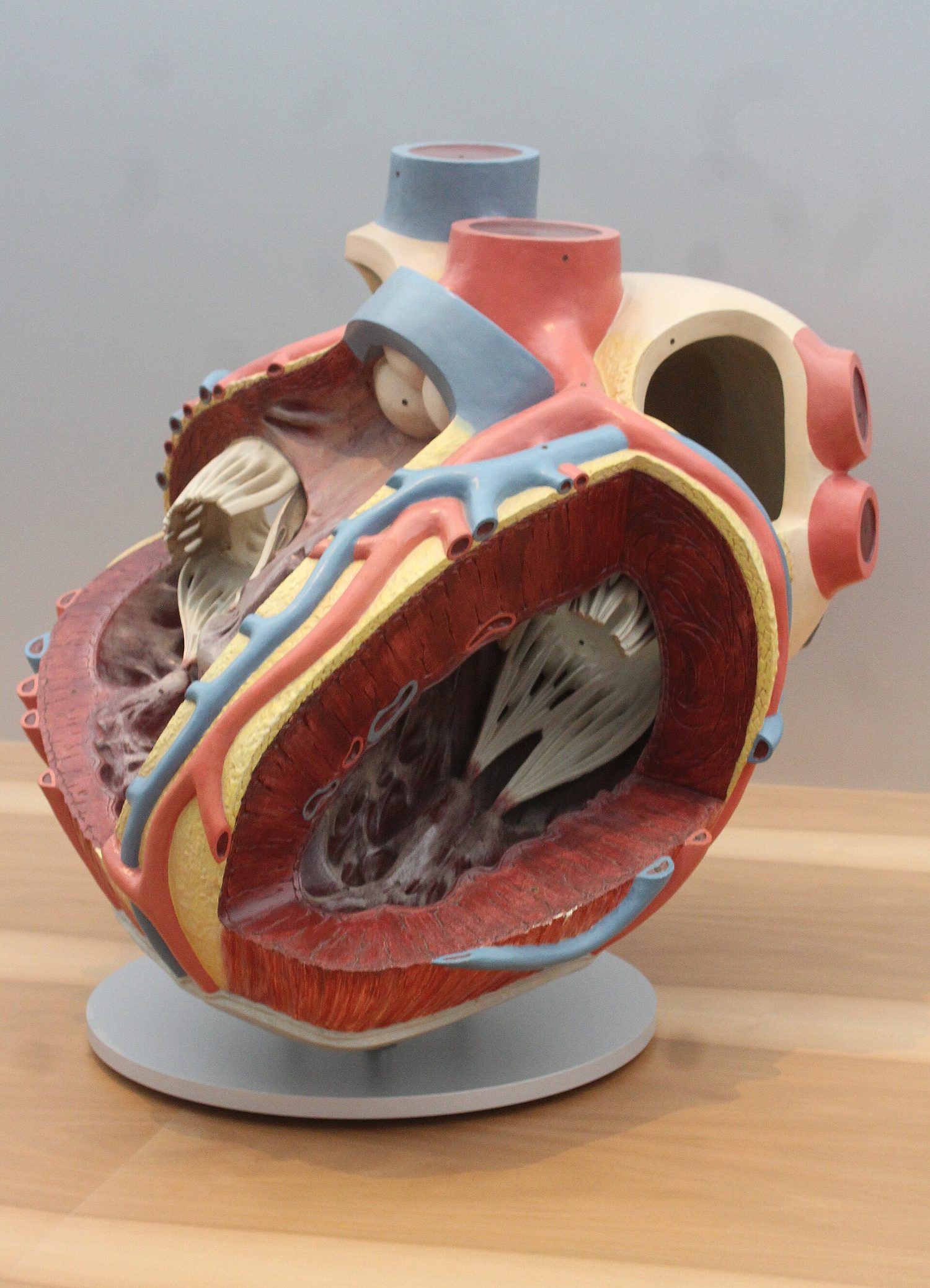
ภาพด้านหน้าของหัวใจแสดง atria และ ventricles ซ้าย-ขวา, septum ระหว่างกัน, วาล์ว, vena cava, pulmonary artery, aorta และ pulmonary vein; ventricle ซ้ายมีผนังหนาที่สุด
ห้องหัวใจสี่ช่อง; ventricle ซ้ายมีผนังหนาที่สุดเพื่อสูบเลือดไปทั่วทั้งร่างกาย
โมเดลกายวิภาคหัวใจมนุษย์ตัดเปิด แสดงห้องหัวใจสี่ช่องและผนังกล้ามเนื้อหนาของ ventricle ซ้าย
หัวใจชิ้นเดียวกันในรูปแบบนิทรรศการ: ห้องหัวใจสี่ช่อง และผนัง ventricle ซ้ายที่คุณมองเห็นได้ว่าเป็นผนังที่หนา hơn

วิธีการเต้นของหัวใจ (ส่วนเสริม)

  1. chambers หัวใจส่วนบน (收缩) และดันเลือดลงสู่ chamber หัวใจส่วนล่าง
  2. chamber หัวใจส่วนล่าง contract และดันเลือดออกไปยังหลอดเลือดแดง; วาล์วจะปิดสนิทเพื่อไม่ให้เลือดไหลย้อนกลับ
  3. กล้ามเนื้อผ่อนคลายและหัวใจเติมเลือดอีกครั้งหนึ่ง

เลือดถูกปั๊มออกจากหัวใจผ่านหลอดเลือดแดง และไหลกลับเข้าสู่หัวใจผ่านหลอดเลือดดำ

อัตราการเต้นของหัวใจและการออกกำลังกาย

คุณสามารถตรวจสอบการทำงานของหัวใจได้โดยใช้เครื่อง ECG, การสัมผัส ชีพจร ในหลอดเลือดแดง, หรือการฟังเสียงวาล์วหัวใจปิด While ออกกำลังกาย อัตราการเต้นของหัวใจ จะเพิ่มขึ้น เพื่อให้เลือดไปเลี้ยงกล้ามเนื้อได้เร็วขึ้น นำออกซิเจนและกลูโคสเพิ่มเติมที่กล้ามเนื้อต้องการมาด้วย

Chain: การออกกำลังกายทำให้กล้ามเนื้อต้องการออกซิเจนและกลูโคสมากขึ้น ดังนั้นอัตราการเต้นของหัวใจจึงเพิ่มขึ้น และเลือดไปยังกล้ามเนื้อได้เร็วขึ้น
ขณะออกกำลังกาย อัตราการเต้นของหัวใจเพิ่มขึ้น sehingga เลือดไปยังกล้ามเนื้อได้เร็วขึ้น

ตัวอย่างวิธีทำ. นักเรียนนับ 18 ครั้งของชีพจรใน 15 วินาที ขณะพัก, และ 33 ครั้งใน 15 วินาที ทันทีหลังการออกกำลังกาย ให้หาอัตราการเต้นของหัวใจทั้งสองกรณีเป็นครั้งต่อนาที หนึ่งนาทีมี 60 ÷ 15 = 4 เท่าของ 15 วินาที ดังนั้นให้คูณด้วย 4 ขณะพัก: 18 × 4 = 72 ครั้งต่อนาที. หลังการออกกำลังกาย: 33 × 4 = 132 ครั้งต่อนาที. การเพิ่มขึ้นนี้ส่งมอบออกซิเจนและกลูโคสเพิ่มให้กับกล้ามเนื้อ ข้อผิดพลาดที่พบบ่อยที่สุดคือการเขียนค่าที่นับได้ใน 15 วินาที ลงไปโดยตรงว่าเป็นอัตราการเต้นของหัวใจ - ต้องปรับขยายให้เท่ากับหนึ่งนาทีเต็มเสมอ

โรคหัวใจหลอดเลือด coronary

โรคหัวใจหลอดเลือด coronary เกิดขึ้นเมื่อหลอดเลือด coronary คับหรืออุดตัน ทำให้กล้ามเนื้อหัวใจได้รับออกซิเจนไม่เพียงพอ ปัจจัยเสี่ยง ได้แก่ อาหารที่มีไขมันสูง, ขาดการออกกำลังกาย, ความเครียด, การสูบบุหรี่, ประวัติครอบครัว (พันธุกรรม), อายุมาก, และการเป็นเพศชาย อาหารที่ดีต่อสุขภาพและการออกกำลังกายอย่างสม่ำเสมอช่วยลดความเสี่ยงได้

Artery coronary ที่แคบลงจากการสะสมของไขมัน ทำให้กล้ามเนื้อหัวใจได้รับออกซิเจนไม่พอ พร้อมแสดงปัจจัยเสี่ยงและการรับประทานอาหารที่ดีต่อการออกกำลังกายอย่างสม่ำเสมอช่วยลดความเสี่ยง
โรคหัวใจหลอดเลือด coronary ทำให้หลอดเลือดแคบลง; ปัจจัยเสี่ยงหลายประการเพิ่มความเสี่ยง และวิถีชีวิตที่ดีลดความเสี่ยงลง
Explore · ⁨สำรวจ⁩

Blood flow through the heart · ⁨การไหลเวียนเลือดผ่านหัวใจ⁩

The four chambers and their valves keep blood moving one way — deoxygenated to the lungs, oxygenated to the body. · ⁨ห้องหัวใจทั้งสี่และวาล์วช่วยให้เลือดไหลทางเดียว — ขาดออกซิเจนไปยังปอด, มีออกซิเจนไปร่างกาย⁩

Explore · ⁨สำรวจ⁩

The heart (double circulation) · ⁨หัวใจ (การไหลเวียนเลือดสองรอบ)⁩

Follow the blood: blue comes back from the body, the right side pumps it to the lungs (it turns red), and the left side pumps it out to the body — a double circulation. · ⁨ติดตามการไหลของเลือด: สีน้ำเงินกลับมาจากร่างกาย ด้านขวาสูบไปปอด (เปลี่ยนเป็นสีแดง) และด้านซ้ายสูบออกไปร่างกาย — การไหลเวียนเลือดสองรอบ⁩

Vocabulary · ⁨คำศัพท์⁩ Train · ⁨ฝึกฝน⁩
English ไทย
circulatory system/ˌsɜːkjʊˈleɪtəri ˈsɪstəm/ ระบบไหลเวียนเลือด
heart/hɑːt/ หัวใจ
valves/vælvz/ วาล์ว
single circulation/ˈsɪŋɡl ˌsɜːkjʊˈleɪʃn/ การไหลเวียนเลือดแบบเดียว
septum/ˈseptəm/ septum
atrium/ˈeɪtrɪəm/ ห้องหัวใจบน
ventricle/ˈventrɪkl/ ห้องหัวใจ
coronary arteries/ˈkɒrənəri ˈɑːtəriz/ หลอดเลือดหัวใจ
oxygenated blood/ˈɒksɪdʒeneɪtɪd blʌd/ เลือดที่มีออกซิเจน
deoxygenated blood/diːˈɒksɪdʒeneɪtɪd blʌd/ เลือดที่ไม่มีออกซิเจน
atrioventricular valves/ˌeɪtrɪˌəʊvənˈtrɪkjʊlə vælvz/ วาล์วระหว่าง心房และ ventricle
semilunar valves/ˌsemɪˈluːnə vælvz/ วาล์วรูปครึ่งวงกลม
contract/ˈkɒntrækt/ สัญญา / Contract
pulse/pʌls/ ชีพจร
heart rate/hɑːt reɪt/ อัตราการเต้นของหัวใจ
coronary heart disease/ˈkɒrənəri hɑːt dɪˈziːz/ โรคหัวใจ Coronary
risk factors/rɪsk ˈfæktəz/ ปัจจัยเสี่ยง
arteries/ˈɑːtəriz/ หลอดเลือดแดง
veins/veɪnz/ หลอดเลือดดำ
capillaries/ˈkæpɪləriz/ หลอดเลือดฝอย
surface area/ˈsɜːfɪs ˈeərɪə/ พื้นที่ผิว
aorta/eɪˈɔːtə/ หลอดเลือดแดงเอออร์ตา
vena cava/ˈviːnə ˈkɑːvə/ หลอดเลือดดำใหญ่ (vena cava)
pulmonary artery/ˈpʌlmənəri ˈɑːtəri/ หลอดเลือดปอด
pulmonary vein/ˈpʌlmənəri veɪn/ หลอดเลือดดำปอด
renal artery/ˈriːnl ˈɑːtəri/ หลอดเลือดไต
renal vein/ˈriːnl veɪn/ หลอดเลือดดำไต
hepatic artery/heˈpætɪk ˈɑːtəri/ หลอดเลือดTpatic
hepatic vein/heˈpætɪk veɪn/ หลอดเลือดTpatic Vein
hepatic portal vein/heˈpætɪk ˈpɔːtl veɪn/ Tpatic Portal Vein
red blood cells/red blʌd selz/ เม็ดเลือดแดง
pigment/ˈpɪɡmənt/ pigment
haemoglobin/ˌhiːməˈɡləʊbɪn/ ฮีโมโกลบิน
white blood cells/waɪt blʌd selz/ เม็ดเลือดขาว
platelets/ˈpleɪtlɪts/ เกล็ดเลือด
clot/klɒt/ ลิ่มเลือด
plasma/ˈplæzmə/ พลาสมา
ions/ˈaɪɒnz/ ไอออน
nutrients/ˈnjuːtrɪənts/ สารอาหาร (nutrients)
urea/juːˈrɪə/ ยูเรีย
hormones/ˈhɔːməʊnz/ ฮอร์โมน (hormones)
carbon dioxide/ˈkɑːbən daɪˈɒksaɪd/ คาร์บอนไดออกไซด์
pathogens/ˈpæθədʒnz/ เชื้อโรค
phagocytes/ˈfæɡəsaɪts/ ฟักโกไซต์
phagocytosis/ˌfæɡəsɪˈtəʊsɪs/ โฟเจโตซิส
lymphocytes/ˈlɪmfəsaɪts/ ลิมโฟไซต์
antibodies/ˈæntɪbɒdiz/ แอนติบอดี
fibrinogen/ˈfɪbrɪnədʒn/ fibrinogen
fibrin/ˈfɪbrɪn/ fibrin
Watch lesson · ⁨ดูบทเรียน⁩
9.3

Blood vessels · ⁨หลอดเลือด⁩

Syllabus · ⁨หลักสูตร⁩
English
Core Supplement
1 Describe the structure of arteries, veins and capillaries, limited to: relative thickness of wall, diameter of the lumen and the presence of valves in veins 4 Explain how the structure of arteries and veins is related to the pressure of the blood that they transport
2 State the functions of capillaries 5 Explain how the structure of capillaries is related to their functions
3 Identify in diagrams and images the main blood vessels to and from the: (a) heart, limited to: vena cava, aorta, pulmonary artery and pulmonary vein (b) lungs, limited to: pulmonary artery and pulmonary vein (c) kidney, limited to: renal artery and renal vein 6 Identify, in diagrams and images, the main blood vessels to and from the liver as: hepatic artery, hepatic veins and hepatic portal vein
ไทย
แก่น เพิ่มเติม
1 อธิบายโครงสร้างของหลอดเลือดแดง หลอดเลือดดำ และหลอดเลือดฝอย โดยจำกัดเฉพาะ: ความหนาสัมบูรณ์ของผนัง, เส้นผ่านศูนย์กลางของช่องภายใน และการมีวาล์วในหลอดเลือดดำ 4 อธิบายความสัมพันธ์ระหว่างโครงสร้างของหลอดเลือดแดงและหลอดเลือดดำกับความดันของเลือดที่ขนส่ง
2 ระบุหน้าที่ของหลอดเลือดฝอย 5 อธิบายความสัมพันธ์ระหว่างโครงสร้างของหลอดเลือดฝอยกับหน้าที่ของมัน
3 ชี้ระบุในแผนภาพและภาพถ่าย หลอดเลือดหลักที่นำเลือดไปและออกจาก: (ก) หัวใจ, จำกัดเฉพาะ: เวนาควา, แอโอตา, หลอดเลือดแดงปอด และหลอดเลือดดำปอด (ข) ปอด, จำกัดเฉพาะ: หลอดเลือดแดงปอด และหลอดเลือดดำปอด (ค) ไต, จำกัดเฉพาะ: หลอดเลือดแดงไต และหลอดเลือดดำไต 6 ชี้ระบุในแผนภาพและภาพถ่าย หลอดเลือดหลักที่นำเลือดไปและออกจากตับ โดยระบุชื่อ: หลอดเลือดแดงตับ, หลอดเลือดดำตับ และหลอดเลือดดำพอร์ทัลตับ

Source: Cambridge International syllabus · ⁨แหล่งที่มา: หลักสูตร Cambridge International⁩

English

There are three kinds of blood vessel. The space inside a vessel is its lumen 管腔.

Vessel Wall Lumen Valves? Job
arteries 动脉 thick, muscular, elastic narrow no carry blood away from the heart, at high pressure
veins 静脉 thin wide yes carry blood back to the heart, at low pressure
capillaries 毛细血管 one cell thick very narrow no let oxygen, glucose and wastes pass between blood and cells

(Supplement) Arteries have thick elastic walls to cope with the high pressure from the heart. Veins have valves because their blood is at low pressure and could otherwise flow backwards. Capillaries are very thin (one cell thick) and very narrow, giving a large surface area 表面积 and a short distance for fast exchange.

Main blood vessels

Connects Artery (carries blood out) Vein (carries blood in)
heart ↔ body aorta 主动脉 vena cava 腔静脉
heart ↔ lungs pulmonary artery 肺动脉 pulmonary vein 肺静脉
↔ kidney renal artery 肾动脉 renal vein 肾静脉

(Supplement) The liver is served by the hepatic artery 肝动脉, the hepatic vein 肝静脉, and the hepatic portal vein 肝门静脉, which brings blood from the gut to the liver.

ไทย

มีหลอดเลือดสามชนิด ช่องว่างภายในหลอดเลือดเรียกว่า lumen

(socket) ผนัง ช่องว่างภายใน มีวาล์ว? หน้าที่
หลอดเลือดแดง หนา, กล้ามเนื้อ, ยืดหยุ่น แคบ ไม่มี พาเลือดออกจากหัวใจ ด้วยความดันสูง
หลอดเลือดดำ บาง กว้าง มี พาเลือดกลับเข้าสู่หัวใจ ด้วยความดันต่ำ
หลอดเลือดฝอย บางเพียงชั้นเซลล์เดียว แคบมาก ไม่มี ให้ออกซิเจน, กลูโคส และของเสียผ่านระหว่างเลือดกับเซลล์

(ส่วนเสริม) หลอดเลือดแดงมีผนังยืดหยุ่นหนาเพื่อรองรับความดันสูงจากหัวใจ หลอดเลือดดำมีวาล์วเพราะเลือดมีความดันต่ำและอาจไหลย้อนกลับได้ หลอดเลือดฝอยมีบางมาก (บางเพียงชั้นเซลล์เดียว) และแคบมาก ทำให้มีพื้นที่ผิวมากและระยะทางสั้นสำหรับการแลกเปลี่ยนสารอย่างรวดเร็ว

ภาพตัดขวางของหลอดเลือดทั้งสาม: หลอดเลือดแดงที่มีผนังกล้ามเนื้อหนาและ lumen แคบ, หลอดเลือดดำที่มีผนังบาง lumen กว้าง และมีวาล์ว, และหลอดเลือดฝอยขนาดเล็กที่มีผนังเพียงชั้นเซลล์เดียว
หลอดเลือดแดง หลอดเลือดดำ และหลอดเลือดฝอยถูกสร้างมาเพื่อทำหน้าที่เฉพาะเจาะจง

หลอดเลือดหลัก

เชื่อมต่อกับ หลอดเลือดแดง (พาเลือดออก) หลอดเลือดดำ (พาเลือดเข้า)
หัวใจ ↔ ร่างกาย เออร์โต เวนาคาวา
หัวใจ ↔ ปอด หลอดเลือดแดงปอด หลอดเลือดดำปอด
↔ ไต หลอดเลือดแดงไต หลอดเลือดดำไต

(ส่วนเสริม) ตับได้รับเลือดจากหลอดเลือดแดงตับ, หลอดเลือดดำตับ และหลอดเลือดดำพอร์ทัลตับ ซึ่งนำเลือดจากลำไส้ไปเลี้ยงตับ

Explore · ⁨สำรวจ⁩

The three blood vessels · ⁨หลอดเลือดสามชนิด⁩

Compare an artery, a vein and a capillary — their walls and lumens suit the pressure and job of each. · ⁨เปรียบเทียบหลอดเลือดแดง หลอดเลือดดำ และหลอดเลือดฝอย — ผนังและรูภายในรองรับความดันและหน้าที่ของแต่ละชนิด.⁩

Vocabulary · ⁨คำศัพท์⁩ Train · ⁨ฝึกฝน⁩
English ไทย
blood vessels/blʌd ˈveslz/ หลอดเลือด
lumen/ˈluːmen/ lumen
9.4

Blood · ⁨เลือด⁩

Syllabus · ⁨หลักสูตร⁩
English
Core Supplement
1 List the components of blood as: red blood cells, white blood cells, platelets and plasma
2 Identify red and white blood cells in photomicrographs and diagrams 5 Identify lymphocytes and phagocytes in photomicrographs and diagrams
3 State the functions of the following components of blood: (a) red blood cells in transporting oxygen, including the role of haemoglobin (b) white blood cells in phagocytosis and antibody production (c) platelets in clotting (details are not required) (d) plasma in the transport of blood cells, ions, nutrients, urea, hormones and carbon dioxide 6 State the functions of: (a) lymphocytes – antibody production (b) phagocytes – engulfing pathogens by phagocytosis
4 State the roles of blood clotting as preventing blood loss and the entry of pathogens 7 Describe the process of clotting as the conversion of fibrinogen to fibrin to form a mesh
ไทย
แก่น เพิ่มเติม
1 รายชื่อองค์ประกอบของเลือด ได้แก่: เซลล์เม็ดเลือดแดง, เซลล์เม็ดเลือดขาว, เพลตเลต และพลาสมา
2 ชี้ระบุเซลล์เม็ดเลือดแดงและเซลล์เม็ดเลือดขาวในภาพจุลทรรศน์และแผนภาพ 5 ชี้ระบุไลโมฟิโลไซต์และฟาโกไซต์ในภาพจุลทรรศน์และแผนภาพ
3 ระบุหน้าที่ขององค์ประกอบเลือดต่อไปนี้: (ก) เซลล์เม็ดเลือดแดงในการขนส่งออกซิเจน, รวมถึงบทบาทของฮีโมโกลบิน (ข) เซลล์เม็ดเลือดขาวในการกินเชื้อโรคโดยฟาโกไซโทซิสและการผลิตแอนติบอดี (ค) เพลตเลตในการสร้างลิ่มเลือด (ไม่จำเป็นต้องรายละเอียด) (ง) พลาสมาในการขนส่งเซลล์เลือด, ไอออน, สารอาหาร, ยูเรีย,ฮอร์โมนและคาร์บอนไดออกไซด์ 6 ระบุหน้าที่ของ: (ก) ไลโมฟิโลไซต์ – การผลิตแอนติบอดี (ข) ฟาโกไซต์ – การกลืนเชื้อโรคด้วยกระบวนการฟาโกไซโทซิส
4 ระบุบทบาทของการเกิดลิ่มเลือด即在ป้องกันการสูญเสียเลือดและการเข้าสู่ของเชื้อโรค 7 อธิบายกระบวนการเกิดลิ่มเลือด即在การเปลี่ยนไฟบริโนเจนเป็นไฟบรินเพื่อสร้างตาข่าย

Source: Cambridge International syllabus · ⁨แหล่งที่มา: หลักสูตร Cambridge International⁩

English

Blood has four parts:

Part Function
red blood cells 红细胞 carry oxygen, using the red pigment 色素 haemoglobin 血红蛋白
white blood cells 白细胞 defend the body against disease
platelets 血小板 help the blood to clot 凝血
plasma 血浆 a liquid that carries blood cells, ions 离子, nutrients 营养物质, urea 尿素, hormones 激素 and carbon dioxide 二氧化碳

Defending the body, and clotting

White blood cells fight pathogens 病原体 (the microbes that cause disease) in two ways:

  • phagocytes 吞噬细胞 carry out phagocytosis 吞噬作用 — they surround and digest the pathogens.
  • lymphocytes 淋巴细胞 make antibodies 抗体, which stick to the pathogens and destroy them.

When you cut yourself, the blood clots to seal the wound. This stops blood loss and stops pathogens getting in. (Supplement) During clotting, a soluble protein called fibrinogen 纤维蛋白原 is changed into threads of fibrin 纤维蛋白. The threads form a net that traps blood cells and makes a solid clot.

ไทย

เลือดประกอบด้วยสี่ส่วน:

ส่วนประกอบ หน้าที่
เม็ดเลือดแดง พาออกซิเจน โดยใช้สี ฮีโมโกลบิน
เม็ดเลือดขาว ป้องกันร่างกายจากโรค
เกล็ดเลือด ช่วยให้เลือดจับตัวเป็นลิ่ม
พลาสมา ของเหลวที่พาเม็ดเลือด ไอออน, สารอาหาร, ยูเรีย, ฮอร์โมน และคาร์บอนไดออกไซด์
หลอดเลือดที่แยกชั้นแล้ว: ชั้นบนเป็นพลาสมาใส ชั้นบางๆ ของเม็ดเลือดขาวและเกล็ดเลือด และชั้นล่างเป็นเม็ดเลือดแดง พร้อมภาพขยายของเม็ดเลือดแดง เม็ดเลือดขาว และเกล็ดเลือด
เลือดประกอบด้วยเซลล์เม็ดเลือดแดง, เซลล์เม็ดเลือดขาว และเกล็ดเลือดที่ลอยอยู่ในพลาสมาเหลว
ภาพจุลินาโนสแกนนิ่งของเม็ดเลือดจริง: เม็ดเลือดแดงรูปดิสก์บุ๋มหลายชิ้น เม็ดเลือดขาวกลมขรุขระสองเม็ด และเศษเกล็ดเลือดเล็กๆ
เม็ดเลือดจริงภายใต้กล้องจุลทรรศน์อิเล็กตรอน: เม็ดเลือดแดง (ดิสก์), เม็ดเลือดขาว และเกล็ดเลือด

การป้องกันร่างกายและการจับตัวเป็นลิ่ม

เม็ดเลือดขาวต่อสู้กับเชื้อโรค (จุลินทรีย์ที่ทำให้เกิดโรค) ได้สองวิธี:

  • แมคโรฟาจ ทำการฟาโกไซโทซิส — โดยล้อมรอบและย่อยสลายเชื้อโรค
  • ไลม์โฟไซต์ สร้างแอนติบอดี ซึ่งยึดติดกับเชื้อโรคและทำลายมัน

เมื่อคุณบาดแผล เลือดจะจับตัวเป็นลิ่มเพื่อปิดบาดแผล สิ่งนี้ช่วยหยุดเลือดไหลและป้องกันเชื้อโรคเข้า (ส่วนเสริม) ระหว่างการจับตัวเป็นลิ่ม โปรตีนที่ละลายน้ำได้ชื่อ ไฟบริโนเจน จะเปลี่ยนเป็นเส้นใยของ ไฟบริน เส้นใยเหล่านี้สร้างตาข่ายกักเม็ดเลือดและทำให้เป็นลิ่มแข็งตัว

Explore · ⁨สำรวจ⁩

Blood component lab · ⁨ห้องปฏิบัติการส่วนประกอบเลือด⁩

Classify blood components by the job they do. · ⁨จำแนกส่วนประกอบเลือดตามหน้าที่⁩

9.4

Exam tips · ⁨ข้อแนะนำสำหรับการสอบ⁩

English
  • Circulatory system = pump (heart) + vessels + valves for one-way flow.
  • Double circulation (mammals): blood passes through the heart twice. The left ventricle wall is thickest because it pumps to the whole body.
  • Arteries carry blood away (thick, high pressure); veins carry it back (valves, low pressure); capillaries are for exchange (one cell thick).
  • Blood = red cells (oxygen, haemoglobin) + white cells (defence) + platelets (clotting) + plasma (transport).
  • Phagocytes engulf pathogens; lymphocytes make antibodies.
ไทย
  • ระบบไหลเวียนเลือด = ปั๊ม (หัวใจ) + หลอดเลือด + วาล์วสำหรับการไหลทางเดียว
  • การไหลเวียนเลือดแบบคู่ (สัตว์เลี้ยงลูกด้วยนม): เลือดไหลผ่านหัวใจสองครั้ง ผนังห้องหัวใจด้านซ้ายหนาที่สุดเพราะปั๊มเลือดไปทั่วร่างกาย
  • หลอดเลือดแดงพาเลือดออก (หนา, ความดันสูง); หลอดเลือดดำพาเลือดกลับ (มีวาล์ว, ความดันต่ำ); หลอดเลือดฝอยใช้สำหรับการแลกเปลี่ยน (บางเพียงชั้นเซลล์เดียว)
  • เลือด = เม็ดเลือดแดง (ออกซิเจน, ฮีโมโกลบิน) + เม็ดเลือดขาว (ป้องกัน) + เกล็ดเลือด (การจับตัวเป็นลิ่ม) + พลาสมา (การขนส่ง)
  • แมค्रोฟาจกลืนกินเชื้อโรค; ไลม์โฟไซต์สร้างแอนติบอดี

Interactive lessons on this topic · ⁨บทเรียนเชิงโต้ตอบสำหรับหัวข้อนี้⁩

Work through it step by step, with instant-check exercises. · ⁨ทำทีละขั้นตอน พร้อมแบบฝึกหัดตรวจสอบผลทันที⁩

Past Papers · ⁨ข้อสอบย้อนหลัง⁩

More topics in IGCSE Biology · ⁨Biology IGCSE⁩ · ⁨หัวข้อเพิ่มเติมใน IGCSE Biology · ⁨Biology IGCSE⁩⁩

Log in or create account · ⁨เข้าสู่ระบบหรือสร้างบัญชี⁩

IGCSE, A-Level & AP