Your heart beats about a hundred thousand times a day, and it has never once stopped to rest. It is not really one pump — it is two, side by side, sharing a…
English narration · English + 中文 subtitles burned in · קריאת קול באנגלית · תרגום אנגלי + סינית שרוף בתוך הסרטון
9.1
The circulatory system · מערכת המחזור הדם
Syllabus · סיילבוס
English
Core
Supplement
1 Describe the circulatory system as a system of blood vessels with a pump and valves to ensure one-way flow of blood
2 Describe the single circulation of a fish
3 Describe the double circulation of a mammal
4 Explain the advantages of a double circulation
עברית
ליבה
תוספת
1 תאר את מערכת הדם כמערכת של כלי דם עם משאבה ושסתומים כדי להבטיח זרימת דם חד-כיוונית
2 תאר את הסירוקולציה היחידה של דג
3 תאר את הסירוקולציה הכפולה של יונק
4 הסבר את היתרונות של סירוקולציה כפולה
Source: Cambridge International syllabus · מקור: הסיילבוס הבינלאומי של קמבריד'ג'
English
The circulatory system 循环系统 carries blood around the body. It has three parts: a pump 泵 (the heart 心脏), a network of blood vessels 血管, and valves 瓣膜 that keep the blood flowing one way only.
Single and double circulation (Supplement)
A fish has a single circulation 单循环: the blood passes through the heart once on each trip around the body.
A mammal 哺乳动物 has a double circulation 双循环: the blood passes through the heart twice on each full trip — once on the way to the lungs, and once on the way to the rest of the body.
The advantage of a double circulation: the blood can be pumped again at high pressure 压力 before going to the body, so it travels faster and delivers oxygen 氧气 quickly.
עברית
מערכת** הדם** מובילה דם לכל הגוף. היא מורכבת משלושה חלקים: משאבה (הלב), רשת של** כלי דם**, ושסתומים שמבטיחים זרימת דם בכיוון אחד בלבד.
זרימה יחידה וכפולה (תוספת)
לדג יש** זרימה יחידה**: הדם עובר דרך הלב** פעם אחת** בכל סיבוב בגוף.
לחיות** סרטן יש** זרימה כפולה: הדם עובר דרך הלב** פעמיים** בכל סיבוב מלא — פעם בדרך לריאות ופעם בדרך שאר הגוף.
היתרון של זרימה כפולה: הדם יכול להיות משופר שוב בלחץ גבוה לפני שהוא מגיע לגוף, כך שהוא נע מהר יותר ומספק** חמצן** במהירות.
*בזרימה כפולה, הדם עובר דרך הלב פעמיים בכל סיבוב סביב הגוף
Explore · חקור
Double circulation · מחזור כפול
Mammals have a double circulation — blood passes through the heart TWICE on each full trip round the body. · ליונקים יש זרימת דם כפולה — הדם עובר דרך הלב פעמיים בכל סיבוב מלא בגוף.
1 Identify in diagrams and images the structures of the mammalian heart, limited to: muscular wall, septum, left and right ventricles, left and right atria, one-way valves and coronary arteries
7 Identify in diagrams and images the atrioventricular and semilunar valves in the mammalian heart
8 Explain the relative thickness of: (a) the muscle walls of the left and right ventricles (b) the muscle walls of the atria compared to those of the ventricles
9 Explain the importance of the septum in separating oxygenated and deoxygenated blood
10 Describe the functioning of the heart in terms of the contraction of muscles of the atria and ventricles and the action of the valves
2 State that blood is pumped away from the heart in arteries and returns to the heart in veins
3 State that the activity of the heart may be monitored by: ECG, pulse rate and listening to sounds of valves closing
4 Investigate and describe the effect of physical activity on the heart rate
11 Explain the effect of physical activity on the heart rate
5 Describe coronary heart disease in terms of the blockage of coronary arteries and state the possible risk factors including: diet, lack of exercise, stress, smoking, genetic predisposition, age and sex
6 Discuss the roles of diet and exercise in reducing the risk of coronary heart disease
עברית
ליבה
תוספת
1 זיהה במخططات ותמונות את מבנה הלב ביונקים, המוגבל ל: קיר שרירי, ספטום, ventricles שמאל וימין, atria שמאל וימין, שסתומים חד-כיווניים ושקיקי לב
7 זיהה במخططات ותמונות את השסתומים האטריאו-ונטריקולריים והשסתומים למינריים בלב היונקים
8 הסבר את העובי היחסי של: (א) הקירות השריריים של הventricles השמאלי והימני; (ב) הקירות השריריים של האtrias בהשוואה לאלו של הventricles
9 הסבר את חשיבות הספטום בבידוד דם מחומצן ודם לא מחומצן
10 תאר את פעולת הלב בתוך התכווצות שרירי האtrias והventricles ופעולת השסתומים
2 ציין שדם מופקע מן הלב באמצעות עורקים וחוזר אל הלב באמצעות ורידים
3 ציין שהפעילות של הלב עשויה להיות ניטור על ידי: ECG, קצב דופק והאזנה לקולות סגירת השסתומים
4 חקור ותיאר את השפעת הפעילות הגופנית על קצב הלב
11 הסבר את השפעת הפעילות הגופנית על קצב הלב
5 תאר מחלת לב כרונית במונחים של חסימת כלי הדם הקורונריים ופרט גורמי הסיכון האפשריים, לרבות: תזונה, חוסר פעילות גופנית, מתח, עישון, נטייה גנטית, גיל ומין
6 דן בתפקידה של התזונה והפעילות הגופנית בהפחתת הסיכון למחלת לב כרונית
Source: Cambridge International syllabus · מקור: הסיילבוס הבינלאומי של קמבריד'ג'
English
The heart is made of muscle 肌肉. A wall called the septum 隔膜 divides it into a left side and a right side. On each side there is an upper chamber, the atrium 心房, and a lower chamber, the ventricle 心室. Valves between the chambers stop the blood flowing backwards. The heart muscle is fed with blood by the coronary arteries 冠状动脉.
(Supplement) The wall of the left ventricle is thicker than the right, because it must pump blood all the way around the body; the right side only pumps blood to the nearby lungs. The atria have thin walls, as they only push blood into the ventricles just below them.
The septum keeps oxygenated blood 含氧血 on the left side apart from deoxygenated blood 缺氧血 on the right side, so the two never mix.
(Supplement) The valves between each atrium and ventricle are the atrioventricular valves 房室瓣. The valves at the exit of each ventricle are the semilunar valves 半月瓣.
How the heart beats (Supplement)
The atria contract 收缩 and push blood down into the ventricles.
The ventricles contract and force blood out into the arteries; the valves snap shut so blood cannot flow back.
The muscle relaxes and the heart fills with blood again.
Blood is pumped away from the heart in arteries and returns to the heart in veins.
Heart rate and exercise
You can check the heart's activity with an ECG, by feeling the pulse 脉搏 in an artery, or by listening to the valves closing. During exercise the heart rate 心率 goes up, so blood reaches the muscles faster, bringing the extra oxygen and glucose they need.
Worked example. A student counts 18 pulse beats in 15 seconds at rest, and 33 beats in 15 seconds straight after exercise. Give both heart rates in beats per minute. A minute holds 60 ÷ 15 = 4 lots of 15 seconds, so multiply by 4. At rest: 18 × 4 = 72 beats per minute. After exercise: 33 × 4 = 132 beats per minute. The rise delivers more oxygen and glucose to the muscles. Writing the 15-second count straight down as the heart rate is the commonest mistake - always scale it up to a full minute.
Coronary heart disease
Coronary heart disease 冠心病 happens when the coronary arteries become narrow or blocked, so the heart muscle cannot get enough oxygen. Risk factors 风险因素 include a diet high in fat, lack of exercise, stress, smoking, family history (genetics), older age, and being male. A healthy diet and regular exercise lower the risk.
עברית
הלב עשוי משריר. קיר הנקרא** ספטום** מחלק אותו לצד שמאל וצד ימין. בכל צד ישנם חלון עליון, האtrium, וחלון תחתון, הventricle. שסתומים בין החלונות מונעים זרימת דם הפוכה. שריר הלב מזווד בדם על ידי** העורקים הקורונריים**.
(תוספת) קיר ה ventricle השמאלי עבה מהימני, כי הוא צריך לשפוך דם לכל אורך הגוף; הצד הימני משפך דם רק לריאות הסמוכות. ל atria יש קירות דקים, כי הם רק דוחפים דם ל ventricles שמ下方יהם.
ה septum שומר על** דם עשיר בחמצן** בצד שמאל בנפרד מ** דם לא-עשיר בחמצן** בצד ימין, כך שהשניים לעולם לא מתערבלים.
(תוספת) השסתומים בין כל atrium ו ventricle הם** השסתומים ה atrioventricular**. השסתומים ביציאת כל ventricle הם** השסתומים ה semilunar**.
ארבעת חללי הלב; לקיר החדר השמאלי יש הקיר העבה ביותר, כדי לשאוב דם לכל הגוף
*אותו לב כמודל מוזיאלי: ארבעה חללים, וקיר החדר השמאלי שהעובי שלו נראה עבה יותר
כיצד הלב פועם (תוספת)
האוזניות מתכווצות ושואבות דם כלפי מטה אל החדרים.
החדרים מתכווצים ושואבים דם החוצה אל הערמונים; השסתומים נסגרים בחוזקה כך שדם לא יזרום אחורה.
השריר רפוי והלב ממלא בדם שוב.
דם משאוב מהלב בערמונים וחוזר ללב בוורידים.
קצב לב ותרגיל גופני
ניתן לבדוק את פעילות הלב באמצעות ECG, על ידי מגע בפולס בערמון, או בהקשבת לסגירת השסתומים. במהלך פעילות גופנית קצב הלב עולה, כך שדם מגיע לשרירים מהר יותר ומביא את החמצן והגלוקוז הנוספים שהם זקוקים להם.
*במהלך פעילות גופנית קצב הלב עולה כך שדם מגיע לשרירים מהר יותר
דוגמה מפורשת. סטודנט סופר 18 פעימות פולס ב-15 שניות במנוחה, ו-33 פעימות ב-15 שניות מיד לאחר תרגיל. נתנו את שני קצבי הלב בפעימות לדקה. לדקה יש 60 ÷ 15 = 4 מחלקות של 15 שניות, ולכן מכפילים ב-4. במנוחה: 18 × 4 = 72 פעימות לדקה. לאחר תרגיל: 33 × 4 = 132 פעימות לדקה. העלייה מספקת יותר חמצן וגלוקוז לשרירים. כתיבת הספירה של 15 שניות ישירות כקצב לב היא הטעות הנפוצה ביותר - יש תמיד להכפיל אותה עד לדקה מלאה.
מחלת לב כתרונית
מחלת לב כתרונית מתרחשת כאשר הערמונים הכתרוניים מצטמצמים או חסומים, כך שאין לשריר הלב מספיק חמצן. גורמי הסיכון כוללים תזונה עשירה בשומן, חוסר בתרגיל גופני, מתח, עישון, היסטוריה משפחתית (גנטיקה), גיל מבוגר והיות גבר. תזונה בריאה ותרגיל גופני קבועים מפחיתים את הסיכון.
*מחלת לב כתרונית מצמצמת את הערמון; מספר גורמי סיכון מעלים אותו ואורח חיים בריא מוריד אותו
Explore · חקור
Blood flow through the heart · זרימת דם דרך הלב
The four chambers and their valves keep blood moving one way — deoxygenated to the lungs, oxygenated to the body. · ארבעת החדרים והשסתומים שלהם שומרים על תנועת הדם בכיוון אחד — דם מועמם לריאות, ודם עשיר בחמצן לגוף.
Explore · חקור
The heart (double circulation) · הלב (זרימה כפולה)
Follow the blood: blue comes back from the body, the right side pumps it to the lungs (it turns red), and the left side pumps it out to the body — a double circulation. · עקבו אחרי הדם: הכחול חוזר מהגוף, הצד הימני משאב אותו לריאות (הוא הופך לאדום), והצד השמאל משאב אותו לגוף — זהו זרימה כפולה.
1 Describe the structure of arteries, veins and capillaries, limited to: relative thickness of wall, diameter of the lumen and the presence of valves in veins
4 Explain how the structure of arteries and veins is related to the pressure of the blood that they transport
2 State the functions of capillaries
5 Explain how the structure of capillaries is related to their functions
3 Identify in diagrams and images the main blood vessels to and from the: (a) heart, limited to: vena cava, aorta, pulmonary artery and pulmonary vein (b) lungs, limited to: pulmonary artery and pulmonary vein (c) kidney, limited to: renal artery and renal vein
6 Identify, in diagrams and images, the main blood vessels to and from the liver as: hepatic artery, hepatic veins and hepatic portal vein
עברית
ליבה
תוספת
1 תאר את מבנה העורקים, הוורידים והמלועיות, במגבלות הבאות: עובי הקיר היחסי, קוטר הלומה ונוכחות שסתומים בוורידים
4 הסבר כיצד המבנה של עורקים ווידים מקשור ללחץ הדם שהם מעבירים
2 פרט את הפונקציות של מלועיות
5 הסבר כיצד המבנה של מלועיות מקשור לפונקציותיהן
3 זיהה בתרשימים ובתמונות את כלי הדם הראשיים אל ועבור: (א) הלב, במגבלות הבאות: הוורה הצרה, האorta, העורק הריאי והווריד הריאי (ב) ריאות, במגבלות הבאות: העורק הריאי והווריד הריאי (ג) כליה, במגבלות הבאות: העורק הכלייתי והווריד הכלייתי
6 זיהה בתרשימים ובתמונות את כלי הדם הראשיים אל ועבור הכבד כ: עורק הכבד, ורידי הכבד וווריד הכבד החלקי
Source: Cambridge International syllabus · מקור: הסיילבוס הבינלאומי של קמבריד'ג'
English
There are three kinds of blood vessel. The space inside a vessel is its lumen 管腔.
Vessel
Wall
Lumen
Valves?
Job
arteries 动脉
thick, muscular, elastic
narrow
no
carry blood away from the heart, at high pressure
veins 静脉
thin
wide
yes
carry blood back to the heart, at low pressure
capillaries 毛细血管
one cell thick
very narrow
no
let oxygen, glucose and wastes pass between blood and cells
(Supplement) Arteries have thick elastic walls to cope with the high pressure from the heart. Veins have valves because their blood is at low pressure and could otherwise flow backwards. Capillaries are very thin (one cell thick) and very narrow, giving a large surface area 表面积 and a short distance for fast exchange.
Main blood vessels
Connects
Artery (carries blood out)
Vein (carries blood in)
heart ↔ body
aorta 主动脉
vena cava 腔静脉
heart ↔ lungs
pulmonary artery 肺动脉
pulmonary vein 肺静脉
↔ kidney
renal artery 肾动脉
renal vein 肾静脉
(Supplement) The liver is served by the hepatic artery 肝动脉, the hepatic vein 肝静脉, and the hepatic portal vein 肝门静脉, which brings blood from the gut to the liver.
עברית
ישנם שלושה סוגים של כלי דם. החלל הפנימי בכלי הוא ה-לומן.
כלי דם
דופן
לומן
שסתומים?
תפקיד
עורקים
עבים, שריריים ואלסטיים
צרים
אין
מובילים דם מרחוק הלב, בלחץ גבוה
ורידים
דקים
רחבים
כן
מובילים דם חזרה ללב, בלחץ נמוך
נימי דם
בעובי תא אחד
מאוד צרים
אין
מאפשרים מעבר של חמצן, גלוקוז ומפגעים בין הדם לתאים
(תוספת) לעורקים יש דפנות אלסטיות עבות כדי להתמודד עם הלחץ הגבוה מהלב. לורידים יש שסתומים מכיוון שהדם בהם נמצא בלחץ נמוך ועלול לזרום אחורה ללא קיום שסתומים אלו. נימי הדם דקים מאוד (בעובי תא אחד) וצרים מאוד, מה שמספק שטח פנים גדול ומרחק קצר להחלפה מהירה.
ערמוניות, ורידים ושיאים הם כל אחד מהם בנוים למטרתם
כלי דם ראשיים
מחבר
ערק (מוביל דם החוצה)
וריד (מוביל דם פנימה)
לב ↔ גוף
אורטה
וורדה תחתונה
לב ↔ ריאות
ערק ריאתי
ורייד ריאתי
↔ כליה
ערק כלוי
ורייד כלוי
(תוספת) הכבד מקבל אספקה דרך העורק הכבדי, הורייד הכבדי והווריד הפורטלי הכבדי, המוביל דם מהמערכת העיכולית לכבד.
Explore · חקור
The three blood vessels · שלושת כלי הדם
Compare an artery, a vein and a capillary — their walls and lumens suit the pressure and job of each. · השוו בין ערוגית, ורה ונימי דם — הקירות והחלל שלהם מותאמים ללחץ ולתפקיד של כל אחד.
1 List the components of blood as: red blood cells, white blood cells, platelets and plasma
2 Identify red and white blood cells in photomicrographs and diagrams
5 Identify lymphocytes and phagocytes in photomicrographs and diagrams
3 State the functions of the following components of blood: (a) red blood cells in transporting oxygen, including the role of haemoglobin (b) white blood cells in phagocytosis and antibody production (c) platelets in clotting (details are not required) (d) plasma in the transport of blood cells, ions, nutrients, urea, hormones and carbon dioxide
6 State the functions of: (a) lymphocytes – antibody production (b) phagocytes – engulfing pathogens by phagocytosis
4 State the roles of blood clotting as preventing blood loss and the entry of pathogens
7 Describe the process of clotting as the conversion of fibrinogen to fibrin to form a mesh
עברית
ליבה
תוספת
1 ספור את רכיבי הדם כ: תאי דם אדומים, תאי דם לבנים, פלטות וסרום
2 זיהה תאי דם אדומים ולבנים בפוטומיקרוגרפים ובתרשימים
3 פרט את הפונקציות של רכיבי הדם הבאים: (א) תאי דם אדומים בהובלת חמצן, כולל תפקיד ההימוגלובין (ב) תאי דם לבנים בפגוציטוז ובייצור אנטיגופים (ג) פלטות בקרישת דם (פרטים אינם נדרשים) (ד) סרום בהובלת תאי דם, יונים, מזון, אוריאה, הורמונים ודיוקسيد הפחמן
6 פרט את הפונקציות של: (א) לימפוציטים – ייצור אנטיגופים (ב) פגוציטים – בליעת מחוללים על ידי פגוציטוז
4 פרט את תפקידי קרישת הדם כמניעת אובדן דם והיכנסות מחוללים
7 תאר את תהליך הקרישה כהמרת פיברינוגן לפיברין ליצירת רשת
Source: Cambridge International syllabus · מקור: הסיילבוס הבינלאומי של קמבריד'ג'
English
Blood has four parts:
Part
Function
red blood cells 红细胞
carry oxygen, using the red pigment 色素haemoglobin 血红蛋白
white blood cells 白细胞
defend the body against disease
platelets 血小板
help the blood to clot 凝血
plasma 血浆
a liquid that carries blood cells, ions 离子, nutrients 营养物质, urea 尿素, hormones 激素 and carbon dioxide 二氧化碳
Defending the body, and clotting
White blood cells fight pathogens 病原体 (the microbes that cause disease) in two ways:
phagocytes 吞噬细胞 carry out phagocytosis 吞噬作用 — they surround and digest the pathogens.
lymphocytes 淋巴细胞 make antibodies 抗体, which stick to the pathogens and destroy them.
When you cut yourself, the blood clots to seal the wound. This stops blood loss and stops pathogens getting in. (Supplement) During clotting, a soluble protein called fibrinogen 纤维蛋白原 is changed into threads of fibrin 纤维蛋白. The threads form a net that traps blood cells and makes a solid clot.
עברית
לדם ארבעה חלקים:
חלק
תפקיד
תאי דם אדומים
מובילים חמצן באמצעות הפיגמנט האדום המוגלובין
תאי דם לבנים
מגנים על הגוף מפני מחלות
צלחות דם
מסייעות לקרישת הדם
פלזמה
נוזל המוביל תאי דם, יונים, חומרי תזונה, אוריאה, הורמונים ופחמן די-אוקסיד
דם מכיל תאי דם אדומים, לבנים ופלטות המופקות בפלזמה נוזליתתאי דם אמיתיים תחת מיקרוסקופ אלקטרוני: תאי אדומות (דיסקים), תאי לבן ופלטינות
הגנה על הגוף והקשירה
תאי לבן נלחמים במחוללי מחלות (המיקרובים הגורמים למחלה) בשתי דרכים:
פאגוציטים מבצעים פאגוציטוזה — הם מקיפים ועוררים את המחוללים.
לימפוציטים מייצרים נוגדנים, הנדבקים למחוללים ומשמידים אותם.
כאשר אתה נפצע, הדם מתקשה כדי לסגור את הפצע. כך נמנע מאובדן דם ומונע כניסת מחוללים. (משלים) במהלך הקשיחה, חלבון מסיס הנקרא פיברינוגן הופך לסיבי פיברין. הסיבים יוצרים רשת שתוחבת תאי דם ומייצרת קרישה מוצקה.
Explore · חקור
Blood component lab · מעבדת רכיבי דם
Classify blood components by the job they do. · סווגו את רכיבי הדם לפי התפקיד שהם מבצעים.
Pick one and the site follows you — notes, papers, videos and practice all open on it. · בחרו נושא אחד והאתר יעקוב אחריו — הערות, מסמכים, וידאו ותרגולים פתוחים בו.
Type to search notes, lessons, code, vocabulary and past-paper questions across every subject. · הקלד כדי לחפש הערות, שיעורים, קוד, אוצר מילים ושאלות מבחנים בכל הנושאים.


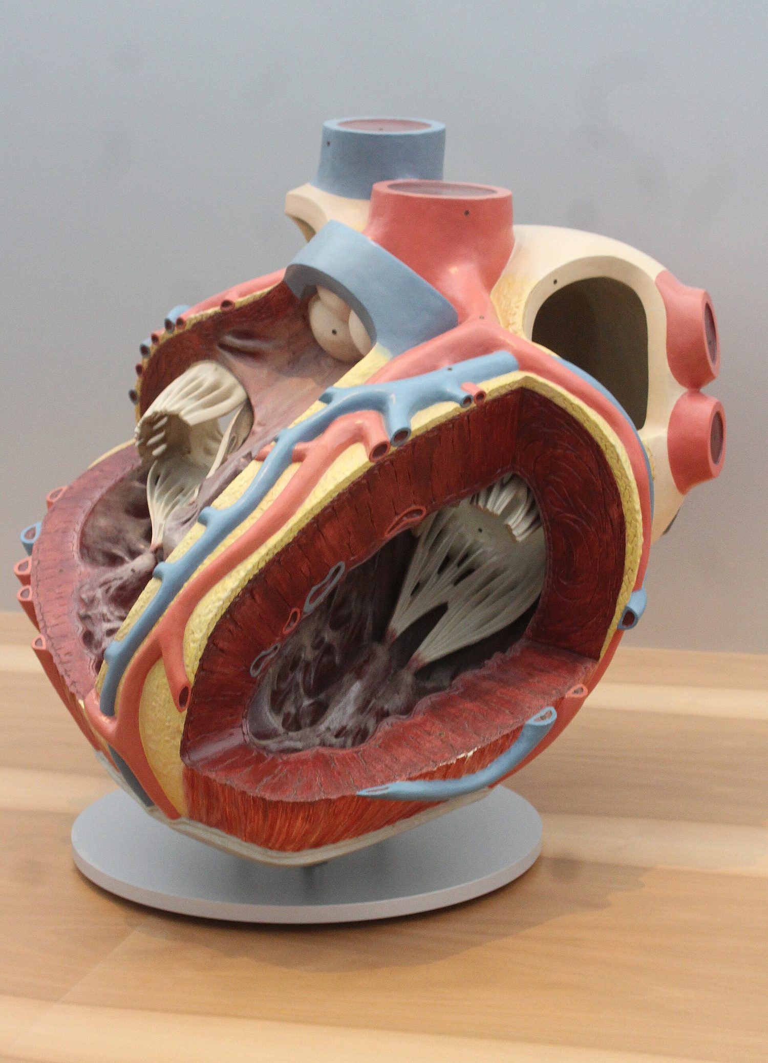 *אותו לב כמודל מוזיאלי: ארבעה חללים, וקיר החדר השמאלי שהעובי שלו נראה עבה יותר
 *במהלך פעילות גופנית קצב הלב עולה כך שדם מגיע לשרירים מהר יותר
 *מחלת לב כתרונית מצמצמת את הערמון; מספר גורמי סיכון מעלים אותו ואורח חיים בריא מוריד אותו

