يتنبض قلبك حوالي مائة ألف مرة في اليوم، ولم يتوقف أبداً للراحة. إنه ليس مضخة واحدة — بل اثنتين، جنباً إلى جنب، يشاركان في…
English narration · English + 中文 subtitles burned in · سرد باللغة الإنجليزية · ترجمة مدمجة بالإنجليزية + الصينية
9.1
The circulatory system · الجهاز الدوري
Syllabus · المنهج
English
Core
Supplement
1 Describe the circulatory system as a system of blood vessels with a pump and valves to ensure one-way flow of blood
2 Describe the single circulation of a fish
3 Describe the double circulation of a mammal
4 Explain the advantages of a double circulation
العربية
أساسي
تكميلي
1 صف الجهاز الدوري كنظام من الأوعية الدموية مع مضخة وصمامات لضمان تدفق الدم في اتجاه واحد
2 صف دوران الدم أحادي المسار في السمك
3 صف دوران الدم ثنائي المسار في الثدييات
4 اشرح مزايا الدوران الدم ثنائي المسار
Source: Cambridge International syllabus · المصدر: منهج كامبريدج الدولي
English
The circulatory system 循环系统 carries blood around the body. It has three parts: a pump 泵 (the heart 心脏), a network of blood vessels 血管, and valves 瓣膜 that keep the blood flowing one way only.
Single and double circulation (Supplement)
A fish has a single circulation 单循环: the blood passes through the heart once on each trip around the body.
A mammal 哺乳动物 has a double circulation 双循环: the blood passes through the heart twice on each full trip — once on the way to the lungs, and once on the way to the rest of the body.
The advantage of a double circulation: the blood can be pumped again at high pressure 压力 before going to the body, so it travels faster and delivers oxygen 氧气 quickly.
العربية
الجهاز الدوري ينقل الدم حول الجسم. يتكون من ثلاثة أجزاء: مضخة (القلب)، وشبكة من الأوعية الدموية، وصمامات تحافظ على تدفق الدم في اتجاه واحد فقط.
الدوران المفرد والمزدوج (إضافي)
للسمكة دوران مفرد: يمر الدم عبر القلب مرة واحدة في كل دورة حول الجسم.
للـثديياتدوران مزدوج: يمر الدم عبر القلب مرتين في كل دورة كاملة — مرة في الطريق للرئتين، ومرة أخرى في الطريق لباقي الجسم.
ميزة الدوران المزدوج: يمكن ضخ الدم مرة أخرى بـضغط عالٍ قبل إرساله للجسم، مما يجعله يسافر بسرعة أكبر ويوصل الأكسجين بسرعة.
*في الدوران المزدوج، يمر الدم عبر القلب مرتين في كل دورة حول الجسم
Explore · استكشف
Double circulation · الدورة المزدوجة
Mammals have a double circulation — blood passes through the heart TWICE on each full trip round the body. · للمثديات دورة دموية مزدوجة — يمر الدم عبر القلب مَرَّتَيْنِ في كل دورة كاملة حول الجسم.
1 Identify in diagrams and images the structures of the mammalian heart, limited to: muscular wall, septum, left and right ventricles, left and right atria, one-way valves and coronary arteries
7 Identify in diagrams and images the atrioventricular and semilunar valves in the mammalian heart
8 Explain the relative thickness of: (a) the muscle walls of the left and right ventricles (b) the muscle walls of the atria compared to those of the ventricles
9 Explain the importance of the septum in separating oxygenated and deoxygenated blood
10 Describe the functioning of the heart in terms of the contraction of muscles of the atria and ventricles and the action of the valves
2 State that blood is pumped away from the heart in arteries and returns to the heart in veins
3 State that the activity of the heart may be monitored by: ECG, pulse rate and listening to sounds of valves closing
4 Investigate and describe the effect of physical activity on the heart rate
11 Explain the effect of physical activity on the heart rate
5 Describe coronary heart disease in terms of the blockage of coronary arteries and state the possible risk factors including: diet, lack of exercise, stress, smoking, genetic predisposition, age and sex
6 Discuss the roles of diet and exercise in reducing the risk of coronary heart disease
7 حدد في المخططات والصور الصمامات بين الأذين والبطين والنصف قمرية في قلب الثدييات
8 اشرح النسبة النسبية للسماكة في: (أ) الجدران العضلية للبطينين الأيسر والأيمن (ب) الجدران العضلية للأذينين مقارنة بتلك الموجودة في البطينين
9 اشرح أهمية الحاجز في فصل الدم المؤكسج عن الدم غير المؤكسج
10 صف عمل القلب من حيث انقباض عضلات الأذينين والبطينين وعمل الصمامات
2 بيّن أن الدم يُضخ خارج القلب عبر الشرايين ويعود إلى القلب عبر الوريد
3 بيّن أنه يمكن رصد نشاط القلب بواسطة: تخطيط كهربية القلب، معدل النبض وسماع أصوات إغلاق الصمامات
4 تحقق وصِف تأثير النشاط البدني على معدل ضربات القلب
11 اشرح تأثير النشاط البدني على معدل ضربات القلب
5 صف مرض القلب التاجي بدلالة انسداد الشرايين التاجية وبيّن عوامل الخطر المحتملة بما في ذلك: النظام الغذائي، قلة ممارسة الرياضة، التوتر، التدخين، الاستعداد الوراثي، العمر والجنس
6 ناقش دور النظام الغذائي وممارسة الرياضة في تقليل خطر الإصابة بمرض القلب التاجي
Source: Cambridge International syllabus · المصدر: منهج كامبريدج الدولي
English
The heart is made of muscle 肌肉. A wall called the septum 隔膜 divides it into a left side and a right side. On each side there is an upper chamber, the atrium 心房, and a lower chamber, the ventricle 心室. Valves between the chambers stop the blood flowing backwards. The heart muscle is fed with blood by the coronary arteries 冠状动脉.
(Supplement) The wall of the left ventricle is thicker than the right, because it must pump blood all the way around the body; the right side only pumps blood to the nearby lungs. The atria have thin walls, as they only push blood into the ventricles just below them.
The septum keeps oxygenated blood 含氧血 on the left side apart from deoxygenated blood 缺氧血 on the right side, so the two never mix.
(Supplement) The valves between each atrium and ventricle are the atrioventricular valves 房室瓣. The valves at the exit of each ventricle are the semilunar valves 半月瓣.
How the heart beats (Supplement)
The atria contract 收缩 and push blood down into the ventricles.
The ventricles contract and force blood out into the arteries; the valves snap shut so blood cannot flow back.
The muscle relaxes and the heart fills with blood again.
Blood is pumped away from the heart in arteries and returns to the heart in veins.
Heart rate and exercise
You can check the heart's activity with an ECG, by feeling the pulse 脉搏 in an artery, or by listening to the valves closing. During exercise the heart rate 心率 goes up, so blood reaches the muscles faster, bringing the extra oxygen and glucose they need.
Worked example. A student counts 18 pulse beats in 15 seconds at rest, and 33 beats in 15 seconds straight after exercise. Give both heart rates in beats per minute. A minute holds 60 ÷ 15 = 4 lots of 15 seconds, so multiply by 4. At rest: 18 × 4 = 72 beats per minute. After exercise: 33 × 4 = 132 beats per minute. The rise delivers more oxygen and glucose to the muscles. Writing the 15-second count straight down as the heart rate is the commonest mistake - always scale it up to a full minute.
Coronary heart disease
Coronary heart disease 冠心病 happens when the coronary arteries become narrow or blocked, so the heart muscle cannot get enough oxygen. Risk factors 风险因素 include a diet high in fat, lack of exercise, stress, smoking, family history (genetics), older age, and being male. A healthy diet and regular exercise lower the risk.
العربية
القلب مصنوع من عضلات. جدار يسمى الحاجز يقسمه إلى جانب أيسر وجانب أيمن. في كل جانب يوجد تجويف علوي هو الأذين، وتجويف سفلي هو البطين. الصمامات بين التجاويف تمنع رجوع الدم. تغذي عضلة القلب بواسطة الشرايين التاجية.
(إضافي) جدار البطين الأيسر أكثر سماكة من الأيمن، لأنه يجب أن يضخ الدم عبر كامل الجسم؛ بينما الجانب الأيمن يضخ الدم فقط إلى الرئتين القريبة. الأذينة لها جدران رقيقة لأنها فقط تدفع الدم إلى البطينات الموجودة أسفلها مباشرة.
الحاجز يحافظ على الدم المؤكسج في الجانب الأيسر منفصلًا عن الدم غير المؤكسج في الجانب الأيمن، بحيث لا يختلط الاثنان أبدًا.
(إضافي) الصمامات بين كل أذين وبطين هي الصمامات الأذينية البطينية. الصمامات عند مخرج كل بطين هي الصمامات الهلالية.
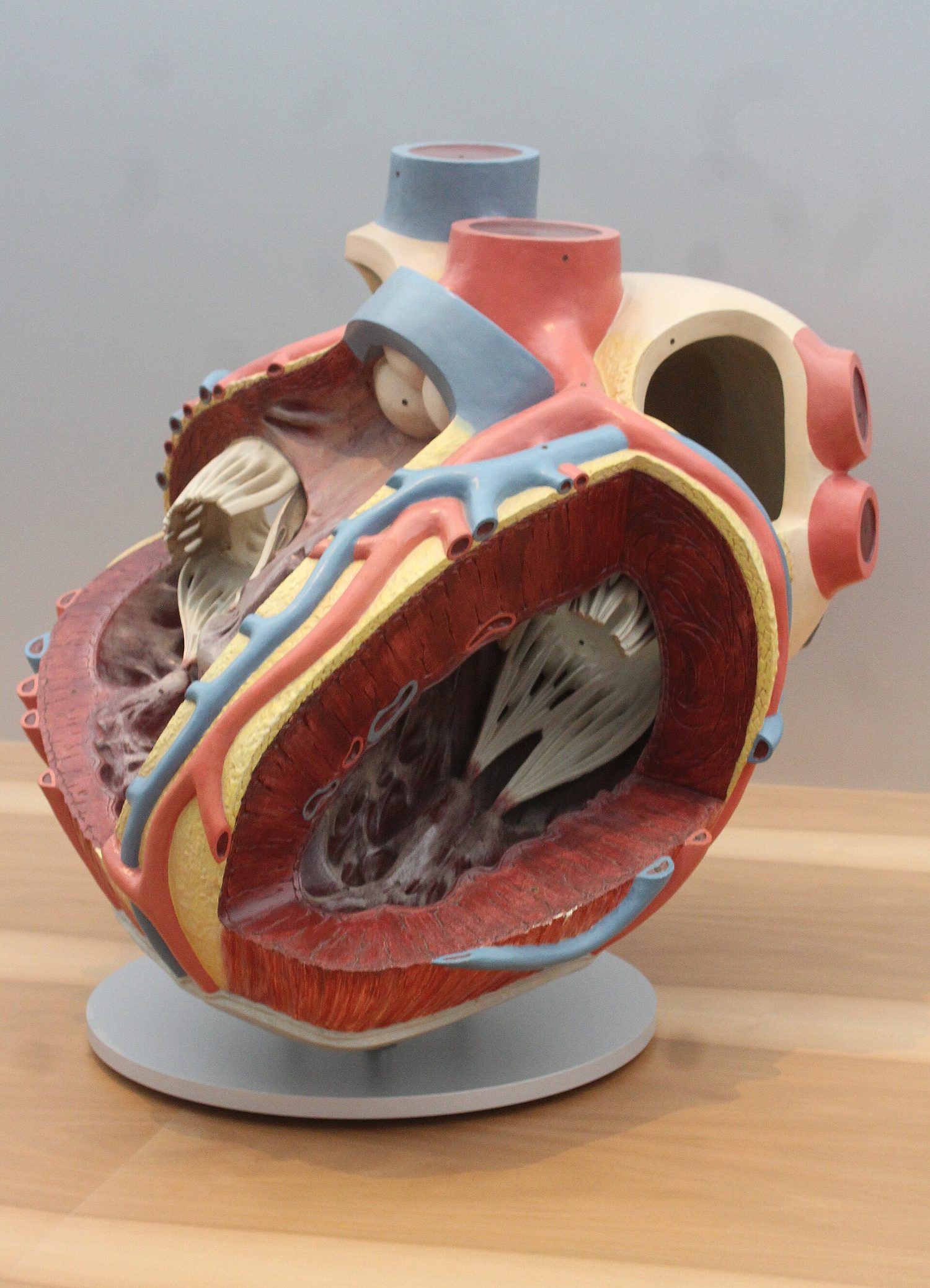
أربعة تجاويف للقلب؛ البطين الأيسر له الجدار الأكثر سماكة لضخ الدم لجميع أنحاء الجسمنفس القلب كنموذج متحف: أربعة تجاويف، وجدار البطين الأيسر الذي يمكنك رؤيته أكثر سماكة
كيف ينبض القلب (إضافي)
تنقبض الأذينة وتدفع الدم لأسفل إلى البطينات.
تنقبض البطينات وتدفق الدم بقوة إلى الشرايين؛ تنغلق الصمامات بإحكام لمنع رجوع الدم.
ترتاح العضلة امتلاء القلب بالدم مرة أخرى.
يدفع الدم بعيداً عن القلب عبر الشرايين ويعود إليه عبر الأوردة.
معدل ضربات القلب والرياضة
يمكنك فحص نشاط القلب باستخدام تخطيط كهربية القلب، أو الإحساس بـالنبض في شريان، أو الاستماع إلى إغلاق الصمامات. أثناء الرياضة يرتفع معدل ضربات القلب، ليصل الدم إلى العضلات بشكل أسرع، محملاً بالأكسجلوز الزائد الذي تحتاجه.
*أثناء الرياضة يرتفع معدل ضربات القلب ليصل الدم إلى العضلات بشكل أسرع
مثال محلول. يعد طالب 18 نبضة في 15 ثانية في وقت الراحة، و33 نبضة في 15 ثانية مباشرة بعد التمارين. أعطِ كلا معدلَي ضربات القلب النبضات في الدقيقة الواحدة.Minute يحتوي على 60 ÷ 15 = 4 مجموعات من 15 ثانية، لذا اضرب في 4. في وقت الراحة: 18 × 4 = 72 نبضة في الدقيقة. بعد التمارين: 33 × 4 = 132 نبضة في الدقيقة. يزيد هذا التوصيل من الأكسجلوز إلى العضلات. الخطأ الأكثر شيوعاً هو كتابة عدد النبضات خلال 15 ثانية مباشرة كمعدل ضربات القلب - يجب دائماً تضخيمه لتصبح دقيقة كاملة.
أمراض القلب التاجية
أمراض القلب التاجية تحدث عندما تصبح الشرايين التاجية ضيقة أو مسدودة، فلا تستطيع عضلة القلب الحصول على ما يكفي من الأكسجلوز. تشمل عوامل الخطر نظاماً غذائياً عالياً بالدهون، قلة ممارسة الرياضة، التوتر، التدخين، التاريخ العائلي (الجينات)، التقدم في العمر، والذكورة. النظام الغذائي الصحي وممارسة الرياضة بانتظام تقلل من خطر الإصابة بها.
*أمراض القلب التاجية تضيق الشريان؛ عدة عوامل خطر ترفع هذا الخطر ونمط الحياة الصحي يقلله
Explore · استكشف
Blood flow through the heart · تدفق الدم عبر القلب
The four chambers and their valves keep blood moving one way — deoxygenated to the lungs, oxygenated to the body. · الأربع حجرات وصماماتها تحافظ على حركة الدم في اتجاه واحد — غير مؤكسج إلى الرئتين، ومؤكسج إلى الجسم.
Explore · استكشف
The heart (double circulation) · القلب (الدورة الدموية المزدوجة)
Follow the blood: blue comes back from the body, the right side pumps it to the lungs (it turns red), and the left side pumps it out to the body — a double circulation. · تابع الدم: يأتي الأزرق من الجسم، يضخه الجانب الأيمن إلى الرئتين (يتحول إلى الأحمر)، ويضخه الجانب الأيسر إلى الجسم — هذه دورة دموية مزدوجة.
1 Describe the structure of arteries, veins and capillaries, limited to: relative thickness of wall, diameter of the lumen and the presence of valves in veins
4 Explain how the structure of arteries and veins is related to the pressure of the blood that they transport
2 State the functions of capillaries
5 Explain how the structure of capillaries is related to their functions
3 Identify in diagrams and images the main blood vessels to and from the: (a) heart, limited to: vena cava, aorta, pulmonary artery and pulmonary vein (b) lungs, limited to: pulmonary artery and pulmonary vein (c) kidney, limited to: renal artery and renal vein
6 Identify, in diagrams and images, the main blood vessels to and from the liver as: hepatic artery, hepatic veins and hepatic portal vein
العربية
أساسي
تكميلي
1 صف بنية الشرايين والأوردة والشعيرات الدموية، مقصورةً على: السماكة النسبية للجدار، قطر التجويف ووجود الصمامات في الأوردة
4 اشرح كيفية ارتباط بنية الشرايين والأوردة بضغط الدم الذي تنقله
2 بيّن وظائف الشعيرات الدموية
5 اشرح كيفية ارتباط بنية الشعيرات الدموية بوظائفها
6 حدد في المخططات والصور الأوعية الدموية الرئيسية المتجهة إلى وبالغة الكبد باعتبارها: الشريان الكبدي، الأوردة الكبدية والوريد البابي الكبدي
Source: Cambridge International syllabus · المصدر: منهج كامبريدج الدولي
English
There are three kinds of blood vessel. The space inside a vessel is its lumen 管腔.
Vessel
Wall
Lumen
Valves?
Job
arteries 动脉
thick, muscular, elastic
narrow
no
carry blood away from the heart, at high pressure
veins 静脉
thin
wide
yes
carry blood back to the heart, at low pressure
capillaries 毛细血管
one cell thick
very narrow
no
let oxygen, glucose and wastes pass between blood and cells
(Supplement) Arteries have thick elastic walls to cope with the high pressure from the heart. Veins have valves because their blood is at low pressure and could otherwise flow backwards. Capillaries are very thin (one cell thick) and very narrow, giving a large surface area 表面积 and a short distance for fast exchange.
Main blood vessels
Connects
Artery (carries blood out)
Vein (carries blood in)
heart ↔ body
aorta 主动脉
vena cava 腔静脉
heart ↔ lungs
pulmonary artery 肺动脉
pulmonary vein 肺静脉
↔ kidney
renal artery 肾动脉
renal vein 肾静脉
(Supplement) The liver is served by the hepatic artery 肝动脉, the hepatic vein 肝静脉, and the hepatic portal vein 肝门静脉, which brings blood from the gut to the liver.
العربية
هناك ثلاثة أنواع من الأوعية الدموية. المساحة داخل الوعاء تسمى 腔 (Lumen).
الوعاء
الجدار
التجويف
صمامات؟
الوظيفة
الشرايين
سميكة، عضلية، مرنة
ضيقة
لا
تنقل الدم بعيداً عن القلب، عند ضغط مرتفع
الأوردة
رقيقة
واسعة
نعم
تنقل الدم إلى القلب، عند ضغط منخفض
الشعيرات الدموية
جدار من خلية واحدة
ضيقة جداً
لا
تسمح بمرور الأكسجين، والجلوكوز والنفايات بين الدم والخلايا
(مكمل) الشرايين لها جدران سميكة مرنة لتتحمل الضغط المرتفع الصادر من القلب. الأوردة تحتوي على صمامات لأن دمها عند ضغط منخفض وقد يتدفق عكسياً بدونها. الشعيرات الدموية رقيقة جداً (خلية واحدة) وضيقة، مما يوفر مساحة سطح كبيرة ومسافة قصيرة للتبادل السريع.
(مكمل) الكبد يُزود بواسطة الشريان الكبدي، والوريد الكبدي، والوريد البابي الكبدي الذي يجلب الدم من الأمعاء إلى الكبد.
Explore · استكشف
The three blood vessels · الأوعية الدموية الثلاثة
Compare an artery, a vein and a capillary — their walls and lumens suit the pressure and job of each. · قارن الشريان والوريد والشعيرة — جدرانها وتجاويها مناسبة للضغط ووظيفة كل منها.
1 List the components of blood as: red blood cells, white blood cells, platelets and plasma
2 Identify red and white blood cells in photomicrographs and diagrams
5 Identify lymphocytes and phagocytes in photomicrographs and diagrams
3 State the functions of the following components of blood: (a) red blood cells in transporting oxygen, including the role of haemoglobin (b) white blood cells in phagocytosis and antibody production (c) platelets in clotting (details are not required) (d) plasma in the transport of blood cells, ions, nutrients, urea, hormones and carbon dioxide
6 State the functions of: (a) lymphocytes – antibody production (b) phagocytes – engulfing pathogens by phagocytosis
4 State the roles of blood clotting as preventing blood loss and the entry of pathogens
7 Describe the process of clotting as the conversion of fibrinogen to fibrin to form a mesh
العربية
أساسي
تكميلي
1 عدّد مكونات الدم باعتبارها: خلايا الدم الحمراء، خلايا الدم البيضاء، الصفائح الدموية والبلازما
2 حدد خلايا الدم الحمراء والبيضاء في الصور المجهرية والمخططات
5 حدد الخلايا الليمفاوية والخلايا البلعمية في الصور المجهرية والمخططات
3 بيّن وظائف مكونات الدم التالية: (أ) خلايا الدم الحمراء في نقل الأكسجين، بما في ذلك دور الهيموجلوبين (ب) خلايا الدم البيضاء في البلعمة وإنتاج الأجسام المضادة (ج) الصفائح الدموية في تخثر الدم (التفاصيل غير مطلوبة) (د) البلازما في نقل خلايا الدم والأيونات والعناصر الغذائية واليوريا والهرمونات وثاني أكسيد الكربون
6 بيّن وظائف: (أ) الخلايا الليمفاوية – إنتاج الأجسام المضادة (ب) الخلايا البلعمية – ابتلاع الممرضات عن طريق البلعمة
4 بيّن أدوار تخثر الدم باعتبارها منع فقدان الدم ودخول الممرضات
7 صف عملية التخثر باعتبارها تحويل الفبرينوجين إلى فبرين لتكوين شبكة
Source: Cambridge International syllabus · المصدر: منهج كامبريدج الدولي
English
Blood has four parts:
Part
Function
red blood cells 红细胞
carry oxygen, using the red pigment 色素haemoglobin 血红蛋白
white blood cells 白细胞
defend the body against disease
platelets 血小板
help the blood to clot 凝血
plasma 血浆
a liquid that carries blood cells, ions 离子, nutrients 营养物质, urea 尿素, hormones 激素 and carbon dioxide 二氧化碳
Defending the body, and clotting
White blood cells fight pathogens 病原体 (the microbes that cause disease) in two ways:
phagocytes 吞噬细胞 carry out phagocytosis 吞噬作用 — they surround and digest the pathogens.
lymphocytes 淋巴细胞 make antibodies 抗体, which stick to the pathogens and destroy them.
When you cut yourself, the blood clots to seal the wound. This stops blood loss and stops pathogens getting in. (Supplement) During clotting, a soluble protein called fibrinogen 纤维蛋白原 is changed into threads of fibrin 纤维蛋白. The threads form a net that traps blood cells and makes a solid clot.
العربية
يتكون الدم من أربعة أجزاء:
الجزء
الوظيفة
كريات الدم الحمراء
تنقل الأكسجين باستخدام الصبغة الحمراء الهيموغلوبين
كريات الدم البيضاء
تحمي الجسم من الأمراض
** الصفائح الدموية**
تساعد الدم على التخثر
البلازما
سائل ينقل خلايا الدم، الأيونات، المغذيات، اليوريا، الهرمونات وثاني أكسيد الكربون
الدم يتكون من خلايا حمراء وخلايا بيضاء وصفيحات محمولة في بلازما سائلة
*خلايا دم حقيقية تحت المجهر الإلكتروني: خلايا حمراء (أقراص)، خلايا بيضاء وصفائح
الدفاع عن الجسم والتخثر
تقاتل كريات الدم الممرضات (الكائنات الدقيقة المسببة للأمراض) بطريقتين:
البلعميات تقوم بالـ البلعمة — حيث تحيط بالممرضات وتهضمها.
الخلايا الليمفاوية تصنع الأجسام المضادة التي ترتبط بالممرضات وتدمرها.
عندما تجرح نفسك، يتخثر الدم لإغلاق الجرح. هذا يمنع فقدان الدم ويمنع دخول الممرضات. (مكمل) أثناء التخثر، يتحول بروتين قابل للذوبان يسمى الفبرينوجين إلى خيوط من الفبرين. تشكل الخيوط شبكة تحتبس خلايا الدم وتصنع جلطة صلبة.
Explore · استكشف
Blood component lab · مختبر مكونات الدم
Classify blood components by the job they do. · صنّف مكونات الدم حسب الوظيفة التي تؤديها.
Pick one and the site follows you — notes, papers, videos and practice all open on it. · اختر واحدًا وسيتبعك الموقع — الملاحظات، الأوراق، الفيديوهات والتدريب جميعها تفتح عليه.
Type to search notes, lessons, code, vocabulary and past-paper questions across every subject. · اكتب للبحث عن ملاحظات، دروس، أكواد، مفردات وأسئلة امتحانات سابقة عبر جميع المواد.


 *أثناء الرياضة يرتفع معدل ضربات القلب ليصل الدم إلى العضلات بشكل أسرع
 *أمراض القلب التاجية تضيق الشريان؛ عدة عوامل خطر ترفع هذا الخطر ونمط الحياة الصحي يقلله

 *خلايا دم حقيقية تحت المجهر الإلكتروني: خلايا حمراء (أقراص)، خلايا بيضاء وصفائح