What this course is, and how to use it
This is the third course in University Medicine: human anatomy 人体解剖学 — the map of the body. Every later clinical course reads this map: where a nerve runs, what a region contains, what one lesion silences.
Scope limit, stated once: a study course, not a medical degree; every case below is fictional, and nothing here licenses or guides treatment. English terms follow Terminologia Anatomica; Chinese terms follow the national standard for medical terms.
- Units 3.1–3.3 are the language and the limbs.
- Units 3.4–3.6 are the trunk cavities: thorax, abdomen, pelvis.
- Units 3.7–3.8 are head, neck and back.
The medicine habit: structure explains symptom — every nerve, vessel and space below is taught with the one lesion that exposes it, and every number (lengths, counts, levels) is stated so you can verify it.
Anatomical language, planes and imaging
Why it matters. Anatomy has a precise language because imprecision has consequences: a "mass on the left" means the patient's left, and a plane is a cut you can point to. Learn the grammar once and every later description is unambiguous.
The idea, with a picture. Position: superior/inferior 上/下, anterior/posterior 前/后, proximal/distal 近侧/远侧. Planes: sagittal 矢状面 (fore-and-aft), coronal 冠状面 (front-and-back), transverse 横断面 (cross-section).

The derivation (why the grammar pays). Radiology reports are plane statements ("axial T2", "coronal CT") plus side statements. A CT slice is a transverse view displayed as if the patient's feet faced you: the patient's right appears on the LEFT of the image. That single convention, applied consistently, is why two readers agree about a lesion without seeing the patient.
Worked example 1 — the directional drill. The elbow is distal to the shoulder and proximal to the wrist ✓; the sternum is anterior to the heart, which is anterior to the oesophagus ✓. Drill until instinct: each pair replaces a sentence of description.
Worked example 2 — clinical link: the wrong side. (Fictional vignette) a tension pneumothorax is decompressed with a needle at "left, second intercostal space, mid-clavicular line". The grammar IS the safety system: side named first, landmark named in two dimensions, depth by space not centimetres — because "left" means the patient's left, always.
Common mistakes. Mixing observer and patient orientation (the X-ray convention above). Mixing the pairs' reference points (medial/lateral are measured from the midline; proximal/distal from where the limb attaches). Reading "coronal" as "crown down" — it is the plane OF the coronal suture, front-to-back.
Summary. Directional pairs, three planes, one display convention (patient's right on image left); the grammar makes every later description unambiguous.
Upper limb
Why it matters. The arm is the most examined region in preclinical anatomy because its wiring is a readable circuit: one plexus, five terminal nerves, each with a motor test and a sensory patch you can point to.
The idea, with a picture. Roots C5–T1 → trunks → cords → five terminal nerves: musculocutaneous 肌皮神经 (elbow flexion), axillary 腋神经 (shoulder abduction), radial 桡神经 (extension), median 正中神经 (precision grip), ulnar 尺神经 (fine finger control).

The derivation (why the plexus condenses). Skin segments (dermatomes 皮节) C5–T1 must map onto a limb that rotates during development: the preaxial border (thumb side, C5–C6) and postaxial (little-finger side, C8–T1) explain why the dermatome strip spirals. The plexus is the remix: one nerve for one movement, drawing whatever roots that movement needs — so nerve lesions follow MOVEMENT patterns, not spinal-segment patterns. That distinction IS the diagnostic method.
Worked example 1 — the circuit read out. Wrist drop = radial (Saturday-night palsy, spiral groove). Ape hand + loss of thumb opposition = median at the carpal tunnel. Claw hand = ulnar at the elbow ✓. Each is a one-line motor test: extend, oppose, spread.
Worked example 2 — clinical link: the pulse and the groove. (Fictional patient) medial elbow pain radiating to the little finger, worse on phone-holding: the ulnar nerve passes BEHIND the medial epicondyle, in a groove you can palpate on yourself — its superficiality there is both the examination (Tinel's tap) and the entrapment. Anatomy you can feel on your own arm is anatomy you keep.
Common mistakes. Learning dermatomes as stripes without the rotation story (they spiral on limbs). Testing muscles by name instead of movement (test extension, not "triceps" first). Forgetting the plexus layers (roots/trunks/cords) exist to explain INJURY PATTERNS, not for their own sake.
Summary. C5–T1 → five terminal nerves; nerve lesions read as movement patterns (radial = extend, median = oppose, ulnar = spread); the ulnar groove is the palpable reminder that superficial anatomy is clinical anatomy.
Lower limb
Why it matters. The leg carries the body for a lifetime: its fascial compartments are pressure vessels, its nerves pass landmarks you can name, and its vessels are the highway between heart and foot.
The idea, with a picture. The thigh: femoral 股神经 (anterior, knee extension), obturator 闭孔神经 (medial, adduction), sciatic 坐骨神经 (posterior → tibial + common peroneal). The leg has four fascial compartments — anterior (dorsiflexion), lateral (eversion), superficial posterior (plantarflexion), deep posterior (inversion + toes).
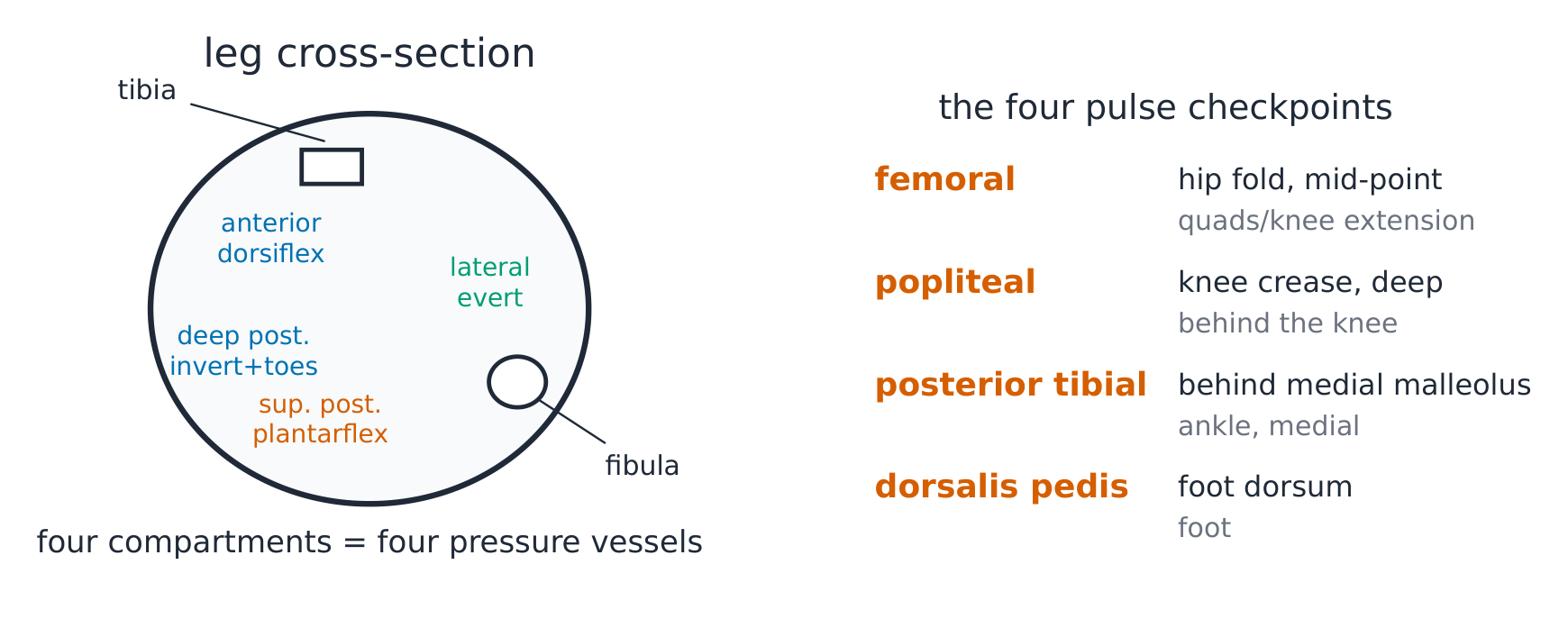
The derivation (why compartments matter). Fascia does not stretch: exercise swells muscle inside an unyielding sleeve, and pressure above capillary perfusion (~30 mmHg) shuts the bed down — compartment syndrome 骨筋膜室综合征. The anatomy (closed sleeves) explains the physiology (pressure) explains the signs (pain on passive stretch, paraesthesia) explains the treatment (open the sleeve). Pulselessness comes late, if at all: a palpable pulse does not exclude compartment syndrome, because the pressure that kills capillaries is far below arterial pressure.
Worked example 1 — the nerve at the neck of the fibula. The common peroneal nerve wraps the fibular neck, subcutaneous: foot drop with preserved inversion ✓ (tibial intact). The site is palpable — your own leg demonstrates why crossing legs or a tight cast strikes exactly this nerve.
Worked example 2 — clinical link: the failing vessel. (Fictional patient) calf pain after 200 m walking, relieved by standing still: the same artery that serves every compartment narrows proximally — claudication distance is the perfusion budget of the whole limb read off one history. Distal pulses by name (femoral, popliteal, posterior tibial, dorsalis pedis) are the limb's four checkpoints.
Common mistakes. Confusing dermatome and nerve-territory maps (they differ in limbs). Forgetting eversion (superficial peroneal) vs inversion (deep posterior/tibial) — the pair that localises a lesion. Testing compartments by poking (the sign is pain on PASSIVE stretch).
Summary. Femoral/obturator/sciatic above the knee; four leg compartments below, each with nerve + disaster; peroneal nerve at the fibular neck = foot drop with intact inversion; pulses at four named points.
Thorax
Why it matters. The thorax is the engine room: heart, great vessels, airways and the oesophagus packed in one split box, with a nerve and two pumps crossing between. Nearly every emergency reads this box.
The idea, with a picture. The mediastinum 纵隔 between the pleural cavities: superior (above the heart), anterior/middle/posterior around it — middle holds the heart and pericardium 心包, posterior holds the descending aorta and oesophagus. The phrenic nerve 膈神经 (C3–5) runs anterior to the lung root (diaphragm motor); the vagus 迷走神经 posterior (parasympathetic to viscera).

The derivation (why the routes diverge). Embryology fixes adult anatomy: the heart descends dragging its pericardium; the phrenic (cervical origin, C3–5) follows the pericardium anteriorly, while the vagus follows the gut tube posteriorly. "Anterior = heart's nerve, posterior = gut's nerve" is a developmental fact that localises tumours: a lung-root mass anteriorly irritates the diaphragm (referred to the shoulder tip, C4 dermatome); posteriorly, it hoarsens the voice (left recurrent laryngeal under the aortic arch).
Worked example 1 — the levels that matter. The trachea bifurcates at the sternal angle (T4/5) ✓; the aortic arch's three branches from right to left: brachiocephalic trunk, left common carotid, left subclavian ✓; the heart's apex beat sits at the left fifth intercostal space, mid-clavicular line ✓ — three numbers that anchor every chest examination.
Worked example 2 — clinical link: the tamponade. (Fictional trauma) hypotension with engorged neck veins and muffled sounds: pericardium is a closed fibrous bag around the heart; 100–200 mL of blood inside it compresses the filling chambers — the anatomy (stiff bag, limited slack) IS the physiology (diastolic collapse) and the procedure (needle below the xiphoid, aimed at the left shoulder).
Common mistakes. Putting the heart mid-line (two-thirds left). Confusing phrenic and vagus at the root (anterior vs posterior). Forgetting the sympathetic chain on the necks of the ribs (the route of a chest-wall infection's spread and the landmark for a nerve block).
Summary. Mediastinum: middle = heart, posterior = aorta + oesophagus; phrenic (C3–5) anterior, vagus posterior; bifurcation, branch order and apex beat are the anchor numbers; a closed pericardium makes small volumes lethal.
Abdomen
Why it matters. The abdomen holds the metabolic organs in a nine-region grid, supplied by three unpaired arteries whose territories explain both referred pain 牵涉痛 and every surgical plane.
The idea, with a picture. Surface: nine regions (four quadrants for speed) — epigastric, umbilical, hypogastric columns flanked by hypochondriac, lumbar and inguinal regions. Visceral supply: coeliac trunk 腹腔干 (foregut to the duodenum), superior mesenteric 肠系膜上动脉 (midgut to the transverse colon), inferior mesenteric 肠系膜下动脉 (hindgut).

The derivation (why pain refers where it does). Viscera report to the spinal segments of their embryological origin, not their adult address: foregut → T5–9 (epigastric), midgut → T10–12 (umbilical), hindgut → L1–3 (hypogastric) ✓. Early, unlocalised visceral pain maps to the midline of that band; only when parieties are involved does it lateralise and sharpen. The referred-pain map is embryology read as symptoms.
Worked example 1 — the classic referrals. (Fictional cases) the appendix: midgut → umbilical dull ache first, then right iliac sharpness as the parietal peritoneum joins in ✓; the gallbladder: foregut → epigastric ache first, then right upper quadrant, and right shoulder-tip pain once the diaphragm is irritated (phrenic, C3–5); the ureter: L1 → groin. One embryological sentence explains three bedside pictures.
Worked example 2 — clinical link: the bleeding territory. (Fictional patient) painless bright-red rectal bleeding from internal haemorrhoids: the inferior mesenteric artery ends as the superior rectal artery, and the anal canal's supply and nerves split at the pectinate line 齿状线 — above it visceral (painless), below it somatic (exquisitely sensitive). Anatomy predicts which bleeds hurt and which do not.
Common mistakes. Quadrants when regions are asked (and vice versa — say which system you use). Forgetting the gut's lymph and venous drainage follows the SAME territories (to the portal system, the coeliac nodes). Mixing renal (T12–L1, lumbar region) with gut pain.
Summary. Nine regions; coeliac/superior/inferior mesenteric = fore/mid/hindgut territories; referred pain T5–9/T10–12/L1–3 to epigastric/umbilical/hypogastric; the pectinate line splits sensation as well as supply.
Pelvis and perineum
Why it matters. The pelvis is the body's floor and its outlet: the bowl of bone, the sling of muscle, and the wiring that shares space with the birth canal and the rectum. Its anatomy explains continence, delivery and the most feared nerve injuries.
The idea, with a picture. Sagittal view: pelvic floor 盆底 = levator ani 肛提肌 + coccygeus, slung from pubis to coccyx; above it the viscera (bladder anterior, uterus/rectum behind), below it the perineum. The pudendal nerve 阴部神经 (S2–4) curves around the ischial spine to supply the perineum — the landmark for a block.

The derivation (why the floor fails). The levator sling carries intra-abdominal pressure for decades; pelvic nerves (S2–4) drive both the bladder's detrusor (parasympathetic) and the external sphincter (somatic via pudendal). Continence is a pressure balance plus two neural switches — so childbirth's stretch (floor) or a spinal lesion (switches) breaks it by two different mechanisms, treated by two different logics.
Worked example 1 — the sacral segments read out. S2–4 = the "pelvic core": detrusor contraction, sphincter control, perineal sensation, and the bulbocavernosus reflex arc ✓. One cone of cord, one exam: a lesion at S2–4 has a signature you can test at the bedside.
Worked example 2 — clinical link: delivery's two injuries. (Fictional delivery) prolonged second stage: the fetal head compresses the pudendal nerve at the ischial spines → transient perineal numbness; a third-degree tear crosses the external sphincter → incontinence risk. Same event, two anatomies, two managements — nerve recovery vs sphincter repair.
Common mistakes. Sexing the pelvis by one feature (the subpubic angle AND the greater sciatic notch shape both differ). Forgetting that the cavernous nerves for erection run beside the prostate (why prostate surgery puts them at risk). Putting the pelvic floor at the outlet (it is the SUPPORT; the perineum is the outlet region below).
Summary. Levator ani floor carrying viscera, perineum below; pudendal (S2–4) around the ischial spine; S2–4 is the testable pelvic core; delivery injures nerve or sphincter by different mechanisms.
Head and neck
Why it matters. Twelve cranial nerves 脑神经 leave the skull through named holes; each hole and each nerve localises a lesion. Head and neck anatomy is the purest structure-localises-disease game in medicine.
The idea, with a picture. The twelve: I smell, II see, III/IV/VI move the eye (VI alone abducts it), V feels the face and chews, VII moves the face and tastes anterior, VIII hears and balances, IX/X swallow and talk (viscera), XI shrugs, XII moves the tongue. Foramina: II through the optic canal, V1 superior orbital fissure, V2 foramen rotundum, V3 foramen ovale, VII/VIII internal auditory meatus, X jugular foramen ✓.

The derivation (why nerves and holes pair up). The skull grew around the brain's outflow: each nerve claimed a channel, and the channel's location fixes what ELSE can be caught there (VII and VIII share the internal auditory meatus — a vestibular schwannoma deafens then paralyses the face in that order). Read the hole, and the overlapping signs follow.
Worked example 1 — the VII vs stroke drill. (Fictional patient) sudden one-sided facial weakness: forehead SPARED = central (upper face has bilateral cortical input) vs forehead INVOLVED = peripheral VII (Bell's pattern) ✓. One muscle test (raise the eyebrows) splits the benign from the emergency.
Worked example 2 — clinical link: the apex at the lung tip. (Fictional smoker) hand wasting, hoarseness, shoulder pain: an apical tumour climbs the cupola and hits, in sequence, the brachial plexus (C8–T1 hand), the recurrent laryngeal (hoarseness) and the sympathetic chain (a drooping eyelid and a small pupil — Horner's). Three systems, one anatomical corner; the corner explains the triad.
Common mistakes. Counting nerves without their holes (learn in pairs). Forgetting VI exits low and long — the false localiser in raised pressure. Splitting IX from X at the bedside: in the gag reflex IX carries the sensation and X the movement, while "aah" and the palate rise test X alone (the uvula swings away from the weak side).
Summary. Twelve nerves, named foramina; VII/VIII share a meatus; forehead-sparing splits central from peripheral VII; the lung apex is a crossroads of plexus, laryngeal nerve and sympathetic.
Back and vertebral column
Why it matters. The spine is a stack of 33 vertebrae shielding a cord, threaded by nerves at 31 pairs of levels, and held by a design that trades stability for movement at predictable cost — the site of most musculoskeletal pain and every cord emergency.
The idea, with a picture. 7 cervical, 12 thoracic, 5 lumbar, 5 sacral (fused), 4 coccygeal (fused) = 33 bones, 24 movable ✓. Curves: cervical and lumbar lordosis, thoracic and sacral kyphosis. The cord ends at L1–2 (adult); below that the cauda equina 马尾 rides in lumbar cistern — the reason lumbar puncture is safe at L3/4.

The derivation (why levels matter twice). In embryos cord and canal match length; the spine grows faster, so adult roots run downward to their exit — the lumbar cord became the cauda equina, and the cone of cord (conus medullaris 脊髓圆锥) sits at L1–2 ✓. Two consequences: a T10 bony injury damages the L1–2 cord segment (the gap between bony and cord level grows down the spine: about one segment in the neck, two in the upper thorax, three or more below T7), and below L2 the canal holds only roots (mass effect there is cauda equina syndrome, not cord).
Worked example 1 — the dermatome ladder. Thumb C6, nipple T4, umbilicus T10, big toe L5, perianal S3–5 ✓ — five rungs that turn a sensory exam into a level read. Combined with the level gap, the ladder names both the segment injured and the bone to image.
Worked example 2 — clinical link: the emergency at the cord's end. (Fictional patient) back pain, saddle numbness, retention: a disc or mass at L4–5 compresses roots of the cauda equina — the anatomical end-state (roots, not cord, S2–4 among them) predicts the exact signs (bladder, bowel, saddle), and decompression is measured in hours. The terminology (cauda equina vs cord) IS the urgency.
Common mistakes. Reading bony level as cord level (the gap grows down the spine). Forgetting the disc's posterior longitudinal ligament is strongest midline — discs herniate posterolaterally onto the NEXT lower root (L4/5 disc → L5 root) ✓. Counting sacral vertebrae as movable (5 fused, 4 fused).
Summary. 33/24 vertebrae, alternating curves; cord ends L1–2, so below is cauda equina; bony level ≠ neural level; five dermatome rungs read the level; saddle symptoms plus retention is the hours-count emergency.