What this course is, and how to use it
This is the fourth course in University Medicine: histology and embryology 组织学与胚胎学. MED03 gave the body's map; this course reads its fabric under the microscope and rewinds to how it was woven — the tissue-level structure that explains organ function, and the developmental sequence whose errors predict the anomalies.
Scope limit, stated once: a study course, not a medical degree; every case below is fictional and nothing here licenses or guides treatment.
- Units 4.1–4.3 are the tissues: preparation, the four basic types.
- Units 4.4–4.5 are organs read as tissue architecture.
- Units 4.6–4.7 are development: fertilisation to birth, and what failure looks like.
The medicine habit: tissue architecture is organ function — every layer, lumen and band below is taught with the physiology it enables and the anomaly its mis-making predicts.
Microscopy and tissue preparation
Why it matters. Histology is not "looking at pictures": the picture is manufactured, and every step of its manufacture can create or destroy the evidence. Reading a slide means knowing what the preparation did.
The idea, with a picture. The sequence: fixation 固定 (formalin cross-links proteins, preserving structure), dehydration 脱水 (alcohols remove water), embedding 包埋 (paraffin supports the tissue), sectioning 切片 (microtome, 3–5 µm sections), staining 染色 (haematoxylin–eosin: H&E, 苏木精-伊红).
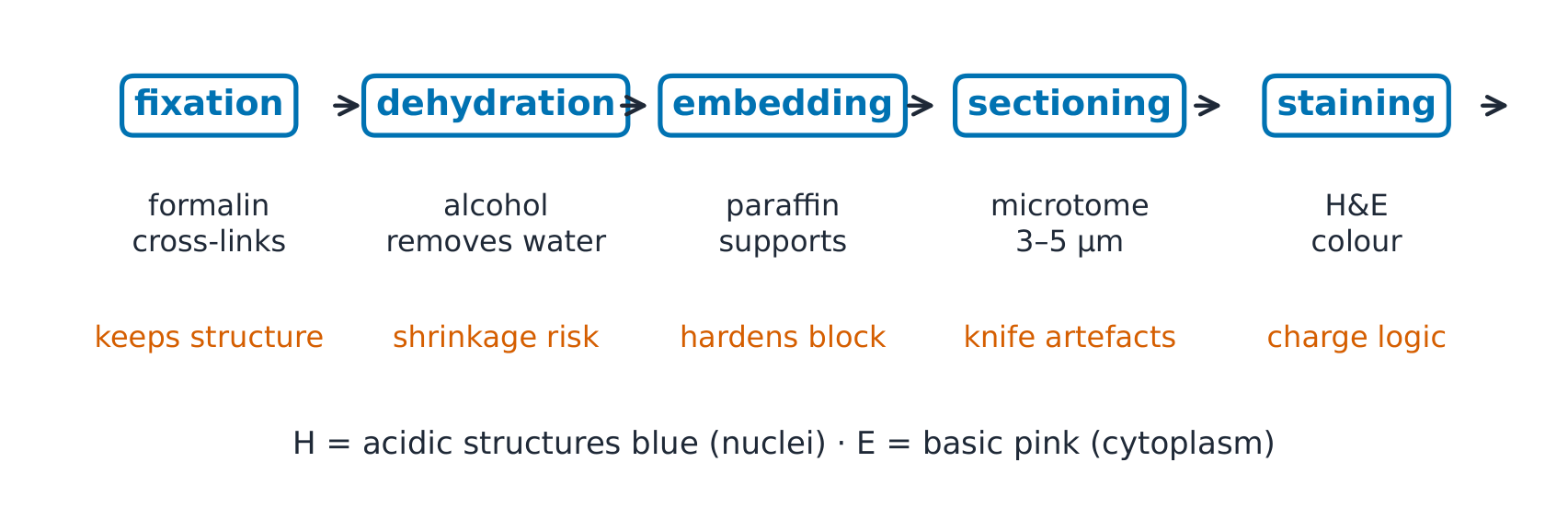
Definitions with numbers. Section thickness 3–5 µm (a cell is 10–30 µm — one section slices through part of a cell). Resolution of the light microscope ~200 nm (half the wavelength of visible light): that limit, not the eye, decides what is visible. Magnification beyond ~1500× is empty — blur enlarged.
The derivation (why H&E works). Haematoxylin is basic and stains acidic structures blue-purple (nuclei: DNA and RNA are acids); eosin is acidic and stains basic structures pink-red (cytoplasm, collagen). The charge pairing is the whole logic: acid-base electrostatics, not magic — and special stains follow the same rule (PAS stains carbohydrates, so basement membranes and glycogen light up magenta).
Worked example 1 — the resolution arithmetic. Two mitochondria 150 nm apart cannot be resolved (below the ~200 nm limit) ✓; a 2 µm bacterium and a 10 µm nucleus both can. The limit explains why electron microscopy (wavelength ~0.005 nm) exists: it buys ~1000× the resolving power, at the cost of killing the specimen.
Worked example 2 — clinical link: the frozen section. (Fictional vignette) a surgeon waits mid-operation for a margin answer: the frozen-section technique skips dehydration/embedding — the tissue is frozen and cut within minutes, stained quickly, read at lower quality. Speed trades artefact: ice crystals tear the tissue, detail is blurred — but a same-hour answer beats a perfect tomorrow answer.
Common mistakes. Treating every gap as a real space (shrinkage during dehydration creates artificial spaces). Reading stain colour as intrinsic colour. Forgetting the section is one plane through a 3-D structure (a tube cut obliquely looks like an oval).
Summary. Fix–dehydrate–embed–section–stain, 3–5 µm sections; light microscopy resolves ~200 nm; H = acidic blue (nuclei), E = basic pink (cytoplasm); frozen sections trade quality for minutes.
Epithelium and connective tissue
Why it matters. Two of the four tissue types carry the body's whole division of labour: epithelium is the boundary workforce (barrier, transport, secretion), connective tissue is the infrastructure (support, transport highway, immune terrain). Organ function is mostly which epithelium sits on which connective tissue.
The idea, with a picture. Epithelium 上皮 is classified by cell layer (simple 单层 one; stratified 复层 many) and cell shape (squamous 扁平, cuboidal 立方, columnar 柱状): simple squamous for exchange (capillary, alveolus), simple cuboidal for transport (tubules), stratified squamous for abrasion (skin, oesophagus). Connective tissue 结缔组织 = cells + extracellular matrix 细胞外基质 (fibres: collagen 胶原, elastic; ground substance), from loose (areolar) to dense regular (tendon) to specialised (bone, blood).

The derivation (structure follows function). Exchange needs minimal diffusion distance: one flat layer (simple squamous, ~0.5 µm) ✓. Abrasion defence needs replaceable depth: many layers (stratified squamous) ✓. Transport and secretion need machinery: tall cells packed with organelles (cuboidal/columnar). The classification grid is not taxonomy for its own sake — each cell of the grid predicts the underlying physiology.
Worked example 1 — reading a slide. A section shows a free surface of tall cells with nuclei in a basal row, on a thin basement membrane, over loose matrix with small vessels: simple columnar epithelium on areolar connective tissue — a transport/absorption surface (intestine pattern). Every clue (apical surface, basal membrane, matrix) is a coordinate in the grid.
Worked example 2 — clinical link: the barrier that fails. (Fictional biopsy) recurrent infections, steatorrhoea: the duodenal biopsy shows flattened villi — the absorptive simple columnar epithelium's surface area has collapsed. Malabsorption is not a "functional" mystery: the exchange architecture itself is gone, and the microscope reads the mechanism.
Common mistakes. Confusing the basement membrane (epithelium's anchor) with the whole matrix. Calling blood "not connective tissue" (it is: cells + matrix = plasma). Forgetting pseudostratified looks layered but IS one layer (all cells touch the basement membrane — respiratory epithelium).
Summary. Layer × shape classifies epithelium; each grid cell predicts function (exchange = simple squamous; abrasion = stratified); connective tissue = cells + collagen/elastic/ground substance, from areolar to tendon to bone and blood.
Muscle and nervous tissue
Why it matters. Movement and control are tissues with visible machinery: the sarcomere's banding IS the contraction mechanism, and the neuron's shape IS its connectivity. Histology here is physiology you can see.
The idea, with a picture. Muscle 肌组织: skeletal (striated, voluntary, multi-nucleate at the periphery), cardiac (striated, involuntary, branched with intercalated discs 闰盘), smooth (unstriated, involuntary, spindle cells). Nervous tissue 神经组织: neurons (cell body, dendrites, axon) plus glia 胶质细胞 (astrocytes support, oligodendrocytes/Schwann cells myelinate, microglia scavenge).

The sarcomere 肌节 derivation. A sarcomere runs Z-line to Z-line. The A-band (anisotropic, dark) is myosin's full length — it does NOT change in contraction. The I-band (isotropic, light) is actin-only; the H-zone is myosin-only within the A-band. As actin slides toward the centre, the I-band and H-zone NARROW and the Z-lines approach — filament lengths never change ✓. The banding you see is the sliding-filament model printed on the tissue.
Worked example 1 — identifying muscle in a section. Peripheral nuclei + long parallel fibres + striations = skeletal ✓. Central nuclei, branching, intercalated discs = cardiac ✓. No striations, spindle cells, central single nuclei = smooth ✓. Three tissues, three fingerprints, no special stains needed.
Worked example 2 — clinical link: myelin read under the scope. (Fictional nerve biopsy) a child with progressive weakness shows "onion bulbs" — concentric Schwann-cell processes around thinly myelinated axons: repeated de/remyelination made visible. The histology names the disease class (demyelinating neuropathy) because glia are as much "nervous tissue" as neurons.
Common mistakes. Expecting the A-band to shorten (it is myosin's constant length). Confusing intercalated discs with striations (discs are junctions between cells, dark lines across the fibre). Treating glia as packing material (they myelinate, buffer, and scar).
Summary. Three muscle fingerprints (peripheral-nuclei striated / branched with discs / unstriated spindle); sarcomere: A constant, I and H narrow, filaments never shorten; glia are functional nervous tissue — myelin pathology is glial pathology.
Organ histology: circulatory, respiratory and digestive
Why it matters. An organ is a tissue arrangement solving a physiological problem — and the three great tube systems (vessels, airways, gut) wear their haemodynamics on their walls. Read the layers and you read the pressures.
The idea, with a picture. Vessels: tunica intima 内膜 (endothelium), media 中膜 (smooth muscle — thick in arteries, thin in veins), adventitia 外膜 (collagen). Airway: the wall thins from bronchus to alveolus 肺泡 where the wall becomes simple squamous over capillaries — the blood–air barrier ~0.5 µm ✓. Gut: mucosa 黏膜 (epithelium + lamina propria + muscularis mucosae), submucosa 黏膜下层, muscularis externa 肌层 (inner circular, outer longitudinal), serosa 浆膜 or adventitia.

The derivation (layers track pressure and traffic). Arteries carry high-pressure pulse: a thick elastic/muscular media smooths it ✓. Veins carry low-pressure volume: thin walls, valves, big lumen ✓. The alveolus trades every defence for distance: no muscle, no glands, one flat cell plus its basement membrane and the capillary wall — exchange optimised to the exclusion of everything else ✓. The gut keeps a muscularis externa because its job is propulsion, plus a mucosa that is the body's most renewable frontline.
Worked example 1 — artery vs vein at a glance. The vessel with the thick media nearly filling the wall and a round lumen is the artery; the floppy wide-lumen vessel with a thin wall is the vein ✓. The histological look IS the pressure record — an artery is "mostly media", a vein is "mostly lumen".
Worked example 2 — clinical link: the barrier that decides gas exchange. (Fictional case) breathless with clear airways: the alveolar wall has thickened (fibrosis pattern) — the 0.5 µm barrier has become several µm, and diffusion length, not airflow, now limits oxygen. The histology explains why the inhaler cannot help: the problem is in the wall, not the tube.
Common mistakes. Calling every inner layer "mucosa" (vessels have intima; mucosa belongs to wet epithelial tubes). Forgetting the gut's two muscle coats run in opposite directions (circular squeezers, longitudinal chasers). Expecting alveoli to look like bronchi (they are the opposite: everything removed).
Summary. Vessel = intima/media/adventitia, media ∝ pressure; gut = mucosa/submucosa/muscularis externa/serosa; alveolus = simple squamous
- basement membrane + capillary, ~0.5 µm total; the layer list is the pressure/traffic profile.
Organ histology: urinary, endocrine, reproductive and skin
Why it matters. The kidney's tubule segments, the endocrine's vascular nets, the skin's stratified armour — each is a recognisable histological machine, and together they cover the urine, the hormones and the barrier: three systems whose diseases are read in biopsies every day.
The idea, with a picture. The nephron 肾单位: glomerulus (fenestrated capillary tuft in Bowman's capsule) → proximal tubule (cuboidal with brush border) → loop of Henle (flat squamous thin limbs, cuboidal thick limb) → distal tubule (cuboidal, no brush) → collecting duct (tall columnar, pale). Endocrine organs = cords or follicles wrapped in fenestrated capillaries 有孔毛细血管 (secreting into blood needs the shortest path). Skin = stratified squamous keratinised 复层扁平角化上皮 on dermis, with the epidermis turning over every ~4 weeks.

The derivation (epithelium predicts tubule function). The proximal tubule reabsorbs ~65 % of the filtrate: it needs surface area — hence a brush border (microvilli) that stains brightly ✓. The loop of Henle's thin limbs let water and ions move passively: flat squamous, minimal machinery ✓; its thick ascending limb pumps salt out and is cuboidal, packed with mitochondria ✓. The collecting duct conveys and fine-tunes: columnar with water channels under hormonal control ✓. You can deduce a segment's physiology from one high-power field.
Worked example 1 — biopsy orientation. A field shows a tuft of capillaries inside a capsule, beside a tubule with a fuzzy violet brush border: glomerulus plus proximal tubule ✓ — renal cortex. The signature pairing names the region without a map.
Worked example 2 — clinical link: the four-week frontline. (Fictional burn vignette) the barrier is lost where the keratinising epithelium died: fluid loss and infection follow immediately, and healing depends on how much basement membrane survived, with the hair follicles and glands below it (they are where new epidermis grows from). One layer's survival decides the prognosis — histology as triage.
Common mistakes. Expecting every kidney tubule to look the same (the segments are the point). Forgetting endocrine cells sit directly ON capillaries (no ducts — that is what "endocrine" means). Calling the dermis "just connective tissue" (it is the vascular, sensory, structural half of skin).
Summary. Nephron segments read as epithelial signatures (brush = proximal; flat = loop; columnar = collecting); endocrine = secretory cells on fenestrated capillaries; skin = keratinised stratified squamous over vascular dermis, ~4-week turnover; basement membrane survival decides regeneration.
Early development: fertilisation to gastrulation
Why it matters. The first three weeks build the body's axes and layers from one cell — with a speed and precision that make them both the most vulnerable and the most instructive period of life. Every later organ trace begins here.
The idea, with a picture. Timeline: fertilisation 受精 (day 0) → cleavage 卵裂 → morula 桑葚胚 (~day 3) → blastocyst 胚泡 (~day 5, implants ~day 6–7) → gastrulation 原肠胚形成 (week 3: forms the three germ layers) → neurulation 神经胚形成 (week 3–4: the neural tube closes).

The derivation (why gastrulation is the pivot). The blastocyst is a ball of cells; the body is three layered germ layers 胚层 with axes — ectoderm 外胚层 (nervous system, epidermis), mesoderm 中胚层 (muscle, bone, blood, kidney, heart), endoderm 内胚层 (gut, lungs' lining, liver, pancreas) ✓. Gastrulation is the reorganisation that CREATES the layers and the axes; everything after is specialisation. The germ-layer map is why a single week-3 field defect predicts a multi-organ syndrome: tissues are neighbours before they are organs.
Worked example 1 — the timeline arithmetic. Fertilisation to implantation ≈ 6–7 days ✓; to gastrulation ≈ 15 days; to neural-tube closure completion ≈ day 28. Why folate matters periconceptionally: the neural tube closes in week 4 after conception — about two weeks after the first missed period, often before the pregnancy is known or the first antenatal visit — so the protective supplement must start before conception ✓.
Worked example 2 — clinical link: the tube that fails to close. (Fictional case) elevated maternal alpha-fetoprotein, ultrasound shows an open spine: neural-tube closure failed at day 26–28. The defect level maps the deficit (lumbar → legs and bladder; cervical → quadriplegia) — development wrote the prognosis in anatomy.
Common mistakes. Mixing up the blastocyst's two parts (the inner cell mass becomes the embryo; the trophoblast becomes the placenta). Treating the germ layers as a list to memorise (they are a spatial map: tube-in-tube-in-body). Counting pregnancy weeks from conception (clinical weeks count from the last period: ~2 weeks ahead).
Summary. Day 0 fertilisation → day 5 blastocyst → week 3 gastrulation makes ectoderm/mesoderm/endoderm → week 4 neural tube; the germ-layer map predicts organ systems and syndrome patterns; closure is complete about two weeks after the first missed period, so folate must start before conception.
Organogenesis and congenital anomalies
Why it matters. Weeks 4–8 turn three layers into recognisable organs — the most error-prone window in human life. Congenital anomaly reasoning is developmental logic read backwards: from a defect, infer the week and the process that failed.
The idea, with a picture. Heart septation 心脏分隔: the heart tube folds (week 4), then septa close the atrial and ventricular holes — the atrial septum's foramen ovale 卵圆孔 must stay open in the fetus (lungs closed) and close at birth ✓. Meanwhile: the palate's shelves fuse (weeks 7–10), the diaphragm closes (week 7), the limb buds pattern out (weeks 4–8).

The derivation (the timing predicts the defect). Organs form in overlapping windows; a teratogen or gene error hits whatever is mid-construction AT THAT WEEK. Weeks 3–4 (neural tube, heart tube): the classic malformation window; weeks 4–8 (septation, palate, limbs): the structural window; after week 8, growth and function mature rather than form ✓. Anomaly reasoning = matching defect to window: defects in organs that were forming in the same weeks (heart septa, palate, limbs) can share one cause, so a cluster is a clue, not a coincidence.
Worked example 1 — the septal arithmetic. The fetal circulation bypasses the lungs: right atrium → foramen ovale → left atrium, and pulmonary artery → ductus arteriosus → aorta ✓. At birth the first breath drops pulmonary resistance, left atrial pressure rises, and the foramen slams shut (functionally immediately, anatomically in months). Patent foramen ovale (persistent in ~25 % of adults) usually matters only when something else forces a right-to-left shunt ✓.
Worked example 2 — clinical link: one cause, several systems. (Fictional child) cleft palate and a heart murmur from a ventricular septal defect. The palate and the ventricular septum were both being built in weeks 4–10, and some causes hit both at once: a teratogen in that window, or a genetic change in a signal both use (the 22q11 deletion is the classic one). So one anomaly is a reason to look for its neighbours: the doctor examining a child with a cleft always listens to the heart.
Common mistakes. Treating "congenital" as "genetic" (most anomalies are multifactorial; many are pure timing). Dating anomalies by birth findings (the error happened weeks earlier). Forgetting the placenta is fetal tissue (its vessels are part of fetal cardiovascular design).
Summary. Weeks 4–8 organogenesis: heart folds then septates, palate fuses, limbs pattern; defects date to their construction windows; the fetal foramen ovale and ductus must be open in utero and close at birth; organs built in the same weeks can share one cause.