Muscles and contraction
Introduit| English | Sens |
|---|---|
| striated muscle/ˈstraɪeɪtɪd ˈmʌsl/ | muscle strié |
| sliding filament model/ˈslaɪdɪŋ ˈfɪləmənt ˈmɒdl/ | modèle des filaments glissants |
| sarcomere/ˈsɑːkəmɪə/ | sarcomère |
| myosin/ˈmaɪəsɪn/ | myosine |
| actin/ˈæktɪn/ | actine |
| sarcoplasmic reticulum/ˌsɑːkəˈplæzmɪk reˈtɪkjʊləm/ | réticulum sarcoplasmique |
| troponin/ˈtrɒpənɪn/ | troponine |
| tropomyosin/ˈtrɒpəmɪɒsɪn/ | tropomyosine |
How muscles contract
- Striated muscle 横纹肌 is made of long fibres.
- Inside, tiny filaments slide over each other to shorten the muscle.
- This is the sliding filament model 肌丝滑动模型.
Muscle structure
- Each fibre is divided into repeating units called sarcomeres 肌节.
- Inside are two filaments: thick myosin 肌球蛋白 and thin actin 肌动蛋白.
- The T-tubules carry the impulse deep into the fibre, and the sarcoplasmic reticulum 肌质网 stores and releases calcium ions.
| Band | What it contains | On contraction |
|---|---|---|
| A band | full length of myosin | stays the same |
| I band | actin only | shorter |
| H zone | myosin only | shorter |
- Z lines mark the ends of each sarcomere. They move closer together as the muscle contracts.
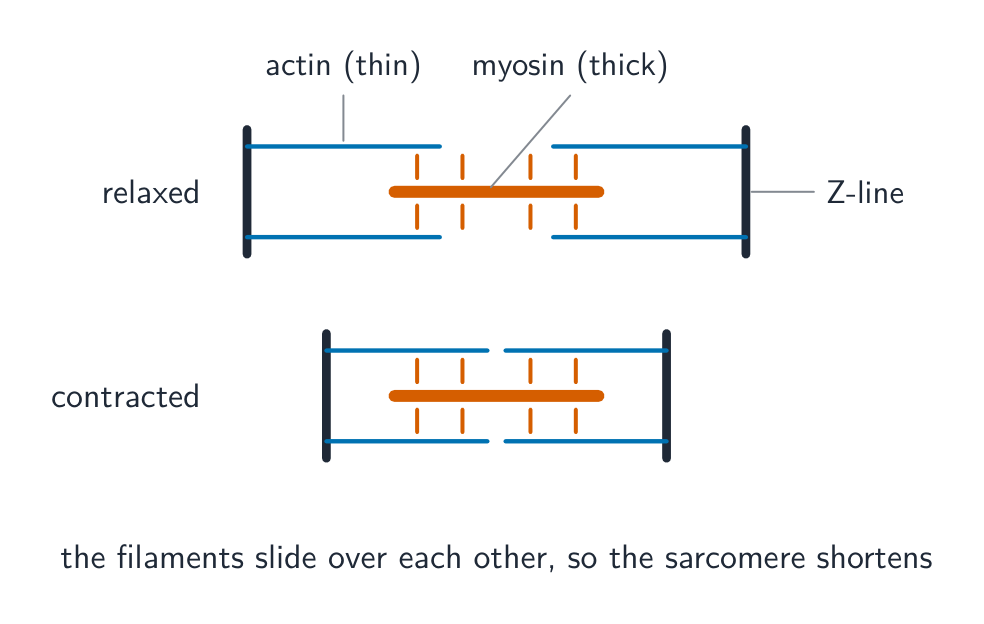
The sliding filament model of muscle contraction
How a muscle contracts
Step through the sliding filament model — calcium uncovers the binding sites, then myosin pulls the actin inwards.
A sarcomere is:
Difficulté 1/5 · 100 pièces + un petit bonus lors de la première réussite aujourd'hui
Muscle fibres are divided into sarcomeres, each holding thick (myosin) and thin (actin) filaments.
The calcium ions for contraction are stored in, and released from, the ______ reticulum.
Difficulté 1/5 · 100 pièces + un petit bonus lors de la première réussite aujourd'hui
When the impulse arrives, the sarcoplasmic reticulum releases Ca²⁺ to trigger contraction.
Which part of a sarcomere stays the same length when the muscle contracts?
Difficulté 1/5 · 100 pièces + un petit bonus lors de la première réussite aujourd'hui
The A band is the length of the myosin filaments, which do not shorten.
From impulse to calcium
- At the neuromuscular junction, acetylcholine depolarises the sarcolemma (the muscle fibre's membrane).
- The depolarisation spreads down the T-tubules, deep into the fibre.
- This makes the sarcoplasmic reticulum release Ca²⁺ ions into the sarcoplasm.
- Ca²⁺ binds to troponin 肌钙蛋白. Troponin changes shape and pulls tropomyosin 原肌球蛋白 away from the myosin-binding sites on actin.
Calcium ions cause contraction by:
Difficulté 1/5 · 100 pièces + un petit bonus lors de la première réussite aujourd'hui
Calcium binds troponin; tropomyosin shifts to expose actin, so myosin heads can attach.
In the sliding filament model, a sarcomere shortens because:
Difficulté 1/5 · 100 pièces + un petit bonus lors de la première réussite aujourd'hui
Myosin heads attach and pull actin inwards (powered by ATP), so the filaments slide and the sarcomere shortens.
Put muscle contraction in order.
Difficulté 3/5 · 200 pièces + un petit bonus lors de la première réussite aujourd'hui
Impulse → Ca²⁺ released → sites uncovered → power stroke → sarcomere shortens.
Match each part to its role in contraction.
Difficulté 2/5 · 140 pièces + un petit bonus lors de la première réussite aujourd'hui
Ca²⁺ exposes the sites, myosin pulls actin (the power stroke), and ATP both powers and releases the heads.
What does calcium allow?
Calcium uncovers the places where myosin attaches.
The cross-bridge cycle and ATP
- A myosin head, carrying ADP and Pi, binds to an exposed site on actin, forming a cross-bridge.
- The head tilts (the power stroke), pulling actin towards the centre of the sarcomere. ADP and Pi are released.
- A new ATP binds to the head, so it detaches from actin.
- The head hydrolyses the ATP and swings back, ready to bind again further along.
- Relaxation: when stimulation stops, Ca²⁺ is actively transported back into the sarcoplasmic reticulum (using ATP), tropomyosin covers the sites again, and the filaments slide apart.
A myosin head needs a new ATP molecule to detach from actin.
Difficulté 1/5 · 100 pièces + un petit bonus lors de la première réussite aujourd'hui
ATP binding releases the head; hydrolysing it re-cocks the head.
Worked example: what shortens in a sarcomere
A relaxed sarcomere is 2.5 µm long; when contracted it is 2.0 µm. The A band stays 1.6 µm. What happened to the I band and H zone, and why?
- The A band (length of myosin) does not change, so the filaments themselves do not shorten. The I band (actin only) and the H zone (myosin only) both get shorter, because actin slides further between the myosin.
- This is the evidence for the sliding filament model: Ca²⁺ exposes binding sites on actin, myosin heads form cross-bridges, pull (power stroke) and detach using ATP, then repeat, so the Z lines move closer.
During contraction, the A band of a sarcomere gets shorter.
Difficulté 1/5 · 100 pièces + un petit bonus lors de la première réussite aujourd'hui
The A band is the length of myosin, which does not change.
In the cross-bridge cycle, ATP is needed for the myosin head to:
Difficulté 1/5 · 100 pièces + un petit bonus lors de la première réussite aujourd'hui
ATP binding releases the head; its hydrolysis re-cocks it.
Worked exam question: rigor mortis
Question (3 marks). A few hours after death, muscles become stiff and cannot be stretched (rigor mortis). Suggest why.
- After death, respiration stops, so no ATP is made (1 mark).
- Without ATP, myosin heads cannot detach from actin, so the cross-bridges stay fixed (1 mark).
- Also, Ca²⁺ cannot be pumped back into the sarcoplasmic reticulum, so the binding sites on actin stay exposed and the filaments are locked together (1 mark).
Why answers lose marks: "ATP is needed for the power stroke". The key point here is that ATP is needed for the head to let go.
Without looking back: describe the roles of Ca²⁺ and ATP in muscle contraction (4 marks).
Two roles for each substance.
You've got it
- striated muscle = fibres divided into sarcomeres (thick myosin + thin actin)
- T-tubules carry the impulse in; the sarcoplasmic reticulum releases calcium
- calcium binds troponin → moves tropomyosin → uncovers actin sites
- cross-bridge cycle: bind → power stroke → ATP binds, head detaches → ATP hydrolysed, head re-cocks
- A band stays the same; I band and H zone shorten; Z lines move closer
- relaxation: Ca²⁺ actively pumped back into the sarcoplasmic reticulum
What does calcium do in contraction?
Difficulté 1/5 · 100 pièces + un petit bonus lors de la première réussite aujourd'hui
Exposed sites let the cross-bridges form.