What this course is, and how to use it
This is the ninth course in University Medicine: neuroscience 神经科学. MED03 named the structures and MED05 gave the cell's electrical grammar; this course traces the circuits and turns them around: given a deficit, find the lesion. Prerequisites: MED03 (anatomy), MED05 (physiology).
Scope limit, stated once: a study course, not a medical degree; every case below is fictional and nothing here licenses or guides treatment.
- Units 9.1–9.4 are the wiring: synapses, sensory, motor and special- sense pathways.
- Units 9.5–9.7 are the systems built on it: autonomic outflow, higher functions, and lesion localisation.
The neuroscience habit: the pathway predicts the lesion — every tract below is learned in two directions, forward (trace it) and backward (localise along it), and every number is something you recompute.
Neurons, synapses and neurotransmitters
Why it matters. The synapse 突触 is the nervous system's decision point — and the target of most neuroactive drugs and toxins. Predicting an effect at the synapse is applied cell biology.
The idea, with a picture. Transmission in six steps: action potential reaches the terminal → voltage-gated Ca²⁺ channels 电压门控钙通道 open → Ca²⁺ influx triggers SNARE-mediated vesicle fusion → transmitter released into the cleft → binds postsynaptic receptors → terminated by reuptake 再摄取 or degradation ✓.

The derivation (why transmitter chemistry predicts the effect). The postsynaptic receptor decides the sign: ionotropic receptors 离子型受体 open channels fast (ms): nicotinic ACh, GABA-A (Cl⁻ influx → inhibition), glutamate AMPA/NMDA (Na⁺/Ca²⁺ → excitation) ✓; metabotropic receptors 代谢型受体 act through G-proteins over hundreds of milliseconds to seconds (muscarinic, adrenergic, most dopamine and serotonin receptors). The receptor, not the transmitter, sets the sign: ACh excites skeletal muscle through nicotinic receptors but slows the heart through M2 receptors. In the adult brain glutamate acts almost only on excitatory receptors and GABA/glycine on inhibitory ones ✓.
Worked example 1 — the NMJ arithmetic. (Fictional lab) one vesicle releases ~5,000–10,000 ACh molecules; one nerve impulse releases ~50–300 vesicles, so 50 × 5,000 = 2.5 × 10⁵ to 300 × 10,000 = 3 × 10⁶ molecules ✓. The end-plate potential this makes is about 3–5 times larger than threshold needs — the safety factor — which is why weakness appears only when about 70–80 % of receptors are blocked ✓.
Worked example 2 — clinical link: three toxins, three steps. (Fictional teaching set) botulinum cleaves SNAREs PREsynaptically → no release → flaccid paralysis; curare blocks the nicotinic receptor POSTsynaptically → flaccid paralysis with normal release; tetanus toxin travels up the motor axon to the spinal cord and blocks release from inhibitory interneurons (glycine and GABA) → unopposed excitation → spastic paralysis ✓. Site of action predicts the sign of the paralysis.
Common mistakes. Calling ACh simply excitatory (the receptor decides: nicotinic excites muscle, M2 slows the heart). Expecting reuptake blockers to change synthesis (SSRIs block the presynaptic serotonin TRANSPORTER, a step after release). Forgetting Ca²⁺ is the trigger, not the fuse: no Ca²⁺ influx, no release, however depolarised the terminal.
Summary. AP → Ca²⁺ → SNARE fusion → cleft → receptor → reuptake or degradation; ionotropic fast (channel), metabotropic slow (G-protein); glutamate excites, GABA/glycine inhibit; NMJ ~5–10k molecules/vesicle; toxin site predicts flaccid (pre- or post-synaptic block) vs spastic (disinhibition).
Somatosensory and pain pathways
Why it matters. Two ascending systems carry the body's sensory news, and they cross at different levels — a sensory deficit's PATTERN names its tract and its side. This is the daily grammar of the neurological examination.
The idea, with a picture. The dorsal column 后索 pathway (fine touch, vibration, proprioception): enters the cord, ascends IPSilaterally, crosses in the MEDULLA ✓. The spinothalamic tract 脊髓丘脑束 (pain, temperature): crosses within 1–2 segments of entry in the CORD, then ascends contralaterally ✓.

The derivation (why the crossing levels differ clinically). Because dorsal-column fibres cross high, a CORD lesion numbs fine touch BELOW the lesion on the SAME side; because spinothalamic fibres cross low, the same lesion numbs pain on the OPPOSITE side ✓ — the Brown-Séquard mirror (9.7). Gate control 闸门控制 adds the modulation: large Aβ fibres activate an inhibitory interneuron that closes the gate on C-fibre pain traffic — rubbing a knock works because Aβ traffic arrives ✓.
Worked example 1 — tracing a hemisection. (Fictional case) left hemicord injury at T10: ipsilateral (left) loss of vibration and proprioception below T10, contralateral (right) loss of pain and temperature starting 1–2 segments below, plus left UMN signs below — each sign traced to one tract and its crossing ✓.
Worked example 2 — clinical link: referred pain by convergence. (Fictional) diaphragmatic irritation felt at the shoulder tip, a referred pain 牵涉痛: both C3–C5 afferents and diaphragmatic visceral fibres converge on the same spinal segments, and the brain misattributes the visceral source to the somatic map — the same arithmetic that refers cardiac pain to the T1–T5 chest and arm ✓.
Common mistakes. Tracing pain pathways as uncrossed (they cross in the cord, at entry). Expecting a spinal LEVEL on the face (facial sensation rides the trigeminal system to the brainstem, not the cord). Putting the pain level at the lesion (spinothalamic fibres ascend 1–2 segments before crossing, so the pain level sits 1–2 segments below).
Summary. Dorsal column: ipsilateral to the medulla, then crossed — fine touch/vibration/proprioception. Spinothalamic: crosses in the cord within 1–2 segments — pain/temperature. Cord lesion → ipsilateral fine touch, contralateral pain; Aβ gates C-fibre traffic; convergence refers visceral pain to the somatic map.
Motor systems
Why it matters. Voluntary movement is a two-neuron system with computers attached — and each level fails with its own signature. Upper motor neuron 上运动神经元 (UMN) vs lower motor neuron 下运动神经元 (LMN) signs are the first fork of every motor examination.
The idea, with a picture. The corticospinal tract 皮质脊髓束: precentral gyrus (Betz cells, layer V) → posterior limb of internal capsule → cerebral peduncle → pyramid, where ~85–90 % cross (the pyramidal decussation 锥体交叉) to the lateral corticospinal tract ✓ → anterior horn lower motor neuron (LMN), which exits to the muscle.

The derivation (why the two neuron classes fail differently). The UMN is a modulatory input; its loss releases the LMN pool from inhibition → spasticity, hyperreflexia, a Babinski sign 巴宾斯基征 (an upgoing big toe), minimal atrophy ✓. The LMN IS the final common path; its loss removes the drive → flaccidity, fasciculations, atrophy, areflexia ✓. The computers tune the loop: basal ganglia scale movement amplitude (dopamine balance; Parkinson = too little → bradykinesia, rigidity, rest tremor; Huntington = striatal loss → chorea), cerebellum times it (ipsilateral incoordination: dysmetria, intention tremor 意向性震颤, dysdiadochokinesia) ✓.
Worked example 1 — one lesion, two sign sets. (Fictional) left-sided cervical cord compression at C5: at the level, LMN signs in C5 muscles (biceps wasting, absent biceps jerk); below it, UMN signs. On which side? The lateral corticospinal tract has ALREADY crossed at the medulla, so the UMN signs appear on the SAME side (left) below C5 ✓: the crossing level is written into the anatomy.
Worked example 2 — clinical link: the tremor fork. (Fictional) a rest tremor that improves with movement plus bradykinesia points to the basal ganglia (Parkinson pattern); a tremor that appears ONLY with intention plus dysmetria points to the cerebellum ✓. Same word — tremor — two circuits, two examinations, two prognoses.
Common mistakes. Expecting corticospinal signs contralateral below a CORD lesion (it crossed at the medulla — contralateral is above the foramen magnum only). Equating spasticity with strength (spasticity is tone, not power). Forgetting the cerebellum is ipsilateral (its tracts double-cross).
Summary. Precentral → internal capsule → pyramids (~85–90 % decussate) → anterior horn → muscle; UMN = spastic, hyperreflexic, Babinski; LMN = flaccid, wasting, areflexic; cord lesions give ipsilateral UMN below (already crossed); basal ganglia scale amplitude, cerebellum times movement (its tracts double-cross to the same side).
Special senses
Why it matters. Vision and hearing are mapped pathways: because the map is preserved end to end, a field defect or an audiogram names the lesion's site with surveyor's precision.
The idea, with a picture. The visual pathway: photoreceptors → retinal ganglion cells → optic nerve 视神经 → optic chiasm 视交叉 (nasal fibres cross) → optic tract → lateral geniculate nucleus → optic radiation → primary visual cortex (V1, calcarine sulcus) ✓.

The derivation (why lesions stamp signatures). Nasal retinal fibres cross at the chiasm and each carries the TEMPORAL visual field: an optic nerve lesion → monocular blindness; a chiasmal lesion → bitemporal hemianopia 偏盲 (loss of half a visual field); a tract/radiation/V1 lesion → contralateral homonymous hemianopia ✓; a temporal-lobe radiation lesion (Meyer's loop, inferior fibres) → contralateral superior quadrantanopia ✓. The map is the lesion detector.
Worked example 1 — reading the fields. (Fictional) driver misses the left half of both visual fields after a stroke: a LEFT homonymous hemianopia (named after the lost half-field) → lesion behind the chiasm on the RIGHT, contralateral to the lost fields ✓. Each word of the field name encodes a side and a level.
Worked example 2 — clinical link: the cochlear map. (Fictional audiogram) air conduction lost, bone conduction preserved (Rinne test 林纳试验 negative, Weber lateralises to the affected ear) → conductive loss (middle ear); air still better than bone (Rinne positive) with both reduced, and Weber lateralising to the BETTER ear → sensorineural (cochlea/nerve) ✓. The cochlea is laid out by pitch, its tonotopy 音调定位 — base transduces high frequencies, apex low (young adult range ~20–20,000 Hz) — so the audiogram's SHAPE localises within the cochlea too. Vestibular twins: three canals in orthogonal planes report rotation; otolith organs report linear acceleration and tilt; nystagmus is the reflex made visible.
Common mistakes. Calling a homonymous hemianopia monocular (it is binocular — same half of EACH field). Reading Rinne without Weber (the pair, not either alone, separates conductive from sensorineural). Forgetting the temporal field projects to the NASAL retina (the crossing that makes chiasmal lesions bitemporal).
Summary. Nerve → chiasm (nasal crosses) → tract → LGN → radiation → V1; nerve = monocular, chiasm = bitemporal, behind = contralateral homonymous, Meyer's loop = superior quadrantanopia; Rinne+Weber split conductive from sensorineural; cochlea tonotopic (base high, apex low, ~20–20,000 Hz).
Autonomic nervous system
Why it matters. The autonomic system runs the viscera through two opposed limbs, and its receptor map is how organ responses — and autonomic drug effects — are predicted rather than memorised.
The idea, with a picture. The sympathetic nervous system 交感神经系统: thoracolumbar outflow T1–L2, short pre-/long postganglionic fibres, ganglia in the paravertebral chain; the adrenal medulla is a modified ganglion releasing ~80 % adrenaline / 20 % noradrenaline ✓. The parasympathetic nervous system 副交感神经系统: craniosacral outflow (CN III, VII, IX, X and S2–S4), long pre-/short postganglionic fibres, ganglia in or near the wall ✓.

The derivation (why receptors predict organ responses). The autonomic code: all preganglionic fibres are ACh/nicotinic; sympathetic postganglionic is noradrenaline (except sweat glands, ACh), parasympathetic postganglionic is ACh/muscarinic ✓. Organ response = receptor expressed: α1 → vasoconstriction, mydriasis; β1 → heart rate and contractility up; β2 → bronchodilation, vasodilation; M2 → heart rate down; M3 → glands, gut motility, miosis, bronchoconstriction ✓. Predict the eye, the airways, the gut — one receptor table, every organ.
Worked example 1 — one agonist, four organs. (Fictional teaching set) a selective β2 agonist: bronchi dilate (asthma logic), skeletal-muscle vessels dilate, skeletal muscle shows a fine tremor, and the heart speeds a little (a reflex to the vasodilation plus some cardiac β2 receptors) ✓ — each line read off the receptor table, no memorisation.
Worked example 2 — clinical link: the lesion that droops. (Fictional) Horner syndrome 霍纳综合征 — ptosis, miosis, anhidrosis — on one side: a sympathetic PATHWAY lesion (anywhere eye-to-spine) with intact parasympathetics ✓. The opposite pupil sign — a fixed dilated pupil with a drooping lid and an eye turned down and out — would implicate the parasympathetic fibres in the III nerve: two drooping lids, two systems.
Common mistakes. Sympathetic = accelerates everything (it CONSTRICTS gut vessels and skin vessels; the response set is receptor-written). Forgetting sweat glands are sympathetic but cholinergic (the classic exception). Assuming the adrenal medulla is innervated postganglionically (it is a ganglion modified to secrete into blood).
Summary. Sympathetic T1–L2 (chain, short pre; adrenal medulla ~80/20 adrenaline/noradrenaline); parasympathetic craniosacral (III, VII, IX, X, S2–S4; wall ganglia). Preganglionic = ACh/nicotinic always; α1 constricts, β1 drives heart, β2 dilates, M2 slows the heart, M3 secretes; Horner = sympathetic lesion; fixed dilated pupil = III.
Higher functions: sleep, memory and language
Why it matters. Sleep, memory and language are network functions — their architecture predicts their disorders, and their localisation is clinical linguistics and electrophysiology applied.
The idea, with a picture. Sleep cycles ~90 minutes, four to six per night: N1 (transition), N2 (sleep spindles, K complexes), N3 slow-wave sleep 慢波睡眠 (delta 0.5–4 Hz, growth hormone, hardest to wake), REM (atonia, vivid dreams, ~20–25 % of the night) ✓. Memory systems: hippocampus → episodic; striatum → procedural/habit; amygdala → emotional tagging; cerebellum → conditioned motor learning ✓. Language: Broca's area 布洛卡区 (inferior frontal, usually left) — production; Wernicke's area 韦尼克区 (superior temporal) — comprehension; the arcuate fasciculus 弓状束 connects them for repetition ✓.
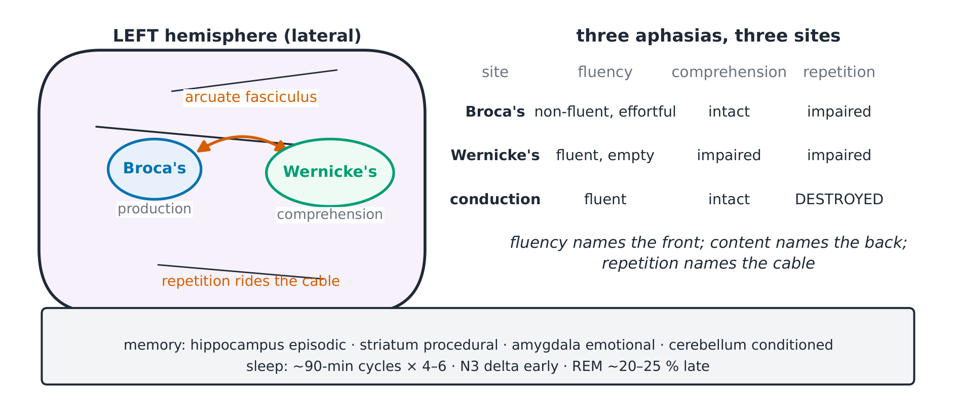
The derivation (why lesion sites name the aphasias). Broca lesion → non-fluent, effortful speech with intact comprehension and intact understanding of own errors; Wernicke lesion → fluent, empty speech with impaired comprehension; arcuate lesion → conduction aphasia 传导性失语: fluent speech, good comprehension, REPETITION destroyed (the cable between them cut) ✓. Fluency names the front; content names the back; repetition names the cable.
Worked example 1 — the night's arithmetic. (Fictional sleep study) 8 hours, five cycles: N3 dominates the first two (slow-wave pressure dissipates), REM episodes lengthen toward morning — early-night sleep loss costs growth hormone, late-night loss costs REM ✓. The architecture, not just the total, is the physiology.
Worked example 2 — clinical link: HM's lesson. (Classic teaching case, no identification needed) bilateral removal of the medial temporal lobes, including most of both hippocampi → profound ANTEROGRADE amnesia with intact procedural learning, while most remote memories survived: the hippocampus writes new episodic memories, the striatum still learns skills ✓ — dissociation is the experiment that separates memory systems.
Common mistakes. Thinking dreams prove REM (they occur in NREM too; vivid recall is REM). Expecting hippocampal lesions to erase OLD memories (consolidated memories live in cortex; the hippocampus writes new ones). Calling all language deficit "aphasia of the left" without checking fluency and repetition first.
Summary. 90-min cycles × 4–6: N3 early (delta, GH), REM late (~20–25 %, atonia); hippocampus episodic, striatum procedural, amygdala emotional, cerebellum conditioned; Broca non-fluent, Wernicke fluent- empty, arcuate = repetition; HM: no new episodic memory, skills intact.
Lesion localisation
Why it matters. Everything above converges here: given a fictional case's signs, name the lesion's level and side — the skill this course was built to teach, one justified step at a time.
The idea, with a picture. The decision map: cortical — higher function deficit (aphasia, seizures, field defects) + contralateral UMN/sensory signs; brainstem — CROSSED findings (ipsilateral cranial nerve + contralateral body); spinal — a sensory LEVEL with UMN signs below and LMN signs at the level; peripheral — LMN/sensory loss in a nerve, root or dermatome distribution, reflex arc gone, no UMN signs ✓.

The derivation (why the map is complete). Each level owns a feature no lower level has: cortex owns higher functions; brainstem owns cranial nerves AND the crossing of tracts (hence ipsilateral face with contralateral body); cord owns the LEVEL (segmental dermatomes) with UMN below; periphery owns individual nerves and roots with their specific territories ✓. One unique feature per level — the map is a partition, not a mnemonic.
Worked example 1 — localise in four steps. (Fictional) right facial weakness (lower face) + right hemiparesis + aphasia: aphasia → left cortex; lower-face-only facial weakness with sparing of the brow → UMN VII → lesion above the VII nucleus; face and body weak on the SAME side (not crossed) → above the brainstem; right-sided weakness → left hemisphere → left middle cerebral artery territory ✓. Each sign climbed one rung; the localisation is the climb, restated.
Worked example 2 — clinical link: the level that moves. (Fictional) burning pain in both feet, absent ankle jerks, normal plantar responses, loss of pain to the knees bilaterally: peripheral pattern (LMN + pain loss without UMN) → a length-dependent polyneuropathy; contrast a cord-compression picture (a LEVEL on the trunk, UMN below) ✓. Symmetry, length dependence and the reflex arc separate periphery from cord.
Common mistakes. Localising from a single sign (the map works on PATTERNS; one sign has many addresses). Forgetting brainstem lesions cross (the single most useful exam clue). Skipping the general examination (wasting and fasciculations together with brisk reflexes and no sensory loss is motor neuron disease — UMN and LMN signs at once, the exception the map must allow for).
Summary. Cortex: higher function + contralateral signs. Brainstem: crossed findings. Cord: sensory level, UMN below, LMN at level. Periphery: nerve/root territories, LMN, no UMN. Four levels, four signatures; localise step by justified step, and say WHY at each rung.