What this course is, and how to use it
This is the second course in University Medicine: medical biochemistry 医学生物化学. MED01 built the cell; this course fills it — the molecules, the enzymes and the fuel flows that keep a human running, and the chemistry that clinical medicine reads every day.
Scope limit, stated once: a study course, not a medical degree; every case below is fictional and nothing here licenses or guides treatment.
- Units 2.1–2.2 are the machinery: proteins and enzyme kinetics.
- Units 2.3–2.4 are energy: glycolysis and the oxidative engine.
- Units 2.5–2.7 are storage and traffic: glycogen, lipids, nitrogen.
- Unit 2.8 integrates: fed, fasting, and diabetes as one ledger.
The medicine habit: mechanism before fact — every enzyme constant, ATP count and fuel flow below is something you recompute, and every clinical link names the broken step.
Proteins and protein structure
Why it matters. Enzymes, transporters, antibodies, receptors — nearly everything a cell does, a protein does it. Structure is not decoration: one level builds on the last, and every level is a place a mutation can break.
The idea, with a picture. Four levels: primary 一级结构 (the amino-acid sequence, read N→C), secondary 二级结构 ($\alpha$-helix, $\beta$-sheet, held by backbone H-bonds), tertiary 三级结构 (the full 3D fold, hydrophobic core), quaternary 四级结构 (subunit assembly).

Definitions with numbers. 20 amino acids; peptide bond ~1.33 Å. The reference protein is haemoglobin 血红蛋白 (Hb): 4 subunits ($\alpha_{2}\beta_{2}$), 4 haem groups, 64.5 kDa, one O₂ each — up to 4 O₂ per molecule.
The derivation (cooperativity). Myoglobin binds O₂ with a simple hyperbola; haemoglobin binds with a sigmoid S形曲线 because each bound O₂ shifts the subunits from the low-affinity T state to the high-affinity R state — binding helps binding: cooperativity 协同效应. The Hill coefficient $n_H \approx 2.8$ (max 4) says "almost fully cooperative". Physiological payoff: full loading in lungs ($pO_{2} \approx 100$ mmHg), about 25 % unloading in resting tissue ($pO_{2} \approx 40$ mmHg) and over 60 % in working muscle ($\approx 20$ mmHg), because the steep middle of the sigmoid sits exactly in that range.
Worked example 1 — the loading arithmetic. Blood carries ~15 g Hb/dL; 64.5 kDa ≈ 64 500 g/mol: $15/64\,500 = 2.33\times10^{-4}$ mol/dL = 233 µmol/dL ×4 sites = 930 µmol O₂/dL ≈ 20.8 mL O₂/dL (22.4 mL per mmol) — the theoretical 1.39 mL O₂ per g Hb. The measured figure is 1.34 mL/g, 20.1 mL O₂/dL ✓, a little lower because some haemoglobin is in forms that cannot carry O₂.
Worked example 2 — clinical link: one charged bead. (Fictional patient) HbS: one Glu→Val at position 6 of the β chain turns a charged surface patch hydrophobic; in the deoxy T state it docks into a pocket on a neighbour, polymerising the Hb and sickling the cell. Primary structure, one bead, changed quaternary behaviour — and vaso-occlusion follows. Sickle cell disease is a primary-structure disease with quaternary consequences.
Common mistakes. Reading "denaturation" as breaking peptide bonds (it unfolds the fold; the sequence survives). Treating all mutations as equally severe (surface conservative changes can be silent; core or active-site ones cannot). Forgetting Hb's ligands (H⁺, CO₂, 2,3-BPG all shift the curve — physiology lives on those shifts).
Summary. Sequence → fold → 3D → assembly; Hb is the reference tetramer with sigmoid, cooperative binding ($n_H \approx 2.8$); one bead (Glu6Val) can rewire quaternary behaviour.
Enzymes and enzyme kinetics
Why it matters. Nearly every diagnosis on a blood panel is an enzyme activity; nearly every drug is an enzyme inhibitor. Kinetics is how biochemistry becomes measurable — and how you will read the lab.
The idea, with a picture. An enzyme lowers the activation barrier and is unchanged by use. The rate curve is Michaelis–Menten 米氏动力学: $v = \frac{V_{max}[S]}{K_m + [S]}$, where $K_m$ 米氏常数 is the substrate concentration at HALF-maximal velocity.

The derivation (why saturation). E + S ⇌ ES → E + P: at low $[S]$, almost all E is free, so $v \approx (V_{max}/K_m)\cdot[S]$ — first order in S. At high $[S]$, every E is in ES: $v$ approaches $V_{max}$ — zero order. The double-reciprocal (Lineweaver–Burk) line $1/v = (K_m/V_{max})\cdot(1/[S]) + 1/V_{max}$ turns the hyperbola into a straight line: y-intercept $1/V_{max}$, x-intercept $-1/K_m$.
Worked example 1 — reading data. An enzyme gives $v = 33$ µmol/min at $[S] = 1$ mM and $v = 60$ at $[S] = 4$ mM. Write $v = V[S]/(K+[S])$ twice and divide: $33(K+1) = 15(K+4)$ ⇒ $K_m = 1.5$ mM, $V_{max} = 33\times2.5 = 82.5$ µmol/min ✓. Check the half point: at $[S] = K_m = 1.5$: $v = 82.5\times1.5/3 = 41.25 = V_{max}/2$ ✓.
Worked example 2 — clinical link: the competitive blocker. (Fictional patient on a statin-like inhibitor) a drug mimics the substrate and occupies the active site (competitive inhibition 竞争性抑制): apparent $K_m$ rises, $V_{max}$ unchanged — overcome by more substrate, and the L-B lines intersect ON the y-axis. Non-competitive binding elsewhere lowers $V_{max}$ and leaves $K_m$ alone (lines meet ON the x-axis). Reading which line moved IS the mechanism.
Common mistakes. Calling $K_m$ an affinity (it is a concentration; small $K_m$ happens to mean high apparent affinity). Fitting two points in your head without checking a third. Expecting inhibitors to change enzyme AMOUNT (induction does; inhibitors change activity).
Summary. $v = V_{max}[S]/(K_m+[S])$; $K_m$ = $[S]$ at half-$V_{max}$; competitive: $K_m$↑ $V_{max}$ same; non-competitive: $V_{max}$↓ $K_m$ same; L-B intercepts identify which.
Bioenergetics and glycolysis
Why it matters. Cells pay for everything in ATP, and the currency exchange is exact: coupled reactions proceed only downhill in free energy. Glycolysis is the oldest, most conserved way to make ATP — and the metabolic anchor of every later unit.
The idea, with a picture. Coupling: an unfavourable reaction runs only when bolted to a favourable one ($\Delta G$ adds). Glycolysis 糖酵解 splits one glucose (6 C) into two pyruvate (3 C): investment phase (2 ATP spent), payoff phase (4 ATP + 2 NADH made), net 2 ATP + 2 NADH ✓ — independent of oxygen.

Definitions with numbers. The three essentially irreversible steps ($\Delta G^{\circ\prime}$ strongly negative): hexokinase (−16.7 kJ/mol), PFK-1 磷酸果糖激酶 (−14.2, the COMMITTED step and main control), pyruvate kinase (−31.4). Without O₂, NAD⁺ is regenerated by lactate dehydrogenase 乳酸脱氢酶: pyruvate + NADH → lactate (fermentation).
The derivation (why PFK-1 is the gate). Control theory says: gate the first committed, irreversible step. Hexokinase is pre-committed (other sugars use it); pyruvate kinase is too late (the investment is already made). PFK-1 is the first step unique to glycolysis — so AMP and ADP activate it; ATP and citrate inhibit it; fructose-2,6- bisphosphate (insulin signal) activates it powerfully. One enzyme sees the cell's whole energy state.
Worked example 1 — the ATP ledger. Per glucose: −2 (investment) +4 (payoff) = net 2 ATP; plus 2 NADH (cytosolic — worth 5 ATP via the malate shuttle or 3 via glycerol phosphate, unit 2.4). Anaerobically the 2 NADH are consumed re-reducing pyruvate to lactate: the anaerobic yield is exactly 2 ATP ✓.
Worked example 2 — clinical link: the gate that will not open. (Fictional patient) muscle cramps within seconds of sprint, no rise in blood lactate: muscle phosphorylase works (glycogen breaks down) but PFK-1 is deficient — sugar phosphates pile up before the gate, the payoff never comes, ATP falls and myoglobin spills. One gate, one exertional syndrome: the regulated step is the disease locus.
Common mistakes. Counting gross ATP where net is asked. Placing regulation at reversible steps (regulation lives at irreversible gates). Forgetting anaerobic NAD⁺ regeneration is WHY lactate forms (not a waste product — a redox necessity).
Summary. Glucose → 2 pyruvate, net 2 ATP + 2 NADH, three irreversible gates with PFK-1 committed; AMP/ADP on, ATP/citrate off; lactate buys NAD⁺ when O₂ is out.
TCA cycle and oxidative phosphorylation
Why it matters. Oxygen exists for one job in the body: to accept electrons at the end of the transport chain. The TCA cycle turns acetyl units into those electrons, and the ATP synthase turns their fall into ~90 % of the body's ATP. This unit is where energy metabolism becomes quantitative.
The idea, with a picture. The TCA cycle 三羧酸循环 (Krebs): acetyl-CoA (2 C) + oxaloacetate (4 C) → citrate (6 C) → two decarbox- ylations return to oxaloacetate, yielding per acetyl: 3 NADH, 1 FADH₂, 1 GTP. The electron transport chain 电子传递链 (inner mitochondrial membrane): complexes I, III, IV pump H⁺ out; the return flow through ATP synthase ATP合酶 makes ATP.
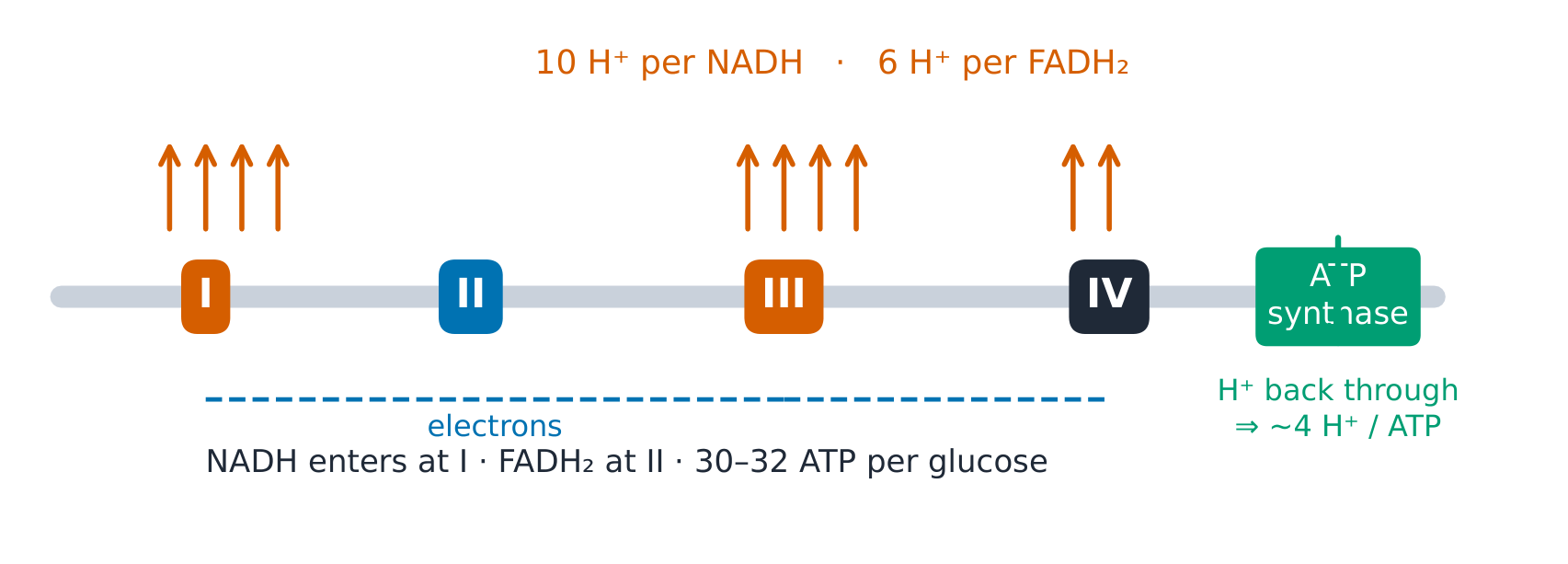
The derivation (the modern ATP count). Per NADH: complexes I+III+IV pump 4+4+2 = 10 H⁺; per FADH₂ (entry at II): III+IV = 6 H⁺. ATP synthase needs ~3 H⁺/ATP plus 1 H⁺ to import Pi: 4 H⁺/ATP. So NADH ≈ 10/4 = 2.5 ATP, FADH₂ ≈ 6/4 = 1.5 ATP ✓ — and per glucose, counting every carrier: 2 (glycolysis ATP) + 2×2.5 (its NADH, malate shuttle) + 2×2.5 (pyruvate dehydrogenase NADH) + 2 GTP + 6×2.5 (TCA NADH) + 2×1.5 = 32 ATP with the malate shuttle, 30 with glycerol phosphate (the older "36–38" used the rounded P/O of 3 and 2).
Worked example 1 — acetyl arithmetic. One glucose → 2 pyruvate → 2 acetyl-CoA (2 NADH from pyruvate dehydrogenase). With pyruvate dehydrogenase, two TCA turns plus entry yield 2 GTP + 8 NADH + 2 FADH₂ ✓ = 2 + 20 + 3 = 25 ATP-equivalents — three quarters of the whole aerobic budget (25 of 32), all from electron pairs destined for O₂.
Worked example 2 — clinical link: the blocked turbine. (Fictional overdose vignette) collapse with bright-red venous blood: cyanide binds complex IV, electrons cannot reach O₂, pumping stops, the gradient collapses and ATP synthase stops (it can even run backwards, spending ATP). The chemistry explains the signs: tissues cannot extract O₂ (venous blood stays red), lactate soars (glycolysis alone remains). Treatments exist to bypass or out-compete the block — mechanism first, antidote second.
Common mistakes. Saying the cycle "makes ATP" directly (it makes GTP + reduced carriers; the chain makes the gradient). Counting CO₂ as from "the acetyl carbons of this turn" (they leave in LATER turns — the cycle mixes its carbons). Using P/O 3 and 2 without saying which convention.
Summary. Acetyl → 3 NADH + 1 FADH₂ + 1 GTP per turn; 10 H⁺/NADH, 6 H⁺/FADH₂, ~4 H⁺/ATP ⇒ 2.5/1.5; 30–32 ATP per glucose (modern count, shuttle-dependent); complex IV is where oxygen earns its keep.
Glycogen and gluconeogenesis
Why it matters. The brain burns ~120 g glucose a day and stores none. Between meals the body lives on two glucose banks with different owners — and in fasting it must make glucose from scratch.
The idea, with a picture. Glycogen 糖原: branched glucose polymer ($\alpha 1\to4$ chains, $\alpha 1\to6$ branch every ~8–12 residues). Liver stores ~100 g — EXPORTS glucose to blood; muscle stores ~400 g — keeps it, burns it locally.

Definitions with numbers. Glycogen phosphorylase cleaves $\alpha1\to4$ (phosphorolysis, no ATP spent); debranching enzyme handles the $\alpha1\to6$ stubs. Gluconeogenesis 糖异生 reverses glycolysis uphill around its three gates — at the cost of 4 ATP + 2 GTP
- 2 NADH per glucose (6 high-energy phosphates, vs glycolysis's net gain of 2).
The derivation (why two banks). Muscle lacks glucose-6-phosphatase: its glucose-6-P is trapped and feeds glycolysis — you cannot export what you cannot dephosphorylate. Liver HAS the phosphatase: glycogen → glucose-1-P → glucose-6-P → free glucose → blood. One enzyme's presence or absence assigns the organ's role; hormones aim it — glucagon 胰高血糖素 (liver only) triggers breakdown, insulin 胰岛素 triggers synthesis.
Worked example 1 — the overnight budget. Liver glycogen ~100 g ≈ 400 kcal; the brain needs ~120 g/day ≈ 480 kcal/day. The bank covers roughly a day's brain use ✓ — after 12–24 h fasting, gluconeogenesis must take over (amino acids from muscle, glycerol from fat, lactate via the Cori cycle).
Worked example 2 — clinical link: the runner who cannot tap the bank. (Fictional) cramps and dark urine after sprinting: muscle phosphorylase is deficient (McArdle pattern) — glycogen is stored but unreachable, ATP falls, myoglobin spills, and the expected lactate rise never comes. The lab IS the mechanism: absent lactate with maximal effort localises the break before the glycolytic gate.
Common mistakes. Treating the two glycogen banks as one (muscle glycogen never becomes blood glucose). Counting gluconeogenesis as "glycolysis backwards for free" (it costs 6 high-energy phosphates). Placing glucagon receptors on muscle (there are none).
Summary. Liver ~100 g exports (has the phosphatase); muscle ~400 g keeps (lacks it); phosphorylase + debrancher break, synthase + branching enzyme build; glucagon/insulin set the sign; gluconeogenesis costs 4 ATP + 2 GTP + 2 NADH per glucose.
Lipid metabolism
Why it matters. Fat is the body's long-term energy warehouse — and its heaviest traffic problem: insoluble in water, lipids need a transport fleet whose failures are the world's most common disease.
The idea, with a picture. β-oxidation β氧化 in mitochondria: each cycle trims 2 C off a fatty acyl-CoA, yielding 1 acetyl-CoA + 1 FADH₂ + 1 NADH. Transport: lipoproteins 脂蛋白 — chylomicrons (intestine → tissues), VLDL (liver → tissues), LDL (cholesterol to tissues — the "bad" carrier), HDL (cholesterol back to liver — the "good" one).

The derivation (the palmitate count). Palmitate (C16): $\beta$- oxidation needs $(16/2) - 1 = 7$ cycles → 8 acetyl-CoA + 7 FADH₂ + 7 NADH. Yield: each acetyl is worth 10 equivalents through the cycle ($3\times2.5 + 1.5 + 1$), so 8 acetyl give 80; the 7 FADH₂ give $7\times1.5 = 10.5$ and the 7 NADH give $7\times2.5 = 17.5$; minus 2 for activation: 106 ATP per palmitate ✓ — against 30–32 per glucose: fat pays several-fold per carbon, because it is almost fully reduced.
Worked example 1 — ketones, the brain's diesel. Sustained fasting: acetyl-CoA from β-oxidation outruns the TCA cycle (OAA diverted to gluconeogenesis), so the liver condenses it to ketone bodies 酮体 (acetoacetate, β-hydroxybutyrate) and exports them — water-soluble, crossing to the brain, worth ~21.5 ATP per β-hydroxybutyrate. By day 3 ketones supply about a third of the brain's energy, and after several weeks up to two-thirds, sparing muscle protein ✓.
Worked example 2 — clinical link: the engine without a spark. (Fictional infant) fasting hypoketotic hypoglycaemia: an acyl-CoA dehydrogenase is deficient, β-oxidation stalls, no ketones can be made AND no ATP to drive gluconeogenesis — the double failure explains BOTH lab findings. The diagnostic signature (low glucose WITH low ketones) is the mechanism read off a blood spot.
Common mistakes. Forgetting the activation cost (−2 equivalents) in ATP counts. Expecting the brain to burn long-chain fatty acids (it oxidises very little fatty acid — ketones are its substitute). Calling LDL "cholesterol" (it is the CARRIER particle).
Summary. β-oxidation: C16 → 8 acetyl-CoA + 7×(FADH₂+NADH) → 106 ATP net; chylomicron/VLDL out, LDL deliver, HDL return; fasting makes the liver a ketone exporter.
Nitrogen metabolism and the urea cycle
Why it matters. Nitrogen cannot be stored: every amino acid not needed today must be deaminated today, and the ammonia released is neurotoxic at micromolar concentrations. Disposal is not optional — it is a five-step conveyor across two compartments.
The idea, with a picture. Amino groups funnel to glutamate 谷氨酸 (transamination 转氨基作用: amino acid + α-ketoglutarate ⇌ keto-acid + glutamate), then oxidative deamination frees NH₃ in the liver mitochondrion. The urea cycle 尿素循环 runs across mitochondrion and cytosol: NH₃ + CO₂ → carbamoyl phosphate → citrulline → (cytosol) arginino- succinate → arginine → urea + regenerated ornithine.
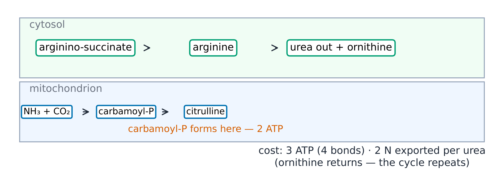
Definitions with numbers. Urea: 2 N per molecule (one from NH₃, one from aspartate), cost 3 ATP (4 high-energy bonds) per urea. Normal blood ammonia is only ~10–35 µmol/L — tight, because the brain has no urea cycle.
The derivation (why the liver, why there). Ammonia detoxication must sit upstream of the systemic circulation: portal blood from the gut drains straight into the liver, which extracts amino acids and ammonia on first pass. The brain and muscle cannot afford the cycle's cost — they export nitrogen as glutamine 谷氨酰胺 (the ammonia taxi) and alanine 丙氨酸 (the carbon-and-nitrogen taxi) for the liver to process. Location IS the safety system.
Worked example 1 — the daily ledger. A typical diet turns over ~70–100 g protein/day ≈ 11–16 g N; excreted almost entirely as urea (25–35 g/day). Cross-check: each urea carries 2 N (28 g of 60 g molecule = 47 % N by mass): 30 g urea × 0.47 ≈ 14 g N ✓ — the urine matches the diet.
Worked example 2 — clinical link: the blocked conveyor. (Fictional newborn) lethargy then coma after first feeds, sky-high ammonia, orotic acid in urine: ornithine transcarbamylase deficiency — the cycle jams at step 2, carbamoyl phosphate spills into pyrimidine synthesis (hence the orotate). Mechanism-first management follows the block: limit nitrogen intake, scavenge ammonia, feed the alternatives — the biochemistry writes the protocol.
Common mistakes. Writing "urea removes ammonia" (it removes TWO nitrogens: NH₃ and aspartate's amino group). Expecting urea-cycle enzymes in brain (liver-only; that is why ammonia hits the brain). Forgetting the ATP cost (nitrogen disposal is expensive: 4 bonds).
Summary. Transamination funnels N to glutamate; the liver cycle (2 compartments, 5 enzymes) builds urea at 3 ATP each, exporting 2 N; glutamine and alanine are the taxis; hyperammonaemia with orotate points to OTC.
Integration of metabolism: fed, fasting and diabetes
Why it matters. Metabolism is one system with a switch. Insulin and glucagon are the lever, the organs are the gears, and diabetes is the switch jammed — the single most instructive integration in medicine.
The idea, with a picture. Fed state 进食状态 (insulin high): glucose into all cells, glycogen filled, extra carbon to fat. Fasting state 空腹状态 (glucagon high): liver glycogenolysis (hours) → gluconeogenesis (days) → ketogenesis (brain adapts). One fuel ledger, three time regimes.

Definitions with numbers. Brain ~120 g glucose/day. Liver glycogen ~100 g (~a day). Ketones cover about a third of brain energy by day 3 of a fast, and up to two-thirds after several weeks. Diagnostic anchor: HbA1c ≥ 6.5 % indicates diabetes — haemoglobin glycation accumulates over the red cell's ~120-day life, integrating glucose like a tape recorder.
The derivation (the switch logic). One variable — is food arriving? — must set EVERY organ's arrows coherently. A hormone pair with opposite actions on the same enzymes (PFK-2, glycogen synthase, phosphorylase, lipase) achieves this: glucagon raises cAMP, so PKA phosphorylates them; insulin lowers cAMP (phosphodiesterase on) and switches on phosphatases, so the same enzymes lose their phosphates. The elegance is that ONE signal, not seven, keeps 4 organs from working at cross purposes — and diabetes shows what cross purposes cost.
Worked example 1 — the timeline of a fast. Hour 0 (meal ends): glucose falls. Hours 0–4: absorbed fuel used, insulin falls. Hours 4–12: liver glycogen drains. Hours 12–24: bank empty, gluconeogenesis ramps (muscle amino acids, glycerol, lactate). Days 1–3: protein sparing begins as ketones rise. Day 3: about a third of brain energy from ketones; weeks: up to two-thirds ✓ — each transition is the previous unit's mechanism, timed.
Worked example 2 — clinical link: the jammed switch, both ways. (Fictional patient) type 1 picture: no insulin, so despite high glucose the cells read FASTING — fat pours out, ketones flood (acidosis, Kussmaul breathing as the lungs compensate), muscle breaks down, urine loses glucose and water (osmotic diuresis, thirst). Every symptom is one switch read by four organs. Type 2 picture: insulin present but resisted — the same ledger, milder arrows, years instead of hours.
Common mistakes. Reading insulin as "lowers glucose" only (it sets the direction of ALL fuels). Treating brain glucose need as negotiable (ketones only partly substitute, and take days). Confusing HbA1c (3-month average) with a glucose reading (minutes).
Summary. Insulin/glucagon = one lever on all organs; fed → store, fast → glycogenolysis → gluconeogenesis → ketones on a ~24 h / 3 day clock; HbA1c ≥ 6.5 % integrates the tape; diabetic ketoacidosis is fasting metabolism running with high glucose.