What this course is, and how to use it
This is the eighth course in University Medicine: medical microbiology and infection 医学微生物学与感染. MED07 built the defences; this course names the attackers — bacteria, viruses, fungi and parasites — and the laboratory methods that tell them apart.
Scope limit, stated once: a study course, not a medical degree; every case below is fictional and nothing here licenses or guides treatment.
- Units 8.1–8.4 are the organisms: classification, bacteria, viruses, fungi and parasites.
- Units 8.5–8.7 are the interaction: transmission, antimicrobials and prevention.
The medicine habit: the organism's biology predicts its disease — every mechanism below is traced from structure or lifecycle to the clinical picture, and every number (doubling times, incubation periods 潜伏期, $R_0$) is something you recompute.
Classification and the diagnostic laboratory
Why it matters. Before any therapy question stands the naming question: what is growing? The diagnostic laboratory is a decision tree turned into instruments, and reading its outputs is a clinical skill.
The idea, with a picture. The tree: specimen → culture on solid media → Gram stain 革兰染色 (positive purple / negative pink) → shape (cocci, bacilli) → catalase/oxidase, then biochemistry, serology, antigen tests and PCR 聚合酶链反应 for the fast lane ✓. Culture remains the reference standard: most bacteria grow visibly in 24–48 h; fastidious or slow organisms demand special media and patience.

The derivation (why Gram staining separates). The stain is chemistry, not colour preference: crystal violet–iodine complexes are TRAPPED by the thick peptidoglycan 肽聚糖 wall of Gram-positive cells (many layers, teichoic acids 磷壁酸 ✓), while Gram-negative walls (1–2 peptidoglycan layers plus an outer membrane) lose the complex at the decolouriser step and take up the pink counterstain. The same chemistry predicts each group's drug susceptibilities (8.6): what stains, targets.
Worked example 1 — reading a report. (Fictional) blood culture at 48 h: Gram-positive cocci in clusters; catalase positive, coagulase positive. The tree walks: clusters → staphylococci; catalase + → not streptococci; coagulase + → Staphylococcus aureus, not the coagulase-negative skin flora ✓. Each step is a fork with one biochemical key — the report's three lines are three forks.
Worked example 2 — clinical link: the fast lane. (Fictional) suspected meningitis: PCR of cerebrospinal fluid can name S. pneumoniae or N. meningitidis within ~1–2 h, while culture needs 24–48 h ✓. The trade: PCR tells you what DNA is present, not what is alive and drug-sensitive — culture plus susceptibilities still follow. Naming fast, acting informed.
Common mistakes. Reading a Gram stain as a species (it narrows to a shape-and-colour class). Expecting PCR to replace culture (viable organism and susceptibilities come only from culture). Forgetting contaminant biology (coagulase-negative staphylococci in ONE of two bottles are more likely skin flora than pathogen ✓).
Summary. Gram stain (thick-wall purple vs thin-wall pink) → shape → biochemistry → serology/PCR; culture 24–48 h remains the reference; PCR names in ~1–2 h but only culture proves viability; report reading = walking the tree fork by fork.
Bacteria: structure, growth and pathogenesis
Why it matters. A bacterium's structure decides both its disease and its therapy: the wall sets the Gram class, the toxins set the picture, and the growth rate sets the pace of everything.
The idea, with a picture. Gram-positive: thick peptidoglycan with teichoic acids, no outer membrane. Gram-negative: thin peptidoglycan PLUS an outer membrane whose lipopolysaccharide 脂多糖 (LPS, endotoxin) faces outward, with porins ✓. Common armament: capsules 荚膜 (defeat phagocytosis — MED07's opsonisation gap), pili/fimbriae (adhesion), flagella (motility), exotoxins 外毒素 (secreted proteins), and the growth curve: lag → log → stationary → death.

The derivation (why doubling time is destiny). $N = N_0 \cdot 2^{t/g}$ with generation time 世代时间 $g$. E. coli doubles in ~20 min: in 10 h that is 30 generations, $2^{30} \approx 10^{9}$ cells from one founder ✓. The tuberculosis bacterium doubles in ~20 h: 10 h is half a generation. Fast growers cause acute 24-hour disease and visible cultures at 24–48 h; slow growers cause weeks-long disease and demand weeks of culture (8.1). The arithmetic is the whole clinical pace.
Worked example 1 — toxin classes, named. (Fictional teaching set) Toxin A blocks protein synthesis in a wound infection; toxin B lyses red cells on a plate: the first behaves like diphtheria's fragment-A enzyme (an ADP-ribosyltransferase that halts elongation), the second like the β-haemolysin family. Mechanism explains the picture: synthesis blockers kill with minimal local destruction (distant organ failure), membrane attackers destroy locally ✓.
Worked example 2 — clinical link: the capsule read. (Fictional) sputum Gram stain: Gram-positive lancet-shaped diplococci, each with a clear halo. The halo IS the capsule (S. pneumoniae) ✓ — the structure that defeats phagocytosis, and the reason people without a spleen are at risk of overwhelming pneumococcal disease. The stain names the virulence factor.
Common mistakes. Calling LPS an exotoxin (it is a wall component released at lysis — endotoxin ✓). Treating all fast growers as pathogens (the gut's commensal E. coli doubles at the same speed). Reading absence of growth as absence of organism (fastidious organisms need their media and their days).
Summary. Wall: thick Gram-positive vs LPS-membrane Gram-negative; virulence: capsule, adhesins, exotoxins; growth $N = N_0 2^{t/g}$ — a 20-min doubler makes $10^{9}$ in 10 h, a 20-h doubler makes half a generation; toxin mechanism predicts the clinical picture; the capsule is visible on the stain.
Viruses
Why it matters. Viruses are genomes in coats — obligate intracellular parasites whose lifecycle IS the disease. The cycle tells you why antivirals must target replication steps and why vaccines (7.4) work so well against them.
The idea, with a picture. Structure: nucleic acid (DNA or RNA) + capsid 衣壳 (symmetric protein shell) ± envelope 包膜 (host-derived membrane studded with viral glycoproteins) ✓. The cycle: attachment → entry → uncoating → replication of genome and proteins → assembly → release. Enveloped viruses bud out (host membrane kept continuous); naked viruses lyse the cell.
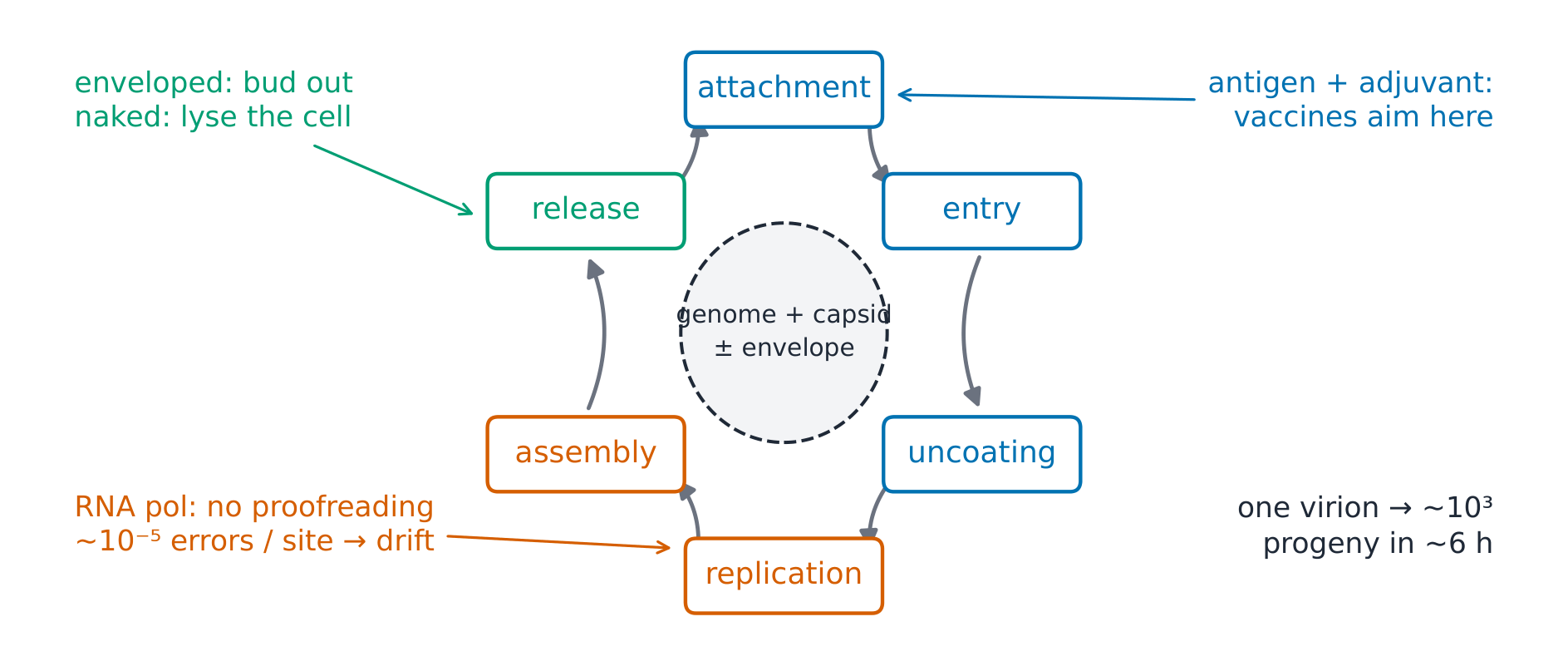
The derivation (why mutation follows genome chemistry). RNA polymerases lack proofreading: influenza's mutation rate is ~$10^{-5}$ substitutions per site per replication, against ~$10^{-8}$–$10^{-6}$ for DNA viruses, whose polymerases proofread — tens to a thousand times lower ✓. Hence antigenic drift 抗原漂移 year by year and updated vaccines. DNA viruses proofread: stable antigens, stable vaccines. Segmented genomes (influenza: 8 segments) enable antigenic shift 抗原转变 — reassortment between human and animal strains — the mechanism behind pandemic influenza.
Worked example 1 — the burst arithmetic. (Fictional) one influenza virion infects a respiratory cell; ~6 h later ~$10^{3}$ progeny bud off ✓; two rounds (12 h) give $10^{6}$. An incubation period of 1–2 days is only ~4–8 cell rounds: viral arithmetic reaches symptoms faster than any culture.
Worked example 2 — clinical link: reading the season's chart. (Fictional public-health data) winter spike of fever and myalgia, incubation 1–2 days, attack rates ~20 % in offices: the pattern of influenza A (short incubation, drift-matched vaccine) — compare the 2–3-week incubation of a DNA virus like varicella, whose outbreaks build over weeks because each generation of cases takes longer. The epidemic curve's slope encodes the virus's cycle speed ✓.
Common mistakes. Treating all RNA viruses as fragile (the envelope decides environmental survival: naked RNA viruses like poliovirus are hardier). Expecting antibacterials to act on viruses (their targets are 8.6's bacteria-specific structures). Forgetting that enveloped viruses carry host membrane — a bud is a disguise as well as an exit.
Summary. Genome (DNA/RNA) + capsid ± envelope; six-step cycle; no proofreading → ~$10^{-5}$ errors per site → drift; 8 segments → shift; one virion → $10^{3}$ in ~6 h; incubation period = the number of rounds.
Fungi and parasites
Why it matters. Eukaryotic pathogens behave differently from bacteria and viruses: dimorphic fungi switch with temperature, protozoa and helminths run complex life cycles through vectors and hosts. These cycles decide geography, diagnosis and whose blood film you are reading.
The idea, with a picture. Fungi: yeasts (single cells, budding), moulds (hyphae), dimorphic fungi 双相真菌 (mould in the environment at 25 °C, yeast in tissue at 37 °C ✓) — opportunists exploit the immunocompromised host along MED07's defect map (T-cell failure → Pneumocystis, Candida; neutrophil failure → Aspergillus). Parasites: protozoa (malaria, amoebae) and helminths (worms). The malaria life cycle: mosquito injects sporozoites 子孢子 → liver schizonts → erythrocytic merozoites 裂殖子 cycling in red cells → gametocytes 配子体 taken up by the next mosquito ✓.

The derivation (why the fever is periodic). Synchronous erythrocytic bursting every 48 h (P. vivax, P. ovale — tertian) or 72 h (P. malariae — quartan) ✓ produces the classic periodic fevers: the parasitaemia's clock IS the symptom's clock. P. falciparum synchronises poorly — fevers less regular — but cytoadherence of infected red cells (sequestration) makes it the killer: the cycle explains both the textbook pattern and the exception's danger.
Worked example 1 — the film read. (Fictional) blood films from a returned traveller: the thick film finds the parasites; the thin film shows ring forms in ~3 % of erythrocytes plus crescent-shaped gametocytes → P. falciparum ✓. The 3 % parasitaemia and the banana gametocyte — morphology names the species and grades severity in one look.
Worked example 2 — clinical link: dimorphism as a geography lesson. (Fictional) a farmer's pneumonia after dust exposure; culture at 25 °C grows a mould, at 37 °C a yeast: a dimorphic fungus (the histoplasma pattern) ✓ — the fungus's thermostat switch doubles as its identity card: the same organism is a mould in the environment and a yeast in you.
Common mistakes. Treating fungi like bacteria (their eukaryotic machinery narrows selective-toxicity options). Expecting all parasites to show in blood films (most need stool, serology or tissue). Forgetting that opportunistic mycoses follow MED07's arms: which defect, which fungus.
Summary. Yeasts/moulds/dimorphs (25 °C mould, 37 °C yeast); opportunists follow the immunodeficiency map; malaria: sporozoite → liver → 48-h blood cycle → gametocyte; fever periodicity = the parasite's clock; falciparum's asynchrony + sequestration = the killer.
Host–pathogen interaction and transmission
Why it matters. An infection is a negotiation between a population of organisms and a host — the outcome (colonisation, carrier state or disease) and the spread between hosts follow rules you can compute.
The idea, with a picture. The chain of infection 感染链: reservoir → exit → transmission route (contact / droplet / airborne / vector / vehicle) → entry → susceptible host ✓; break any link and transmission stops. The basic reproduction number 基本再生数 $R_0$: average secondary cases from one case in a fully susceptible population — measles ~12–18, influenza ~1.3, ancestral SARS-CoV-2 ~2.5–3 ✓ (MED07's herd threshold $1 - 1/R_0$ builds on this).

The derivation (why $R_0$ and the serial interval set epidemic speed). Early in an epidemic, cases multiply by $R_0$ every serial interval (measles ~10–12 days, influenza ~2–3 days) ✓. Doubling time $t_d \approx \mathrm{SI} \times \log 2 / \log R_0$: influenza ($R_0 \approx 1.3$, SI 2–3 d) doubles in ~5–8 days; measles ($R_0 \approx 15$, SI ~11 d) would double in ~2.5–3 days in a susceptible population. The same equation explains why isolation slows spread (it lowers the effective $R$) and why short-interval outbreaks outrun contact tracing.
Worked example 1 — the attack-rate arithmetic. (Fictional wedding) 120 guests ate the buffet, 54 developed gastroenteritis within 48 h: attack rate 罹患率 = 54/120 = 45 % ✓; stratified by dish, 85 % of seafood eaters vs 10 % of non-eaters fell ill → the vehicle is named. One fraction, stratified once, is an outbreak investigation's first page.
Worked example 2 — clinical link: breaking the chain, link by link. (Fictional ward outbreak of diarrhoeal illness) the reservoir (cases plus contaminated environment) → exit (stool) → route (hands and surfaces) → entry (mouth) → susceptible hosts (ward patients). Hand hygiene breaks the route; cohorting breaks the susceptible-host link; each intervention maps onto a named link of the chain — the diagram is the outbreak plan, drawn ✓.
Common mistakes. Equating $R_0$ with severity or case-fatality (transmissibility and virulence are different axes). Using $R_0$ (intrinsic) where the effective $R$ (behaviour, immunity) is the operative number. Forgetting carriers: reservoirs need not be sick — chronic typhoid carriers drive transmission ✓.
Summary. Chain of five links; $R_0$ sets epidemic pace jointly with the serial interval ($t_d \approx \mathrm{SI} \cdot \log 2 / \log R_0$); stratified attack rates name vehicles; carriers make reservoirs silent; interventions = links broken.
Antimicrobials and resistance
Why it matters. Every antibacterial exploits a difference between bacterial and human cells; every resistance mechanism is the bacterium closing that difference. Mechanism-first thinking (no regimens, per the course plan) explains both the drugs and the laboratory's susceptibility report.
The idea, with a picture. Selective toxicity 选择性毒性 needs bacteria-specific targets: the wall (β-lactams bind penicillin-binding proteins, blocking peptidoglycan cross-linking ✓), the ribosome (bacterial 30S/50S subunits vs human 40S/60S), nucleic- acid enzymes (DNA gyrase, RNA polymerase), and folate synthesis (bacteria make it; humans import it) ✓.

The derivation (why resistance is inevitable arithmetic). Mutation supplies resistance at ~$10^{-7}$–$10^{-9}$ per cell per generation; an overnight broth culture holds ~$10^{9}$ cells per mL, so every culture carries mutants against any single drug ✓. Combination approaches exist BECAUSE two simultaneous independent mutations are the product of probabilities (~$10^{-14}$–$10^{-18}$). Horizontal gene transfer 水平基因转移 (plasmids, transposons) then spreads the wins between species — chemistry gave the targets; population genetics sets their shelf life.
Worked example 1 — reading a susceptibility report. (Fictional) S. aureus from an abscess: β-lactamase positive, susceptible to flucloxacillin. The lab's two lines trace to 8.2's wall: β-lactamase chemically OPENS the β-lactam ring (a hydrolysis), while an altered target (PBP2a) is what makes a true methicillin-resistant strain — different mechanisms, different drugs, both named by the report ✓.
Worked example 2 — clinical link: the stewardship logic. (Fictional ward) rising resistance in unit infections tracked to broad spectrum use: narrow the drug to the reported susceptibility and the selective pressure on the ward's flora falls. The mechanism chain: drug pressure → selection of the resistant fraction (the $10^{-7}$ arithmetic) → stewardship narrows the pressure. No regimens — the principle IS the mechanism ✓.
Common mistakes. Treating resistance as all-or-nothing (susceptibility is graded and reported in categories). Expecting new targets to defeat resistance permanently (the $10^{-9}$ clock restarts for every new molecule). Forgetting that host immunity is the drug's partner (the defences of MED07 do most of the clearing).
Summary. Targets: wall (PBPs), ribosomes, gyrase/polymerase, folate — all bacteria-specific; resistance arises at $10^{-7}$–$10^{-9}$ per cell per generation plus horizontal transfer; combination and stewardship exist because of the probability arithmetic; reports name the mechanism, not just the organism.
Infection prevention and control
Why it matters. Prevention is applied transmission knowledge: every precaution level, sterilisation method and screening programme is a link of 8.5's chain, deliberately cut. The cheapest infection is the one that never happens.
The idea, with a picture. Precaution levels: standard precautions 标准预防 (every patient: hand hygiene, gloves for anticipated exposure) plus transmission-based additions — contact (gowns, single room), droplet (masks within ~1–2 m), airborne (fitted respirator, negative-pressure room) ✓. The decontamination ladder: cleaning → disinfection → sterilisation 灭菌 (steam 121–134 °C, or chemical for heat-labile instruments) ✓.

The derivation (why the route sets the level). Droplets (> 5 µm) fall within ~1–2 m: a mask at that distance breaks the chain ✓. Airborne particles (≤ 5 µm) stay suspended for hours and ride air currents: only a fitted respirator plus room ventilation breaks that link. Match the level to the PHYSICS of the route: over-isolation wastes, under-isolation leaves the chain open.
Worked example 1 — the vaccination arithmetic. (Fictional clinic) staff influenza vaccine uptake rises from 40 % to 90 %: if every vaccinated worker were protected, the ward's susceptible-staff fraction would fall from 60 % to 10 % — a six-fold drop (with a vaccine that protects 60 %, it falls from 76 % to 46 %), on top of each vaccinated worker's own reduced risk ✓. Herd thinking (7.4) applied to the staff who meet every reservoir: prevention is a population calculation.
Worked example 2 — clinical link: the outbreak clock. (Fictional) a surgical-site infection cluster presenting on post-operative days 5–7: the timing names the acquisition window (the theatre or the first 48 h of aftercare), the organism's incubation arithmetic (8.2) dates the breach, and the response follows the chain — reservoir (case or environment), route (contact), entry (the wound). One clock, the whole investigation ✓.
Common mistakes. Treating standard precautions as optional when the patient looks well (the reservoir is often silent). Using airborne precautions for droplet organisms (measles and tuberculosis are airborne; influenza is managed with droplet precautions ✓). Confusing disinfection with sterilisation (spores survive disinfection).
Summary. Standard always; add contact/droplet/airborne to match the route's physics (> 5 µm falls in ~1–2 m, ≤ 5 µm drifts for hours); sterilisation for invasive instruments; vaccination is herd arithmetic; the incubation clock dates every breach.