Production and use of X-rays · X射线的产生与使用
| English | 中文 | Pinyin · 拼音 |
|---|---|---|
| tungsten/ˈtʌŋstn/ | 钨 | wū |
| target/ˈtɑːɡɪt/ | 靶 | bǎ |
| anode/ˈænəʊd/ | 阳极 | yáng jí |
| cathode/ˈkæθəʊd/ | 阴极 | yīn jí |
| thermionic emission/ˌθɜːmɪˈɒnɪk ɪˈmɪʃn/ | 热电子发射 | rè diàn zi fā shè |
| kinetic energy/kɪˈnetɪk ˈenədʒi/ | 动能 | dòng néng |
| photons/ˈfəʊtɒnz/ | 光子 | guāng zi |
| Bremsstrahlung/ˈbremʃtrɑːlʊŋ/ | 轫致辐射 | rèn zhì fú shè |
| hardness/ˈhɑːdnəs/ | 硬度 | yìng dù |
| characteristic/ˌkærɪktəˈrɪstɪk/ | 特征 | tè zhēng |
| sharpness/ˈʃɑːpnəs/ | 清晰度 | qīng xī dù |
| contrast/ˈkɒntræst/ | 对比度 | duì bǐ dù |
| attenuation/əˌtenjuːˈeɪʃn/ | 衰减 | shuāi jiǎn |
| contrast medium/ˈkɒntræst ˈmiːdɪəm/ | 造影剂 | zào yǐng jì |
| half-value thickness/hɑːf ˈvæljuː ˈθɪknəs/ | 半值厚度 | bàn zhí hòu dù |
| computed tomography/kəmˈpjuːtɪd təˈmɒɡrəfi/ | 计算机断层扫描 | jì suàn jī duàn céng sǎo miáo |
A machine that wastes ninety-nine per cent of its power
- An X-ray tube run at $75\ \text{kV}$ and $30\ \text{mA}$ takes in $2250\ \text{W}$. About $99\%$ of that becomes heat in a lump of tungsten the size of a coin.
- Left alone, the target's temperature would climb at over $1000\ \text{K}$ per second and melt in a few seconds.
- That is why exposures are brief and the anode spins. The remaining one per cent is the picture.
- This lesson is how X-rays are produced, what sets their penetrating power, and how they make an image.
一台把百分之九十九的功率浪费掉的机器
- 一支在 $75\ \text{kV}$、$30\ \text{mA}$ 下工作的 X 射线管吸收 $2250\ \text{W}$。其中约 $99\%$ 变成一小块硬币大小的钨里的热。
- 若不管它,靶的温度会以每秒一千多开尔文的速度上升,几秒钟内就熔化。
- 这就是曝光时间很短、阳极要旋转的原因。剩下的百分之一才是那张片子。
- 这一课讲 X 射线怎么产生、什么决定它的穿透力,以及它怎样成像。
Production
- The four-mark answer runs in four moves, and each is a mark.
- One. Electrons are emitted from a heated filament (the cathode 阴极) by thermionic emission 热电子发射.
- Two. They are accelerated through a high p.d., tens of kV, across an evacuated tube towards a metal target 靶, the anode 阳极, usually tungsten 钨.
- Three. They decelerate rapidly when they strike the target.
- Four. The kinetic energy 动能 lost is emitted as X-ray photons 光子, this is Bremsstrahlung 轫致辐射 or braking radiation, with most of it becoming heat.
Heat one end, brake at the other
产生
- 四分的答案分四步走,每一步一分。
- 一。电子由受热的灯丝(即阴极(cathode))通过热电子发射(thermionic emission)射出。
- 二。它们在几十千伏的高电压下,在抽真空的管中被加速射向金属靶(target),即阳极(anode),通常是钨(tungsten)。
- 三。它们撞上靶时急剧减速。
- 四。损失的动能(kinetic energy)以 X 射线光子(photons)的形式放出,这就是轫致辐射(Bremsstrahlung),即"刹车辐射",而其中大部分变成了热。

一端加热,另一端刹车
X-rays are produced when fast electrons: · 当快速电子时产生X射线:
The sudden deceleration on the target gives out X-ray photons (Bremsstrahlung). · 在靶上的突然减速释放出X射线光子(轫致辐射)。
Most of the electrons' kinetic energy in an X-ray tube becomes heat, not X-rays. · X射线管中大多数电子的动能转化为热量,而非X射线。
Only a small fraction becomes X-rays — which is why the target must be cooled. · 只有小部分转化为X射线——这就是为什么靶必须冷却的原因。
Two controls, two different jobs
- Intensity, the energy per unit area per second, is set by the number of electrons hitting the target per second, so it is controlled by the filament current. A hotter filament emits more electrons.
- Hardness 硬度, the penetrating power, is set by the photon energies, so it is controlled by the accelerating p.d. A larger p.d. gives higher-energy, shorter-wavelength, more penetrating X-rays.
- Keep them apart. A question asking how to make the beam more penetrating is not answered by turning up the current.
- A metal filter removes the softest X-rays, which would be absorbed in the patient's skin without ever reaching the detector.
两个旋钮,两件不同的事
- 强度,即单位面积单位时间的能量,取决于每秒撞上靶的电子数,所以由灯丝电流控制。灯丝越热射出的电子越多。
- 硬度(hardness),即穿透能力,取决于光子能量,所以由加速电压控制。电压越大,X 射线能量越高、波长越短、穿透力越强。
- 要分清楚。问怎样让射线束更有穿透力的题,靠调大电流是答不出来的。
- 一片金属滤片滤掉最软的 X 射线,它们本来只会被患者的皮肤吸收,根本到不了探测器。
Match each control to what it changes. · 将每个控制参数与其改变的内容相匹配。
A question asking how to make the beam more penetrating is not answered by turning up the current. · 询问如何提高光束穿透力的问题不能通过增大电流来回答。
The minimum wavelength
- The most energy one photon can carry is the whole kinetic energy of one electron, lost in a single event:
- This is the short-wavelength cut-off, and it depends only on the accelerating p.d., never on the target metal or the current.
- Doubling the p.d. halves the minimum wavelength.
最短波长
- 一个光子能携带的最大能量是一个电子的全部动能,在一次事件中全部损失掉:
- 这就是短波截止,它只取决于加速电压,与靶的金属和电流都无关。
- 电压加倍,最短波长减半。
The minimum X-ray wavelength from a tube at accelerating voltage $V$ is: · 加速电压为 $V$ 的X射线管产生的最小X射线波长是:
From $hf_{\max} = eV$ and $c = f\lambda$: $\lambda_{\min} = \dfrac{hc}{eV}$. · 从 $hf_{\max} = eV$ 和 $c = f\lambda$:$\lambda_{\min} = \dfrac{hc}{eV}$。
A higher accelerating voltage gives a ____ minimum wavelength. · 更高的加速电压给出 ____ 的最小波长。
$\lambda_{\min} = \dfrac{hc}{eV}$, so a larger $V$ gives a smaller $\lambda_{\min}$ (more energetic X-rays). · $\lambda_{\min} = \dfrac{hc}{eV}$,因此更大的 $V$ 给出更小的 $\lambda_{\min}$(能量更高的X射线)。
Worked example: the cut-off at 75 kV
- Electrons are accelerated through $75\ \text{kV}$. Find the maximum photon energy and the minimum wavelength.
- Maximum energy $= eV = 75\ \text{keV} = 75000 \times 1.60\times10^{-19} = 1.2\times10^{-14}\ \text{J}$.
- $\lambda_{\text{min}} = \dfrac{hc}{eV} = \dfrac{(6.63\times10^{-34})(3.00\times10^{8})}{1.2\times10^{-14}} = 1.7\times10^{-11}\ \text{m}$.
- The maximum photon momentum is $p = E/c = 4.0\times10^{-23}\ \text{N s}$.
- Note that $eV$ in joules needs the p.d. in volts, not kilovolts. That factor of a thousand is the usual casualty.
例题:75 kV 处的截止
- 电子经 $75\ \text{kV}$ 加速。求最大光子能量和最短波长。
- 最大能量 $= eV = 75\ \text{keV} = 75000 \times 1.60\times10^{-19} = 1.2\times10^{-14}\ \text{J}$。
- $\lambda_{\text{min}} = \dfrac{hc}{eV} = \dfrac{(6.63\times10^{-34})(3.00\times10^{8})}{1.2\times10^{-14}} = 1.7\times10^{-11}\ \text{m}$。
- 最大光子动量是 $p = E/c = 4.0\times10^{-23}\ \text{N s}$。
- 注意用焦耳算 $eV$ 时电压要用伏,不是千伏。那个一千倍是常见的牺牲品。
Electrons are accelerated through 75 kV. What is the minimum X-ray wavelength, in units of 1e-11 m? · 电子经过 75 kV 加速。最小X射线波长是多少(单位为 1e-11 m)?
lambda_min = hc/(eV) = 1.7e-11 m. Use volts, not kilovolts. Doubling the p.d. halves the minimum wavelength, and the target metal makes no difference to it. · lambda_min = hc/(eV) = 1.7e-11 m。使用伏特,而非千伏。加倍电势差会使最小波长减半,且靶材料对其没有影响。
Reading the spectrum
- The spectrum is continuous because each electron may lose any fraction of its energy, in one deceleration or several, so photons of every energy up to the maximum appear.
- It has a sharp cut-off at $\lambda_{\text{min}}$ because no photon can carry more than one electron's energy, $eV$.
- The sharp peaks are characteristic 特征 of the target metal: an incoming electron knocks out an inner electron, an outer one falls into the vacancy, and the photon carries the difference between those two levels.
- So the curve's shape has three separate causes, and a question about it wants all three named.
A cliff, a hump, and two spikes
读懂谱
- 谱是连续的,因为每个电子可以在一次或多次减速中损失任意比例的能量,所以直到最大值为止的各种能量的光子都会出现。
- 它在 $\lambda_{\text{min}}$ 处有一个陡峭的截止,因为没有光子能携带超过一个电子的能量 $eV$。
- 那些锐利的峰是靶金属的特征(characteristic):入射电子打出一个内层电子,一个外层电子落入空位,光子带走的正是那两个能级之差。
- 所以这条曲线的形状有三个各自独立的原因,考它的题要把三个都说出来。
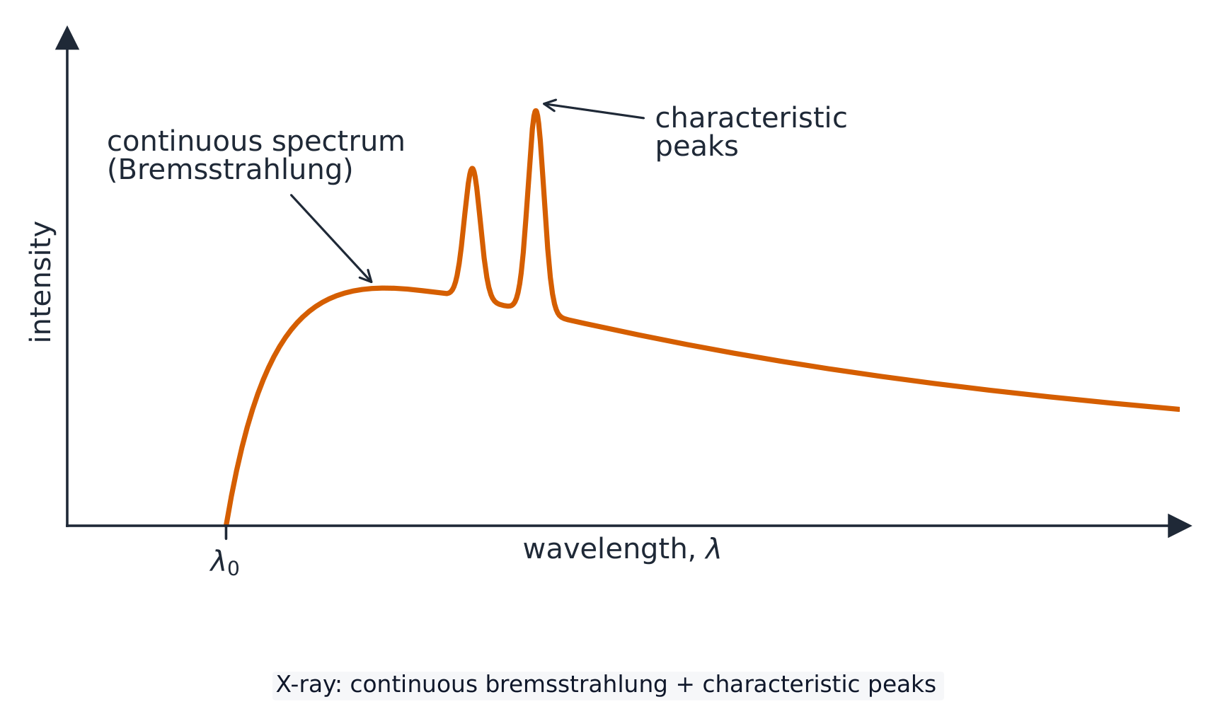
一道断崖,一个鼓包,两根尖刺
X-ray production route · X射线产生流程
Follow electrons from cathode to X-ray photons. · 追踪电子从阴极到X射线光子的过程。
Why does an X-ray spectrum have the shape it does? Select all · 所有 that apply. · 为什么X射线谱具有这种形状?选择 所有 适用项。
The peaks belong to the target's energy levels, so only changing the target moves them. The p.d. moves the cut-off. · 峰值属于靶的能级,因此只有更换靶材才会移动它们。电势差移动的是截止点。
Sharpness and contrast are different things
- Sharpness 清晰度 is how well defined the edges are. It improves with a small source spot, a still patient, and the detector close to the patient.
- Contrast 对比度 is the difference in degree of blackening between neighbouring regions, produced by a difference in attenuation 衰减.
- The two are independent: an image can be sharp with poor contrast, or blurred with strong contrast.
- Where two soft tissues attenuate similarly, a contrast medium 造影剂 such as barium is swallowed or injected to outline one of them.
Same beam, very different shadows
清晰度与对比度是两回事
- 清晰度(sharpness)是边缘有多分明。它随焦点小、患者不动、探测器靠近患者而改善。
- 对比度(contrast)是相邻区域变黑程度之差,由衰减(attenuate)的差别造成。
- 两者是独立的:一张片子可以很清晰而对比度差,也可以很模糊而对比度强。
- 当两种软组织的衰减相近时,就吞服或注射钡剂这样的造影剂(contrast medium)把其中一个勾勒出来。
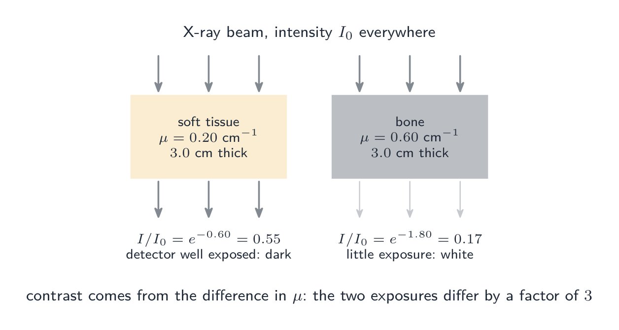
同一束射线,截然不同的影子
An X-ray image with excellent sharpness must also have good contrast. · 一张清晰度极佳的X射线图像也必须具有良好的对比度。
They are independent. Sharpness is edge definition, set by the source size and geometry; contrast is a difference in attenuation between tissues. · 它们是独立的。清晰度是边缘定义,由源尺寸和几何形状决定;对比度是组织间衰减的差异。
Worked example: why bone shows white
- X-rays cross $3.0\ \text{cm}$ of soft tissue ($\mu = 0.20\ \text{cm}^{-1}$) in one region and $3.0\ \text{cm}$ of bone ($\mu = 0.60\ \text{cm}^{-1}$) in another. Compare the transmitted intensities.
- Soft tissue: $I/I_0 = e^{-0.20\times3.0} = e^{-0.60} = 0.55$.
- Bone: $I/I_0 = e^{-0.60\times3.0} = e^{-1.80} = 0.17$.
- The tissue region receives more than three times the exposure, so bone appears white and tissue dark.
- The exposure ratio is $e^{(\mu_{\text{bone}} - \mu_{\text{tissue}})x}$, which grows with thickness: contrast improves for thicker structures, though the total attenuation grows too.
例题:骨为什么显白
- X 射线在一处穿过 $3.0\ \text{cm}$ 软组织($\mu = 0.20\ \text{cm}^{-1}$),在另一处穿过 $3.0\ \text{cm}$ 骨($\mu = 0.60\ \text{cm}^{-1}$)。比较透射强度。
- 软组织:$I/I_0 = e^{-0.20\times3.0} = e^{-0.60} = 0.55$。
- 骨:$I/I_0 = e^{-0.60\times3.0} = e^{-1.80} = 0.17$。
- 软组织区域接受的曝光是骨区域的三倍多,所以骨显白、组织显暗。
- 曝光之比是 $e^{(\mu_{\text{bone}} - \mu_{\text{tissue}})x}$,它随厚度增大:结构越厚对比度越好,尽管总衰减也随之增大。
The attenuation law
- Higher-energy X-rays penetrate further, meaning a smaller $\mu$; bone has a much larger $\mu$ than soft tissue.
- For a thickness, take logs: $x = \dfrac{1}{\mu}\ln\dfrac{I_0}{I}$. The half-value thickness 半值厚度 is $x_{1/2} = \ln 2/\mu$, exactly like a half-life.
- Through two layers the exponentials multiply, so add the exponents: $I = I_0 e^{-(\mu_1 x_1 + \mu_2 x_2)}$. Never add the thicknesses or the coefficients, and note the order of the layers makes no difference.
- Read the question: "what fraction is absorbed" is $1 - e^{-\mu x}$, not $e^{-\mu x}$.
衰减律
- 能量越高的 X 射线穿透越远,也就是 $\mu$ 越小;骨的 $\mu$ 远大于软组织。
- 求厚度就取对数:$x = \dfrac{1}{\mu}\ln\dfrac{I_0}{I}$。半值厚度(half-value thickness)是 $x_{1/2} = \ln 2/\mu$,与半衰期一模一样。
- 穿过两层时指数因子相乘,所以要把指数相加:$I = I_0 e^{-(\mu_1 x_1 + \mu_2 x_2)}$。绝不能把厚度或系数相加,而且两层的先后次序不影响结果。
- 读清题目:"被吸收的比例"是 $1 - e^{-\mu x}$,不是 $e^{-\mu x}$。
Bone looks white on an X-ray image because it: · 骨骼在X射线图像上呈白色,因为它:
Dense, high-$Z$ bone absorbs more ($I = I_0 e^{-\mu x}$ with large $\mu$), casting a stronger shadow. · 高密度、高$Z$的骨骼吸收更多($I = I_0 e^{-\mu x}$ 配合大的 $\mu$),投下更强的阴影。
A beam crosses 2.0 cm of P (mu = 0.35 /cm) then 1.5 cm of Q (mu = 0.90 /cm). What fraction of the intensity is transmitted? · 一束光穿过 2.0 cm 的 P (μ = 0.35 /cm),然后穿过 1.5 cm 的 Q (μ = 0.90 /cm)。透射强度分数是多少?
The exponentials multiply, so add the exponents: e^-(0.70 + 1.35) = e^-2.05 = 0.13. Never add the thicknesses or the coefficients, and the order of the layers makes no difference. · 指数相乘,所以相加指数:e^-(0.70 + 1.35) = e^-2.05 = 0.13。永远不要相加厚度或系数,且层的顺序无关紧要。
Computed tomography
- A computed tomography 计算机断层扫描 (CT) scan builds a three-dimensional image, and the syllabus wants the construction described in order.
- One. Many X-ray images of the same section are taken from different angles around the patient.
- Two. A computer combines them into one two-dimensional image of that section.
- Three. The process is repeated for many sections along an axis.
- Four. The two-dimensional sections are combined into a three-dimensional image, which can be rotated or sliced on screen. The dose is far higher than a single radiograph, which is the trade-off.
计算机断层扫描
- 计算机断层扫描(computed tomography,CT)建立三维图像,而考纲要求按顺序描述这个过程。
- 一。围绕患者,从不同角度对同一断层拍摄许多张 X 射线图像。
- 二。计算机把它们合成该断层的一张二维图像。
- 三。沿一条轴对许多断层重复这一过程。
- 四。把这些二维断层合成三维图像,可以在屏幕上旋转或切开。代价是剂量远高于单张平片。
A CT scan takes X-ray images from many angles to build a 3-D image. · CT扫描从多个角度拍摄X射线图像以构建3D图像。
Rotating the tube and detectors and reconstructing the slices separates overlapping tissues into a 3-D picture. · 旋转管子和探测器并重建切片,将重叠的组织分离成3D图像。
Put the construction of a CT image in order. · 按顺序排列CT图像的构建过程。
Angles first, then sections. The dose is far higher than a single radiograph, which is the trade-off for the 3-D view. · 先角度,后切片。剂量远高于单张放射照片,这是为了获得3D视图而做出的权衡。
Marks that slip away
- Filament current sets intensity, accelerating p.d. sets penetrating power. Do not swap them.
- $\lambda_{\text{min}}$ depends only on the p.d., not on the target or the current.
- Sharpness and contrast are independent. A question about blurred edges is not answered by talking about attenuation.
- Two layers: add the exponents $\mu_1 x_1 + \mu_2 x_2$.
- Match the units of $\mu$ and $x$, and check whether the question wants what gets through or what is absorbed.
容易丢掉的分
- 灯丝电流定强度,加速电压定穿透力。不要弄反。
- $\lambda_{\text{min}}$ 只取决于电压,与靶和电流无关。
- 清晰度和对比度是独立的。问边缘模糊的题,不能用衰减来回答。
- 两层:把指数 $\mu_1 x_1 + \mu_2 x_2$ 相加。
- 让 $\mu$ 和 $x$ 的单位一致,并看清题目要的是透过的还是被吸收的。
You've got it
- X-rays are produced when electrons from a heated cathode, accelerated through a high p.d., decelerate rapidly in a metal target, mostly as heat
- $\lambda_{\text{min}} = hc/(eV)$ is the cut-off set by the accelerating p.d. alone, with characteristic peaks from the target metal on a continuous curve
- sharpness is edge definition, contrast is the difference in attenuation, and they are independent
- $I = I_0 e^{-\mu x}$, with half-value thickness $\ln 2/\mu$ and exponents that add through successive layers; CT combines many angles into a section and many sections into a 3-D image
你掌握了
- X 射线产生于来自受热阴极、经高电压加速的电子在金属靶中急剧减速,其中大部分变成热
- $\lambda_{\text{min}} = hc/(eV)$ 是仅由加速电压决定的截止,连续曲线上还叠着来自靶金属的特征峰
- 清晰度是边缘分明程度,对比度是衰减之差,两者相互独立
- $I = I_0 e^{-\mu x}$,半值厚度为 $\ln 2/\mu$,逐层穿过时指数相加;CT 把多个角度合成一个断层、把多个断层合成三维图像