O coração
Introduzido| English | Significado |
|---|---|
| atria/ˈeɪtrɪə/ | Átrios |
| ventricle/ˈventrɪkl/ | Ventrículo |
| cardiac cycle/ˈkɑːdiæk ˈsaɪkl/ | Ciclo cardíaco |
| systole/ˈsɪstəʊl/ | Sístole |
| diastole/ˈdaɪəstəʊl/ | Diástole |
| sinoatrial node/sɪˈnəʊtrɪəl nəʊd/ | Nó sinusal |
| pacemaker/ˈpeɪsmeɪkə/ | Marcapasso |
| atrioventricular node/ˌeɪtrɪˌəʊvənˈtrɪkjʊlə nəʊd/ | Nó atrioventricular |
| Purkyne tissue/ˈpɜːkaɪn ˈtɪʃuː/ | Tejido de Purkinje |
The pump that never rests
- Your heart beats about 100 000 times a day, and it never stops to rest.
- It is really two pumps side by side: the right side sends blood to the lungs, the left side to the rest of the body.
- It also sets its own rhythm: heart muscle is myogenic 肌源性的, so it contracts without any signal from the brain.
Chambers, valves and vessels
- The two upper chambers are the atria 心房; the two lower chambers are the ventricles 心室. A wall of muscle, the septum 隔膜, keeps the two sides apart, so oxygenated and deoxygenated blood never mix.
- The atrioventricular (AV) valves 房室瓣 lie between each atrium and its ventricle (the tricuspid on the right, the bicuspid or mitral on the left). The semilunar valves 半月瓣 lie at the bases of the aorta and the pulmonary artery.
- Valves only let blood flow one way. Tendons hold the AV valves, so they cannot turn inside out when the ventricles contract.
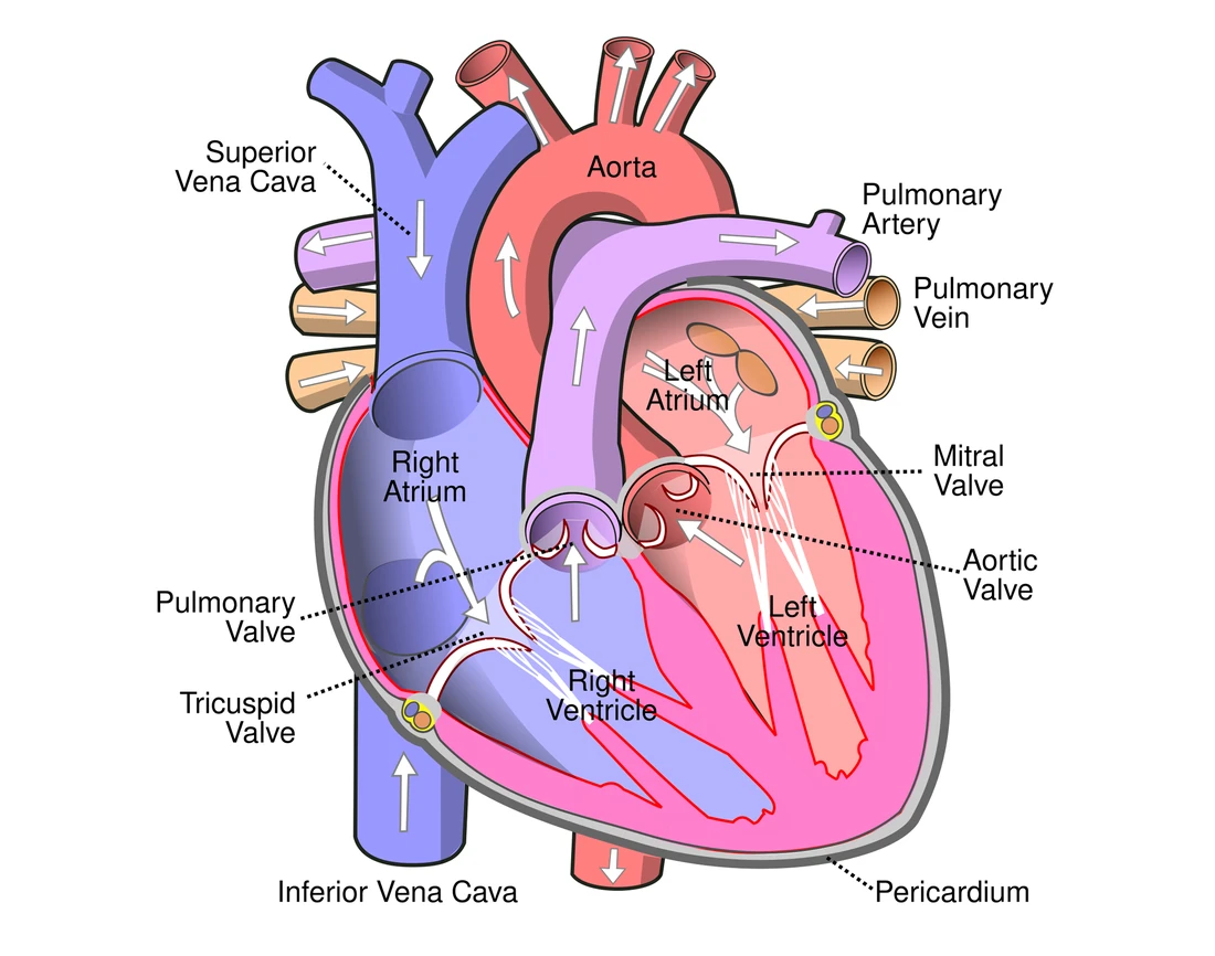
In a heart diagram, the heart's right side is on your left, as if you face the person.
Explore the heart
Tap each part. The right side pumps blood to the lungs; the thicker-walled left side pumps it round the whole body.
Why the walls differ in thickness
| Chamber | Wall | Pumps blood to |
|---|---|---|
| atria | thin | the ventricles below |
| right ventricle | thick | the lungs |
| left ventricle | thickest | the whole body |
- The atria push blood only a short way, at low pressure. The right ventricle pumps a short circuit; a very high pressure would damage the thin lung capillaries.
- The left ventricle has the most muscle, so it contracts with the most force. It makes the high pressure needed to push blood all round the body.
- Exam point: say pressure and resistance, not just "further". The body circuit has many more vessels, so its resistance is greater. The left ventricle must make a higher pressure to overcome it.
Ventricles have thicker walls than atria because they:
Dificuldade 1/5 · Moedas 100 + pequeno bônus na primeira conclusão hoje
Atria only push blood into the ventricles below; ventricles pump blood out, so need thick muscular walls.
The thickest, most muscular chamber, which pumps blood around the whole body, is the left ______.
Dificuldade 1/5 · Moedas 100 + pequeno bônus na primeira conclusão hoje
The left ventricle drives the systemic circulation (whole body), so it needs the most muscle.
Which ventricle has the thicker wall, and why?
The systemic circuit needs the higher pressure.
The cardiac cycle 心动周期 in three stages
- One heartbeat is one cardiac cycle. Systole 收缩期 means contraction; diastole 舒张期 means relaxation. At 75 beats per minute, one cycle lasts 0.8 s.
| Stage | What happens | Valves |
|---|---|---|
| atrial systole (≈0.1 s) | atria contract; the last blood enters the ventricles | AV open, semilunar closed |
| ventricular systole (≈0.3 s) | ventricles contract; blood is forced into the arteries | AV closed, semilunar open |
| diastole (≈0.4 s) | all chambers relax; blood from the veins fills the heart | AV open, semilunar closed |
The cardiac cycle
Step through one heartbeat — atria contract, then ventricles contract, then everything relaxes and refills.
In the cardiac cycle, systole is when the heart muscle:
Dificuldade 1/5 · Moedas 100 + pequeno bônus na primeira conclusão hoje
Systole = contraction (pressure rises); diastole = relaxation and filling.
Match each stage of the cardiac cycle.
Dificuldade 2/5 · Moedas 140 + pequeno bônus na primeira conclusão hoje
Atria contract first, then ventricles, then everything relaxes; the valves keep blood moving one way.
Match each stage of the cardiac cycle to the state of the valves.
Dificuldade 2/5 · Moedas 140 + pequeno bônus na primeira conclusão hoje
The AV valves close only while the ventricles contract; the semilunar valves are open only while blood leaves the ventricles.
Reading the pressure graph
- One rule explains every valve: a valve is pushed open when the pressure behind it is higher than the pressure in front of it. It is pushed shut when the pressure in front is higher.
- 1: ventricle pressure rises above atrial pressure, so the AV valve closes. 2: ventricle pressure rises above aortic pressure, so the semilunar valve opens and blood leaves.
- 3: the ventricle relaxes and its pressure falls below the aorta's, so the semilunar valve closes. 4: ventricle pressure falls below atrial pressure, so the AV valve opens and the ventricle fills again.
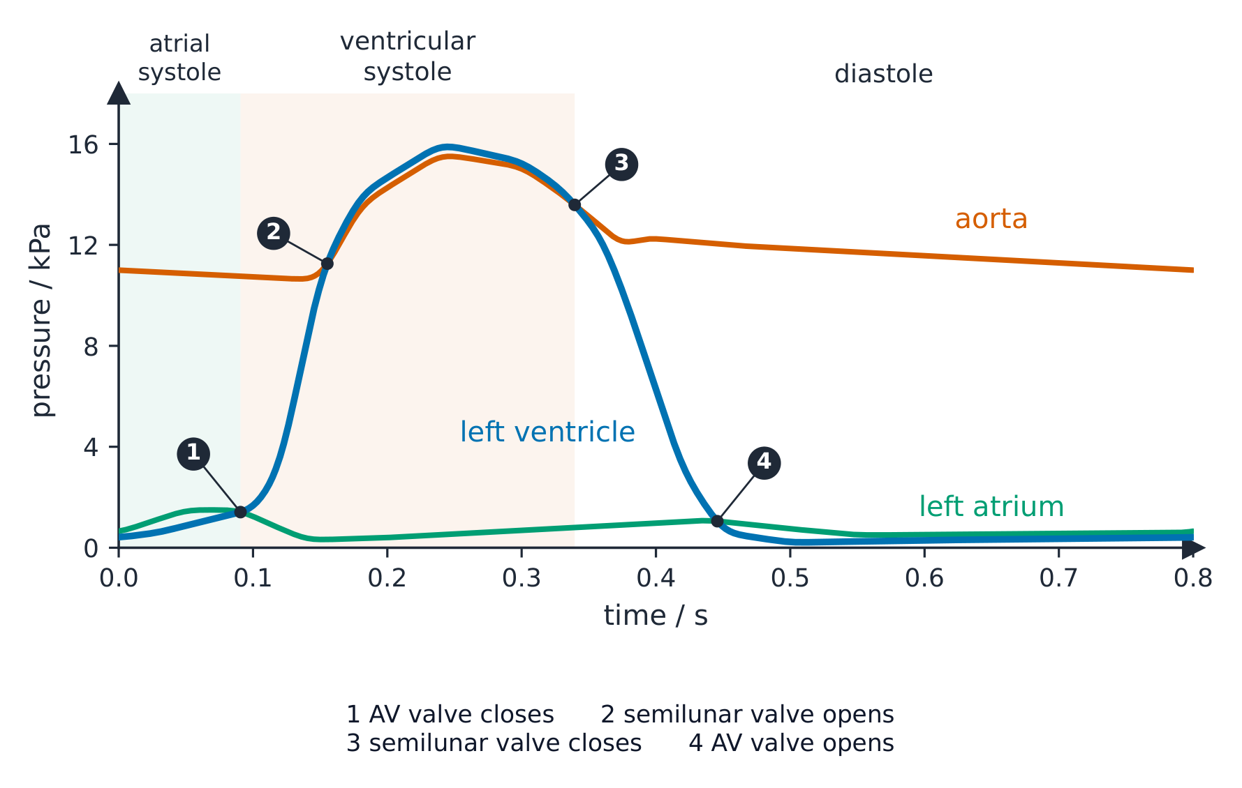
Blood leaves the ventricle only between points 2 and 3, while the semilunar valve is open.
The semilunar valves stop blood flowing back into the ventricles from the arteries.
Dificuldade 1/5 · Moedas 100 + pequeno bônus na primeira conclusão hoje
When the ventricles relax, the semilunar valves (in the aorta and pulmonary artery) shut to stop backflow.
During ventricular systole, left ventricle pressure rises from 8 kPa to 12 kPa. Aortic pressure is 10 kPa. What happens when ventricle pressure passes 10 kPa?
Dificuldade 1/5 · Moedas 100 + pequeno bônus na primeira conclusão hoje
The pressure behind the semilunar valve (ventricle) is now higher than in front of it (aorta), so it is pushed open.
How the heartbeat is controlled
- 1 SAN 窦房结 (sinoatrial node, in the right atrium wall) is the pacemaker 起搏点. It sends a wave of excitation across both atria, so the atria contract.
- 2 A band of non-conducting tissue between the atria and ventricles stops the wave from passing straight down. 3 The only way through is the AVN 房室结 (atrioventricular node). It delays the wave by about 0.1 s, so the atria finish emptying before the ventricles contract.
- 4 Purkyne tissue 浦肯野组织 carries the wave down the septum to the apex (the bottom of the heart). 5 The wave then spreads up the ventricle walls, so the ventricles contract from the apex upwards and squeeze blood up into the arteries.
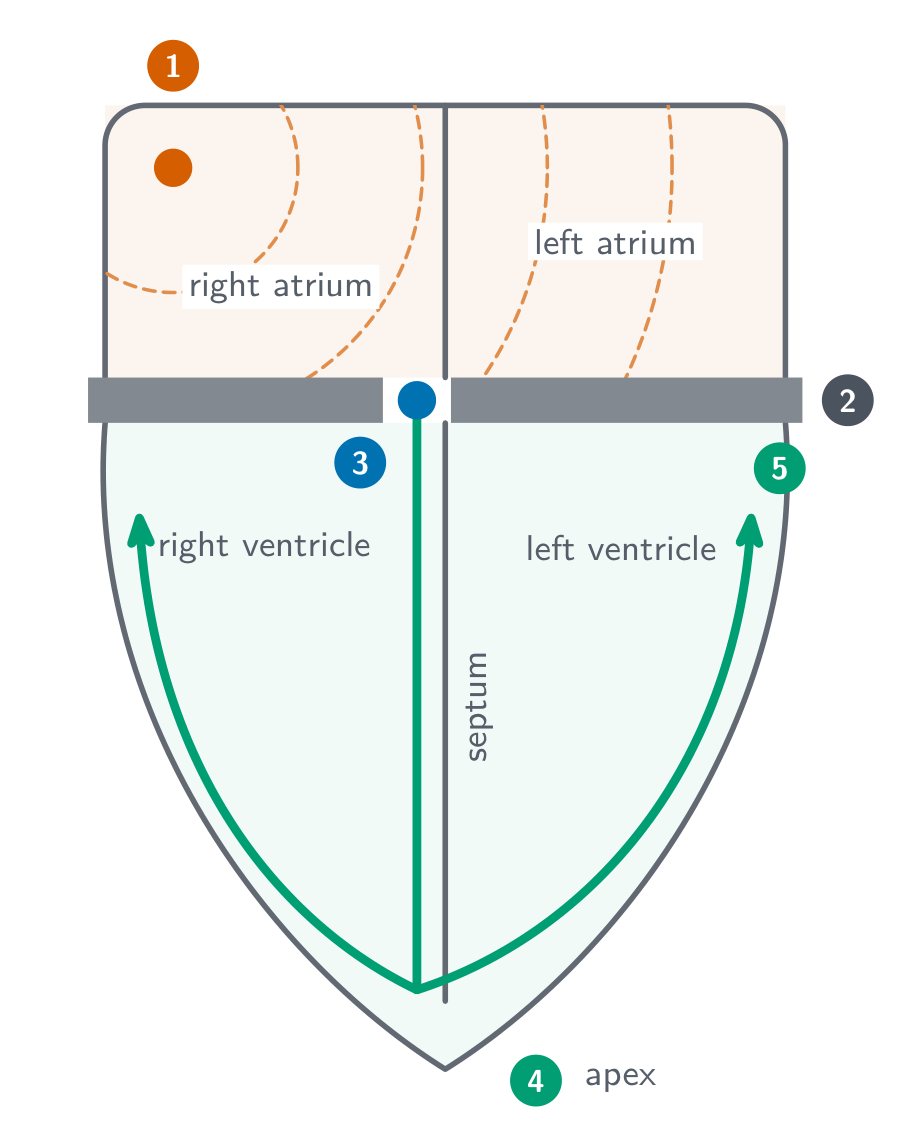
The pacemaker that sets the heart's rhythm is the:
Dificuldade 1/5 · Moedas 100 + pequeno bônus na primeira conclusão hoje
The SAN in the right atrium generates the wave of excitation that triggers each heartbeat.
Put the electrical conduction pathway in the correct order.
Dificuldade 3/5 · Moedas 200 + pequeno bônus na primeira conclusão hoje
SAN (pacemaker) → AVN (delay) → Purkyne tissue (ventricles contract from the bottom up).
What would happen if the band of non-conducting tissue between the atria and ventricles were missing?
Dificuldade 1/5 · Moedas 100 + pequeno bônus na primeira conclusão hoje
The wave would spread straight into the ventricles with no AVN delay, so they would contract before the atria had emptied into them.
Worked exam question: data from the pressure graph
Question (4 marks). Use the pressure graph above. (a) State the time at which the semilunar valve opens and the time at which it closes. (b) Calculate the heart rate. (c) Explain why the semilunar valve closes at 0.34 s.
- (a) Opens at about 0.15 s (point 2), closes at about 0.34 s (point 3). Read times straight off the x-axis where the lines cross.
- (b) One cycle lasts 0.8 s. Heart rate = 60 ÷ 0.8 = 75 beats per minute (1 mark for the working, 1 for the answer).
- (c) The ventricle relaxes, so its pressure falls below the pressure in the aorta. Blood starts to flow back, which pushes the valve shut and prevents backflow into the ventricle.
Mark-scheme trap: "the valve closes because the ventricle has emptied" gains no mark. The marks are for comparing two pressures.
One cardiac cycle lasts 0.6 s. What is the heart rate, in beats per minute?
Dificuldade 2/5 · Moedas 140 + pequeno bônus na primeira conclusão hoje
60 ÷ 0.6 = 100 beats per minute.
Worked exam question: explain the control
Question (5 marks). Explain how the heartbeat is started and coordinated.
Model answer (one mark per point, up to 5):
- Heart muscle is myogenic; the SAN acts as the pacemaker.
- The SAN sends a wave of excitation over the atria, so they contract.
- Non-conducting tissue stops the wave from reaching the ventricles directly.
- The AVN passes it on after a delay, so the atria empty before the ventricles contract.
- Purkyne tissue carries the wave down the septum to the apex.
- The ventricles contract from the apex upwards, pushing blood out into the arteries.
Why answers lose marks: "the SAN sends a signal to the AVN" with no mention of the atria contracting misses a point. Each structure needs what it does and why it matters.
Why does the AVN delay the impulse?
Dificuldade 1/5 · Moedas 100 + pequeno bônus na primeira conclusão hoje
The ventricles must fill before they contract.
Without looking back: explain how the heartbeat is started and coordinated (5 marks).
Each structure needs what it does and why it matters.
You've got it
- atria thin; ventricles thick; left ventricle thickest, to make the high pressure that overcomes the body circuit's resistance
- AV valves (atrium → ventricle) and semilunar valves (ventricle → artery) give one-way flow
- cycle = atrial systole → ventricular systole → diastole; at 0.8 s per cycle the heart rate is 75 beats per minute
- a valve opens when the pressure behind it is higher, and closes when the pressure in front is higher
- control: SAN → atria contract → non-conducting tissue → AVN delay → Purkyne down the septum → ventricles contract apex upwards
Which ventricle has the thicker wall?
Dificuldade 1/5 · Moedas 100 + pequeno bônus na primeira conclusão hoje
The left side supplies the whole body.