Microscopy and tissue preparation
導入済み| English | 意味 |
|---|---|
| fixation/fɪkˈseɪʃn/ | fixation |
| dehydration/ˌdiːhaɪˈdreɪʃn/ | The removal or loss of water from a substance or organism. |
| embedding | embedding |
| sectioning | sectioning |
| staining | staining |
| resolution/ˌrezəˈluːʃn/ | The ability to distinguish fine detail or closely separated features. |
The picture is manufactured
- A histology slide is not a photograph of tissue. It is made, step by step, and each step can create or destroy evidence.
- To read a slide, you must know what the preparation did to it.
Five steps to a slide
- Fixation 固定 (formalin cross-links proteins), dehydration 脱水 (alcohols remove water) and embedding 包埋 (paraffin supports the tissue).
- Sectioning 切片 (a microtome cuts 3–5 µm slices) and staining 染色, usually with haematoxylin and eosin (H&E).
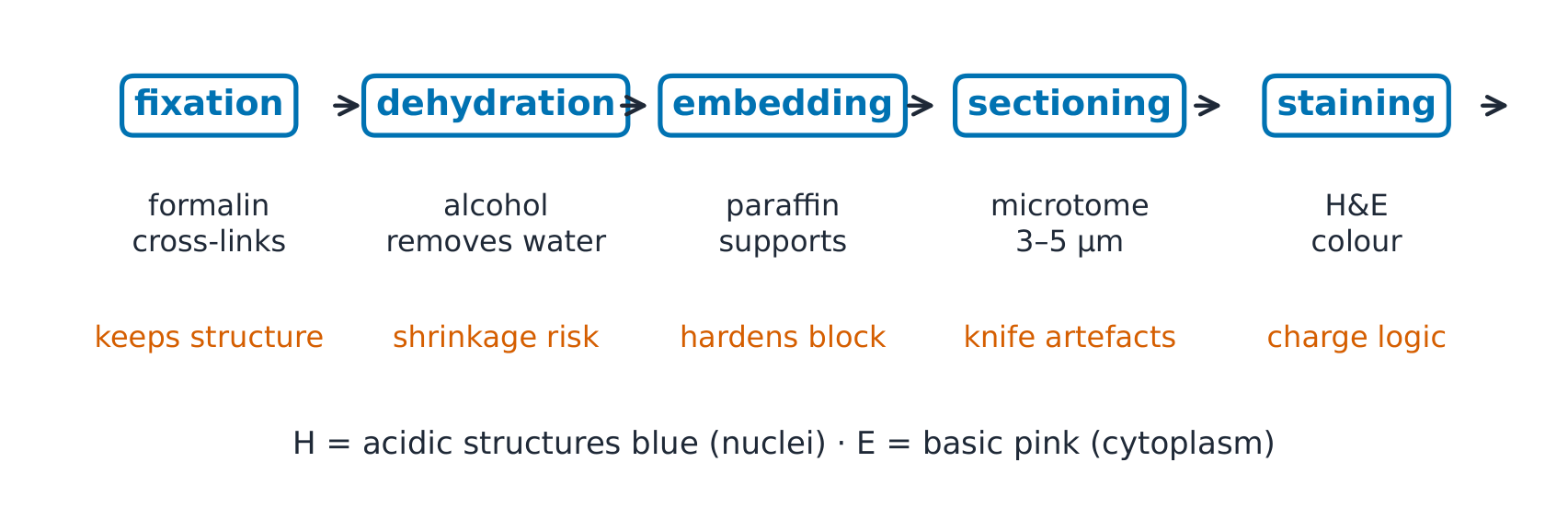
Put the steps of tissue preparation in order.
Difficulty 3/5 · 200 coins + a small bonus on first completion today
Fix to preserve, remove water so paraffin can enter, embed for support, cut thin sections, then stain.
What can be seen
- A section is 3–5 µm thick but a cell is 10–30 µm across, so each section cuts through only part of a cell.
- The light microscope's resolution 分辨率 is about 200 nm, half the wavelength of visible light. Magnifying beyond about 1500× only enlarges the blur.
A cell is 20 µm across. How many 4 µm sections does it take to cut all the way through it?
Difficulty 2/5 · 140 coins + a small bonus on first completion today
20 / 4 = 5, so any one section shows only a fifth of the cell; a nucleus may be missing from a cell's section.
Why H&E works
- Haematoxylin acts as a basic dye and stains acidic structures blue-purple: nuclei, full of DNA and RNA. Eosin is acidic and stains basic structures pink: cytoplasm and collagen.
- The logic is charge pairing. Special stains follow the same idea: PAS stains carbohydrates magenta, so basement membranes and glycogen light up.
Why do nuclei stain blue-purple with H&E?
Difficulty 1/5 · 100 coins + a small bonus on first completion today
Acidic nucleic acids bind the basic dye; basic cytoplasmic proteins bind the acidic eosin.
Worked example: the resolution limit
- Two mitochondria 150 nm apart cannot be told apart, because they are closer than 200 nm. A 2 µm bacterium and a 10 µm nucleus are easy to see.
- The electron microscope's wavelength is about 0.005 nm, so it resolves about 1000 times finer detail, but the specimen must be dead and in a vacuum.
Two structures are 100 nm apart. Can a light microscope show them as separate?
Difficulty 1/5 · 100 coins + a small bonus on first completion today
Resolution is set by the wavelength of light, about 200 nm; more magnification only enlarges the blur. An electron microscope is needed.
Worked example: the frozen section
- (Fictional vignette) A surgeon waits, mid-operation, to know if the tumour's edge is clear. The tissue is frozen and cut within minutes, skipping dehydration and embedding.
- Ice crystals tear the tissue and detail is blurred, but an answer within the hour beats a perfect answer tomorrow.
A frozen section gives the same quality of detail as a paraffin section, only faster.
Difficulty 1/5 · 100 coins + a small bonus on first completion today
Ice crystals tear the tissue and detail is blurred; the frozen section trades quality for an answer within minutes.
Three common slips
- Every gap is real. Shrinkage during dehydration creates false spaces.
- Stain colour is the tissue's colour. Colour comes from the dye chemistry.
- Forgetting the cut is one plane. A tube cut at an angle looks like an oval.
Match each appearance to its cause.
Difficulty 2/5 · 140 coins + a small bonus on first completion today
An artefact you can name is an artefact you can discount.
Recap
- Fix, dehydrate, embed, section (3–5 µm), stain.
- Light microscopy resolves about 200 nm.
- H stains acidic nuclei blue; E stains basic cytoplasm pink. Frozen sections trade quality for speed.