Cell measurements and scale
Introduit| English | Sens |
|---|---|
| magnification/ˌmæɡnɪfɪˈkeɪʃn/ | grossissement |
| resolution/ˌrezəˈluːʃn/ | résolution |
What would explain this observation?
- A cell can look larger on a screen without changing its real size. Two photographs at different zoom settings cannot be compared by eye alone.
Build the model
- Eukaryotic cells contain a nucleus.
- Prokaryotic cells have genetic material but no membrane-bound nucleus.
- A bacterial cell is a living cell; a virus depends on a host cell to reproduce.
- magnification 放大倍数: Image length divided by actual length.
- resolution 分辨率: Ability to distinguish two close points.

Which comparison of two cell images is reliable?
Difficulté 1/5 · 100 pièces + un petit bonus lors de la première réussite aujourd'hui
Reliable comparison needs a shared real scale. Converting each image length with its own scale bar gives real sizes that can be compared; screen lengths alone depend on zoom, which differs between the images.
Match each technical term to its precise meaning.
Difficulté 2/5 · 140 pièces + un petit bonus lors de la première réussite aujourd'hui
Use the definitions to distinguish related quantities and processes.
Choose evidence that can test it
- A scale bar provides a known real distance in the same image.
- Convert the image length and real length to the same unit before dividing.
- Magnification is a ratio and has no unit.
In the lab: Focus a prepared slide at low power first. Move to a higher power and use fine focus. Make a clear line drawing, label structures with straight lines, and record the scale rather than shading the image.
Which two habits make the investigation or model in this case more defensible?
Difficulté 2/5 · 140 pièces + un petit bonus lors de la première réussite aujourd'hui
Stating the conditions and assumptions lets others repeat the work exactly, and recording the supporting evidence lets them check the conclusion. Hiding disagreeing results removes the evidence that could challenge a wrong conclusion, and one measurement can never establish a wider claim.
Work from known quantities
- Known: a cell image is 30 mm long and represents a 50 micrometre cell.
- Use magnification = image size / actual size.
- Convert 30 mm to 30,000 micrometres.
- Magnification = 30,000 / 50 = 600.
- A separate 10 mm scale bar representing 20 micrometres gives 10,000 / 20 = 500, and that magnification applies to every object in its image.
Your turn: An image is 12 mm long; the cell is 40 micrometres long. Calculate magnification.
Enlarge the image, preserve the real size
A 30 mm image of a 50 μm cell has magnification ×600. Resize the cell image and its 20 μm scale bar together, convert units consistently, and recover the same real cell size.
An image is 12 mm long; the cell is 40 micrometres long. Calculate magnification.
Difficulté 2/5 · 140 pièces + un petit bonus lors de la première réussite aujourd'hui
The result is 300 . Known: image length 12 mm; actual length 40 micrometres. Use magnification = image size / actual size with both lengths in the same unit. Convert 12 mm to 12,000 micrometres. Magnification = 12,000 / 40 = 300. The image is 300 times the real cell length; the ratio has no unit.
Check the conclusion and its limits
- A nucleus is not the only cell structure.
- Do not claim bacteria have no DNA, or that more magnification always means better resolution.
Not true: Every bacterial cell has a membrane-bound nucleus. Bacterial cells are prokaryotic: their genetic material lies free in the cytoplasm, not inside a membrane-bound nucleus. Only eukaryotic cells have a true nucleus.
- Eukaryotic cells contain a nucleus.
- A scale bar provides a known real distance in the same image.
- Watch for: A nucleus is not the only cell structure.
Every bacterial cell has a membrane-bound nucleus.
Difficulté 1/5 · 100 pièces + un petit bonus lors de la première réussite aujourd'hui
Bacterial cells are prokaryotic: their genetic material lies free in the cytoplasm, not inside a membrane-bound nucleus. Only eukaryotic cells have a true nucleus.
Image length divided by actual length: write the technical term.
Difficulté 1/5 · 100 pièces + un petit bonus lors de la première réussite aujourd'hui
magnification means Image length divided by actual length.
An image is 18 mm long and the cell is 25 micrometres long. What is the magnification, and why does a bacterial cell not have a membrane-bound nucleus?
Convert 18 mm × 1000 = 18,000 micrometres. Magnification = image length / actual length = 18,000/25 = 720, with no unit. Bacterial DNA is not enclosed by a nuclear envelope.
The SL written case shows a cell image 18 mm long for an actual cell length of 25 micrometres. What is the magnification?
Difficulté 1/5 · 100 pièces + un petit bonus lors de la première réussite aujourd'hui
Convert 18 mm to 18,000 micrometres, then divide by 25: magnification is 720 times, a dimensionless ratio.
Write from a calibrated image measurement
- In the SL written case, an image is 18 mm long and the cell is 25 micrometres long. Convert before dividing: $18\times1000/25=720$ times magnification. This differs from the earlier 12 mm, 40 micrometre case, which gives 300. A large displayed photograph does not itself establish a large actual cell.
- This route develops A2.2 Cell structure through microscopy, typical cellular organization, functions, comparisons and exceptions. Numerical datasets are invented teaching examples. Schematic diagrams simplify structures; the two real micrographs below must be interpreted from visible evidence and their stated scale information, rather than treated as perfectly labelled textbook models.
Connect cell theory to a testable prediction
- Organisms consist of one or more cells; cells are basic structural and functional units; present-day cells arise from pre-existing cells. If an unfamiliar living organism falls within this model, examination at adequate resolution should reveal cellular organization. This deduction guides an investigation but does not replace the observation or settle every question about the earliest origin of cells.
- Typical cells have a plasma membrane, aqueous cytoplasm, hereditary DNA and ribosomes for making proteins. Connect these structures to exchange, chemical reactions, information and synthesis. Specialized mature cells can lose some structures, so a typical model needs qualifications. A virus depends on host cellular machinery and is not a cell; a bacterium is a living cell despite lacking a membrane-bound nucleus.
Prepare and focus a light-microscope image
- For an approved temporary mount, use a thin specimen in a suitable liquid and lower the coverslip at an angle to reduce trapped air. Folded or thick tissue can hide features in overlapping layers. A stain improves contrast for particular structures but can alter or kill cells; an air bubble or dark stain deposit is not automatically a nucleus.
- Start with the lowest-power objective, locate and centre the specimen, then change to higher power and use fine focus. Keep the objective clear of the slide and adjust illumination. Total optical magnification is eyepiece power multiplied by objective power: a 10-times eyepiece and 40-times objective give 400 times. Higher magnification only helps if useful detail is resolved.
Use a scale bar that belongs to the same image
- A photograph's 10 mm scale bar represents 20 micrometres. Its printed magnification is $10\,000/20=500$. A cell measuring 25 mm on that photograph is $25\,000/500=50$ micrometres long. The same result follows from the ratio $25/10\times20=50$. Do not use a magnification label from a different photograph or screen setting.
- Resize an image and its attached scale bar together and their ratio remains valid; a printed magnification label may become incorrect. Keep original images and record the objective and calibration. An uncalibrated photograph can support structural observations but cannot establish cell size merely from its online pixel width. Magnification is a dimensionless length ratio, not a unit of cell size.
In one photograph, a 10 mm scale bar represents 20 micrometres. A cell measures 25 mm on the same photograph. Find its actual length.
Difficulté 2/5 · 140 pièces + un petit bonus lors de la première réussite aujourd'hui
The cell is 25/10 = 2.5 times the bar length, so actual length is 2.5 × 20 = 50 micrometres. This uses the bar in the same image, rather than a magnification label from another image.
Calibrate arbitrary graticule divisions
- An eyepiece graticule has arbitrary divisions. In an invented calibration at one objective, 40 divisions span 100 micrometres on a stage micrometer. One division therefore represents $100/40=2.5$ micrometres. A cell spanning 18 divisions is $18\times2.5=45$ micrometres long. Convert the divisions into distance before reporting the cell's size.
- Recalibrate at a different objective because each division then covers a different actual distance. In another invented setting, 50 divisions span 100 micrometres, so a 15-division object is $15\times100/50=30$ micrometres long. Neither 15 nor 50 alone is the object's size. Calibration establishes the relationship needed for the conversion.
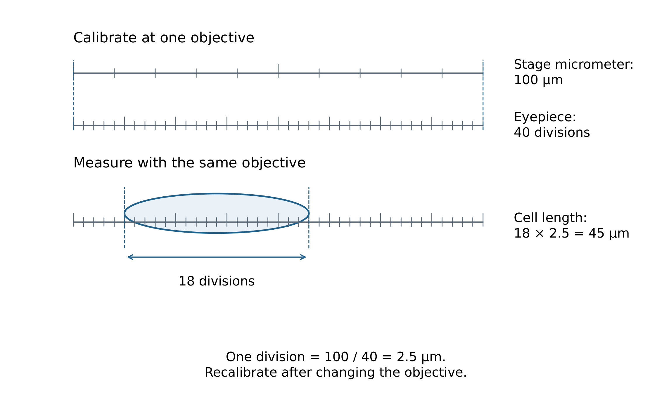
At a new objective setting, 50 graticule divisions span 100 micrometres. A cell spans 15 divisions. Find cell length.
Difficulté 2/5 · 140 pièces + un petit bonus lors de la première réussite aujourd'hui
Each division represents 100/50 = 2 micrometres. The cell length is 15 × 2 = 30 micrometres. Recalibration is needed when objective settings change.
Choose resolution and labelling for the question
- Resolution is the ability to distinguish close points separately; magnification only enlarges the image. Conventional light microscopy resolves roughly 0.2 micrometres under favourable conditions, but optics, contrast and preparation matter. TEM reveals internal details in thin sections; SEM mainly reveals surface form. Conventional electron-microscope preparation does not show an undisturbed living cell, and added colour is not natural specimen colour.
- Fluorescence locates suitable labels; immunofluorescence uses antibody binding and needs specificity controls. Freeze fracture exposes fracture faces, often within membranes, rather than simply photographing the outside of an intact cell. Cryogenic electron microscopy examines rapidly frozen material and still needs interpretation of preparation and reconstruction. Select a method for the needed evidence instead of assuming the largest magnification is always best.
Read a Gram-positive bacterial model
- A typical Gram-positive bacterium has a thick peptidoglycan wall outside its plasma membrane. The wall supports the cell and helps resist osmotic bursting; the membrane regulates exchange. Its DNA-containing nucleoid lacks a nuclear envelope. Cytoplasmic 70S ribosomes synthesize proteins. “No nucleus” does not mean no DNA, no organization or inability to respire.
- Represent the main chromosome as circular in this model, while recognizing variation among bacteria. Plasmids, capsules and flagella are not compulsory features of every species. “Naked DNA” distinguishes the model from eukaryotic histone packaging, not an absolute absence of DNA-associated proteins. A blurred outline without a visible nucleus is insufficient evidence by itself to classify an unknown cell.
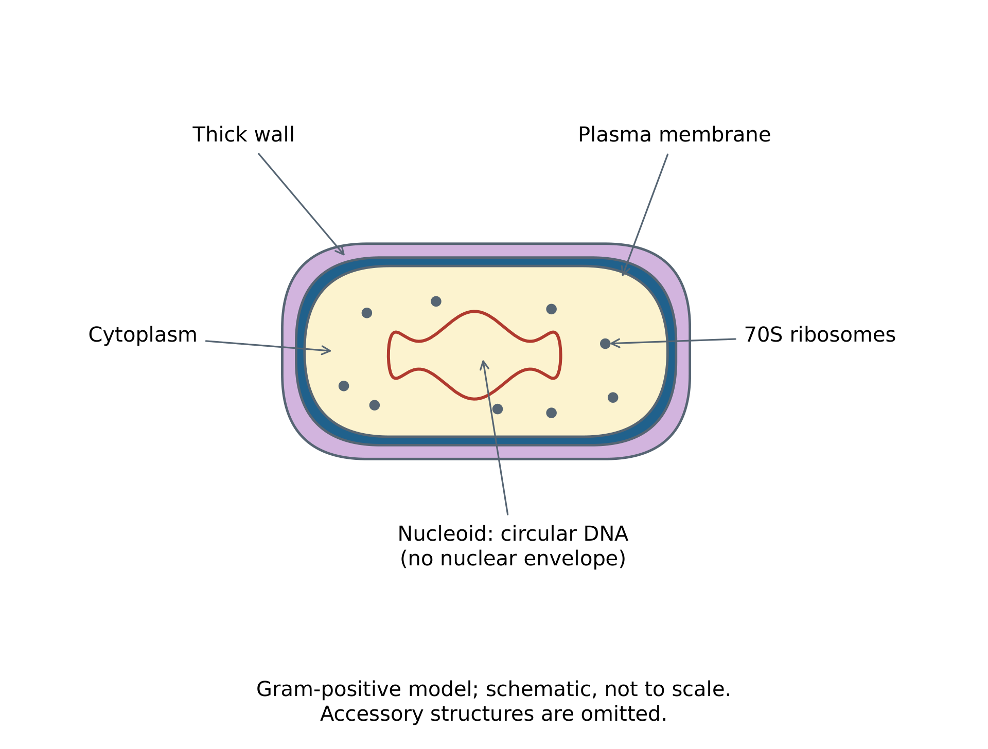
Which statement fits the typical Gram-positive bacterial model?
Difficulté 1/5 · 100 pièces + un petit bonus lors de la première réussite aujourd'hui
The wall supports the cell, the membrane regulates exchange and the nucleoid contains DNA without a nuclear envelope. Ribosomes make proteins; no membrane-bound nucleus does not imply no DNA or no metabolism.
Identify compartments by structure and function
- A typical eukaryotic cell has a double nuclear envelope with pores and DNA associated with histones. Cytoplasmic 80S ribosomes are not membrane-enclosed organelles. Mitochondria have a double envelope and inner-membrane folds called cristae, with matrix inside the inner membrane. Chloroplasts have internal thylakoid membranes and surrounding stroma. Use several recognition clues together in a micrograph.
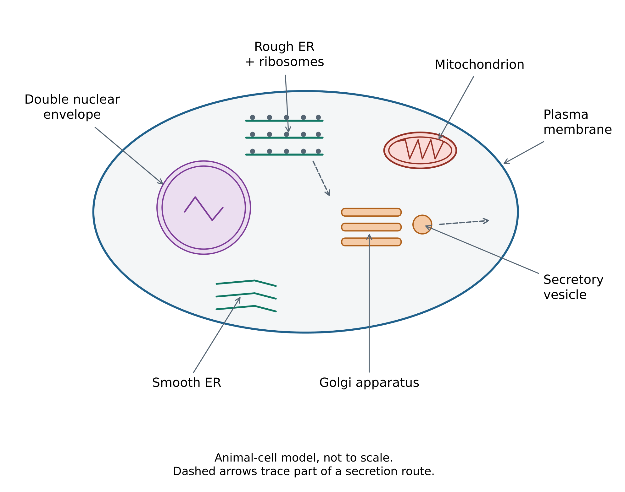
- Rough ER carries attached ribosomes and participates in synthesis and processing of proteins entering the secretory pathway; smooth ER participates in lipid synthesis and other cell-specific roles. Golgi stacks modify, sort and package products. Lysosomes support intracellular digestion, but a round dark profile alone may not establish one. A large plant sap vacuole is bounded by the tonoplast and contributes to storage and turgor.
Follow a secreted protein through a connected pathway
- For a protein entering the secretory pathway, ribosomes associated with rough ER synthesize the polypeptide; ER processing and transport vesicles connect to the Golgi. Further sorting packages material into vesicles, which can fuse with the plasma membrane during exocytosis. A nucleus stores information and is a site of transcription; it does not directly assemble the polypeptide on a ribosome inside the nucleus.
- Cytoskeletal microtubules and microfilaments help organize the cell, move material and participate in division. Microvilli extend the surface and are supported by microfilaments, increasing exchange area. They are not cilia. Cilia and eukaryotic flagella commonly move cells or surrounding fluid and have microtubule-based internal organization. Similar-looking projections can therefore have different structures and roles.
Which is an appropriate simplified route for a protein entering the secretory pathway?
Difficulté 1/5 · 100 pièces + un petit bonus lors de la première réussite aujourd'hui
Ribosomes associated with rough ER synthesize the entering polypeptide. Vesicle transport connects ER, Golgi sorting and eventual exocytosis. Transcription in the nucleus is distinct from polypeptide synthesis on ribosomes.
Explain how one cell performs life's processes
- In a freshwater Paramecium, cilia support movement and help bring food towards an oral region. Food vacuoles support digestion; enzyme-controlled metabolism uses nutrients and releases energy. The cell responds to stimuli and grows by making cellular material. These coordinated activities occur within one cell, rather than being divided among miniature multicellular organs.
- Contractile vacuoles collect and expel excess water entering from dilute surroundings, contributing to homeostasis. Metabolic wastes such as carbon dioxide leave the cell; excretion differs from removal of undigested food. Cell division continues the lineage. A contractile vacuole is not a tiny kidney, and a food vacuole is not a multicellular stomach: relate each structure to its actual cellular function.
Compare plant, animal and fungal cells with qualifications
- Typical animal cells lack a wall; plant walls contain mainly cellulose; fungal walls contain chitin and other polysaccharides. All have plasma membranes. Photosynthetic plant cells contain chloroplasts, while many root and bulb cells do not. A large sap vacuole is common in mature plant cells. Fungal cells are not plants merely because both can have walls.
- Onion bulb epidermis reveals adjoining cell boundaries and often nuclei, but is unsuitable for assuming every plant cell has visible chloroplasts. Cilia, flagella and centrioles vary with lineage, cell type and life stage; one textbook sketch is not a universal presence–absence rule. The photograph below has no calibrated scale bar, so use it for visible structural observations rather than a size calculation.
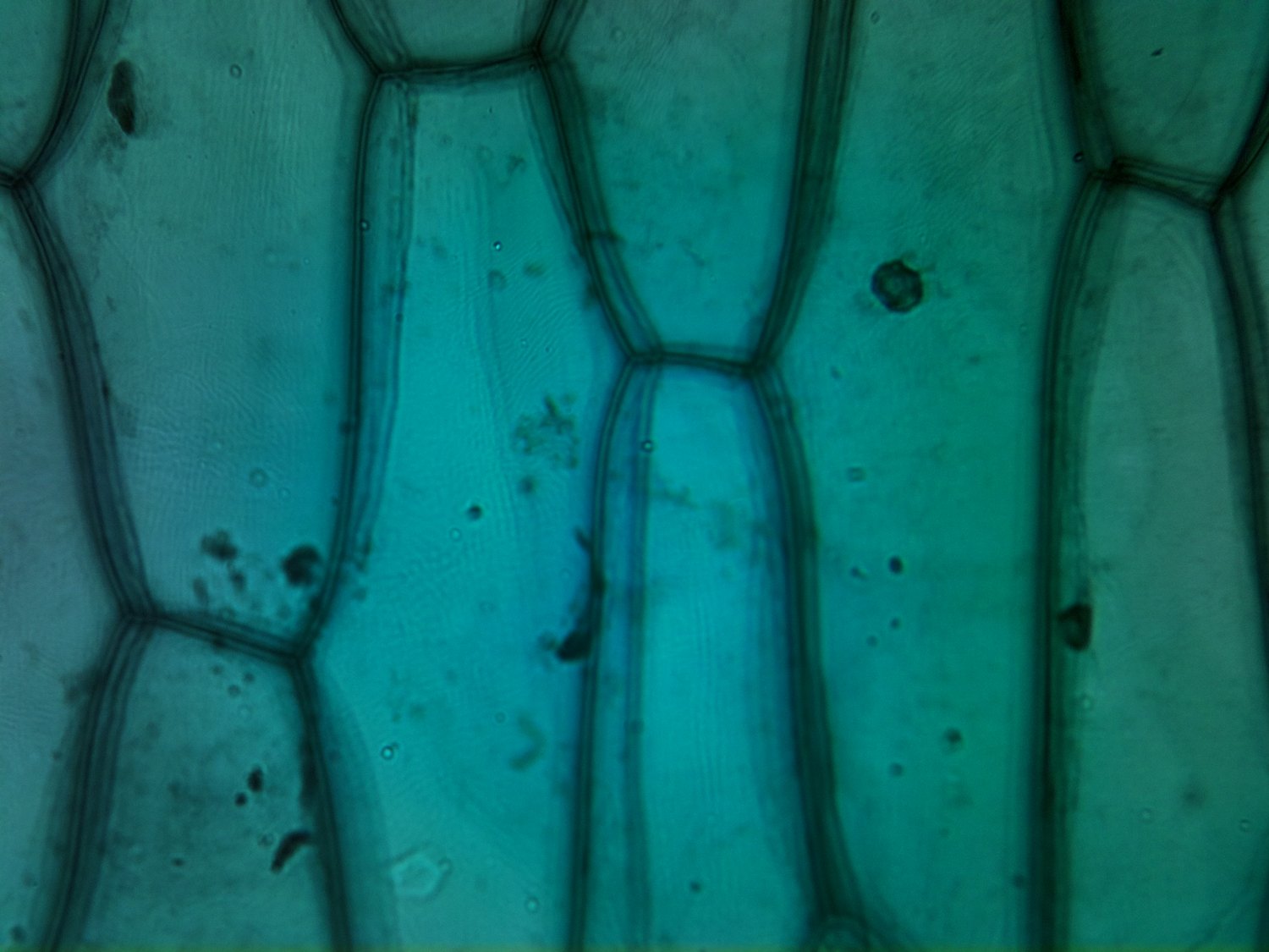
Photo: loganrickert, CC BY 2.0, original source.

Use atypical cells to refine the typical model
- A skeletal muscle fibre is multinucleate after fusion of precursor cells. An aseptate fungal hypha can have many nuclei in shared cytoplasm without the usual cross-wall partitions. Mature mammalian red blood cells lack nuclei and mitochondria, creating space for haemoglobin while limiting repair and normal cell division. The mammalian qualification matters: not all vertebrate red blood cells are anucleate.
- A mature phloem sieve tube element also loses its nucleus and many organelles, yet remains living with companion-cell support. A mature xylem vessel element is different: its conducting structure is dead. These examples do not make microscopy useless; they show why a cell's developmental history, type and function must accompany a typical structural description.
Draw the evidence in a section rather than every remembered organelle
- Identify the method and scale before interpreting a micrograph. A thin section through a three-dimensional object can produce different profiles depending on the cutting plane. The mitochondrial image below shows boundaries and inner-membrane folds; use its original scale bar rather than the displayed pixel width when estimating length. Absence in one section can mean a feature was missed, unresolved or altered during preparation.
- Make a large clear line drawing with continuous outlines and relative proportions based on the observed image. Use ruled label lines ending on the intended structure without crossings or decorative shading. Annotate functions separately from visible shapes. Do not add chloroplasts to an onion bulb cell simply because they occur in a generic plant-cell diagram.
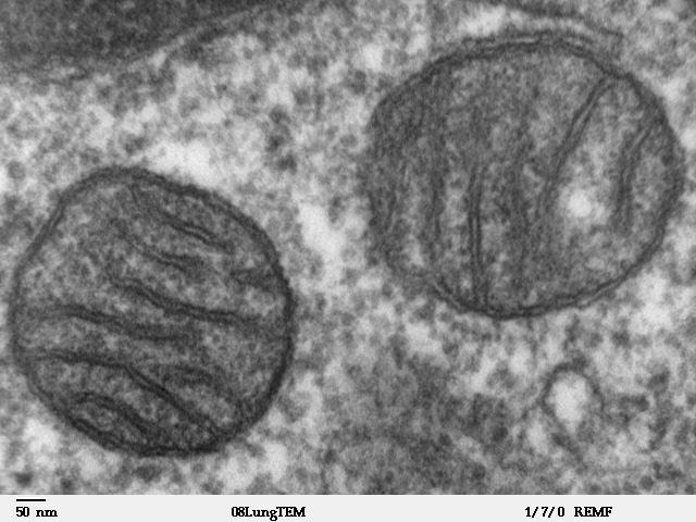
Photo: Louisa Howard, public domain, original source.

Evaluate identification using several clues
- A wall plus a large vacuole and suitable plastids can support plant-cell identification; a wall alone also occurs in fungi and many prokaryotes. A nucleus with membrane-bound organelles supports eukaryotic organization, but a specialized mature cell may have lost a nucleus. Combine visible structures, scale, preparation and biological context rather than classifying from one feature in isolation.
- For a quantitative answer, state the image or calibration used, convert units and check the result's biological scale. For an explanation, connect the structure to the process it enables. For an evaluation, name what the method cannot resolve or what the preparation might change. More magnification, more labels or a more colourful diagram do not automatically supply stronger evidence.
A dark round profile appears in one thin electron-microscope section. Evaluate a claim that it must be a lysosome and that every absent structure is missing from the whole cell.
A structural interpretation depends on method, scale and multiple clues; absence in a section is not automatically whole-cell absence.
Classify organelles using the stated convention
- An organelle is a discrete cell subunit adapted to a function; a membrane is not required around every organelle. In this course's convention, nuclei, ribosomes, vesicles and the plasma membrane count as organelles, while cytoplasm, cytoskeleton and the cell wall do not. State the convention instead of calling every useful cell component an organelle.
- Organelle content depends on cell type and state. An enzyme-secreting cell can have extensive rough ER and Golgi, whereas mature mammalian red blood cells lack a nucleus and mitochondria. Shape and abundance support structure–function explanations but do not by themselves measure the complete activity of a cell.
Separate fractions without assuming perfect purity
- Homogenize tissue to release contents in a suitable cold buffered medium. Cooling slows unwanted reactions, buffering limits pH changes and appropriate osmotic conditions help preserve organelles. Avoid membrane-disrupting detergent when intact organelles are needed. These precautions reduce damage rather than guaranteeing every structure survives.
- Centrifugation produces a sedimented pellet and an overlying supernatant. Carry the supernatant to the next stronger or longer spin to collect another fraction. Sedimentation depends on size, density, shape, medium and time. A pellet is an experimental fraction, not automatically one perfectly purified organelle type.
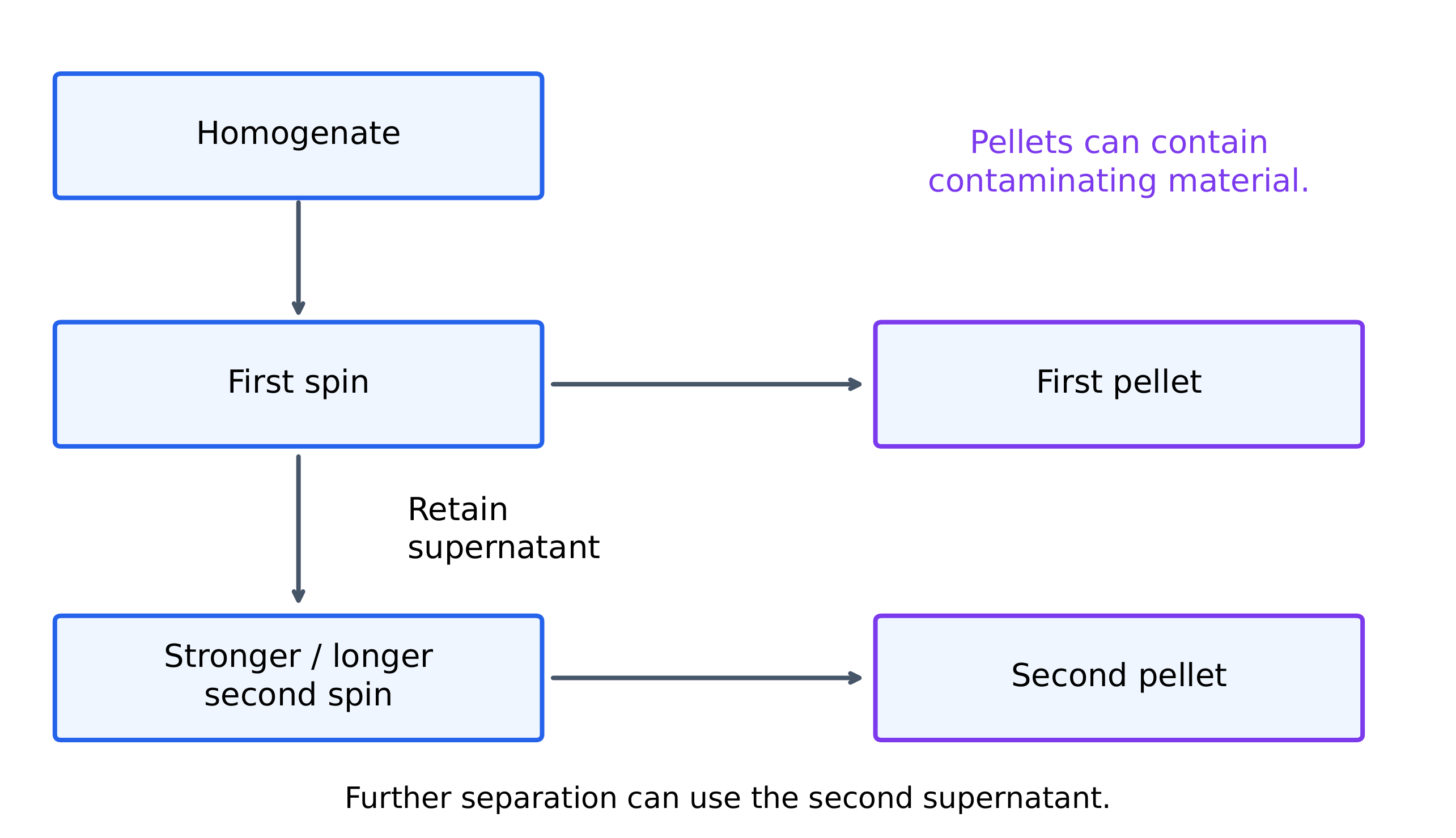
After collecting the first centrifugation pellet, what is carried forward for the next stronger or longer spin in the described fractionation sequence?
Difficulté 1/5 · 100 pièces + un petit bonus lors de la première réussite aujourd'hui
Sequential fractionation removes a pellet and spins the retained supernatant again. Sedimentation conditions and contamination mean fraction identity needs microscopy and marker evidence.
Distinguish enrichment from recovery
- In an invented marker assay, the second pellet contains 82% of the starting mitochondrial marker, leaving 18% in other fractions. If it also contains nuclear and cytosolic markers, it is not pure mitochondria. Test fractions with microscopy and suitable markers; broken structures and trapped material can contaminate a pellet.
- In a separate invented assay, starting activity is 30 units in 15 mg protein, giving 2 units per mg. A fraction has 20 units in 2 mg, giving 10 units per mg. Specific activity rises fivefold, while total activity recovery is only $20/30\times100=66.7\%$. An enriched fraction can lose substantial starting material; compare assays under equivalent conditions.
An invented starting extract has 40 enzyme units in 10 mg protein. A fraction has 18 units in 1.5 mg protein, assayed comparably. What is the fold enrichment in specific activity?
Difficulté 2/5 · 140 pièces + un petit bonus lors de la première réussite aujourd'hui
Starting specific activity = 40/10 = 4 units per mg; fraction = 18/1.5 = 12. Enrichment = 12/4 = 3-fold. Activity recovery is a separate value, 18/40 × 100 = 45%, and enrichment does not prove purity.
Export an RNA copy rather than the gene
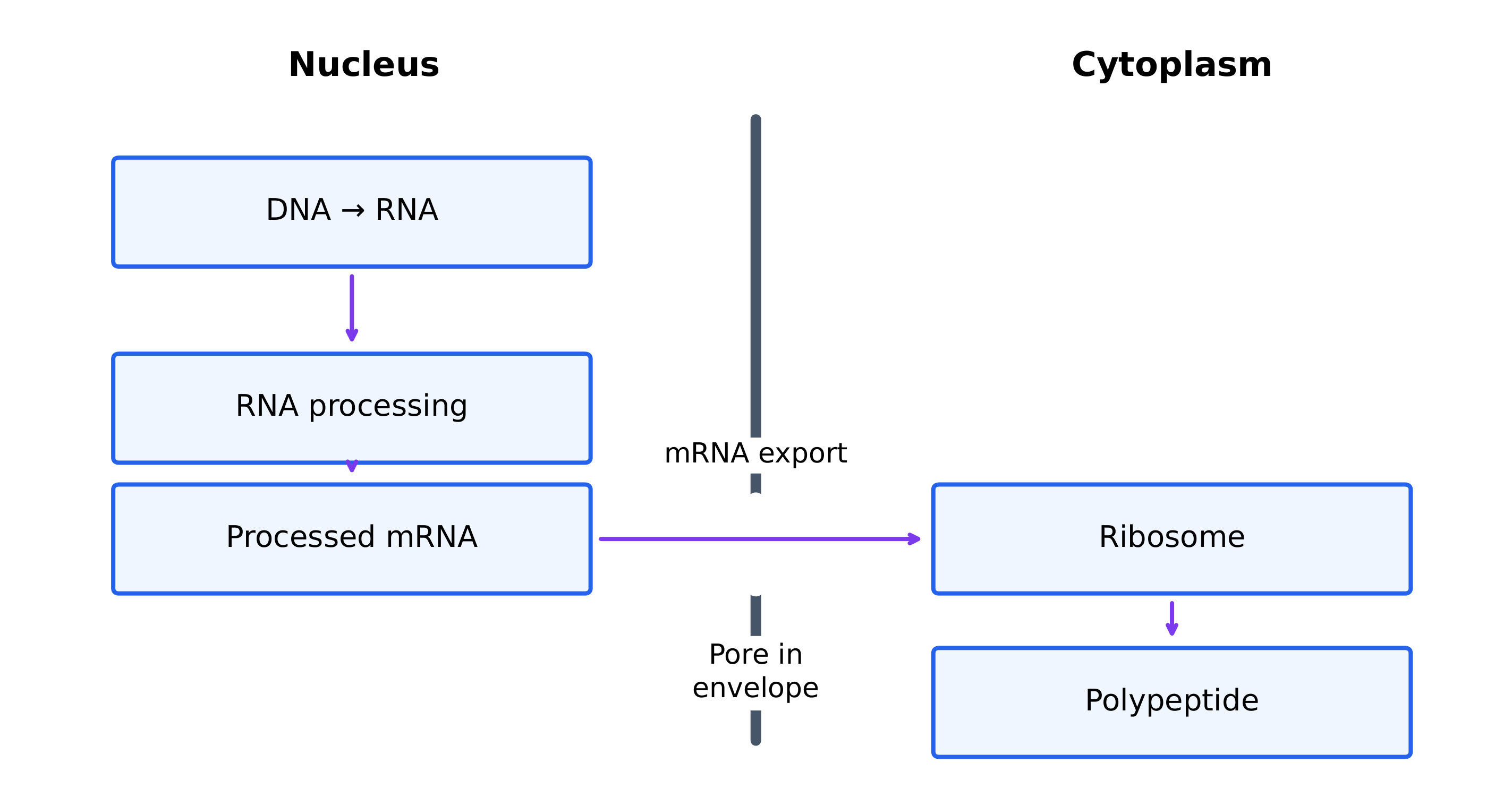
- In a typical eukaryotic protein-coding example, transcription produces RNA inside the nucleus. Processing can remove introns and join exons, with a cap and poly(A) tail commonly added. Processed mRNA exits through pores and is translated at ribosomes outside the nucleus. Nuclear separation allows processing and checking before cytoplasmic translation.
- In an invented timing model, transcription finishes at minute 3, processing at 5 and export at 6. This transcript cannot undergo cytoplasmic translation at minute 2. Prokaryotes lack the nuclear boundary, so translation can begin while transcription continues; this does not mean they lack DNA, ribosomes or every form of RNA processing.
Relate local concentration to compartment volume
- A compartment can concentrate an enzyme and its substrates while separating their conditions from the rest of the cell. In an invented example, 12 femtomoles in 4 picolitres gives 3 millimoles per litre; the same amount in 12 picolitres gives 1 millimole per litre. The smaller volume has three times the concentration, not three times the total amount.
- Higher concentration alone does not guarantee an optimal rate: enzyme properties, transport, cofactors and conditions matter. Boundaries also cost material and energy to maintain. A useful explanation identifies the conditions a compartment preserves and the reactions those conditions support, rather than assuming every extra membrane is automatically beneficial.
An invented compartment contains 12 femtomoles in 4 picolitres. What is its concentration in millimoles per litre? One femtomole is 10⁻¹⁵ mol and one picolitre is 10⁻¹² L.
Difficulté 2/5 · 140 pièces + un petit bonus lors de la première réussite aujourd'hui
(12 × 10⁻¹⁵)/(4 × 10⁻¹²) = 0.003 mol per litre = 3 millimoles per litre. Tripling the volume at the same total amount reduces concentration to one third.
Contain digestion while permitting useful exchange
- Lysosomes maintain acidic conditions suitable for their digestive enzymes, while the surrounding cytosol has different conditions. Their membranes help contain activity without sealing off all exchange. The same cell can therefore support reactions that require different local environments.
- A phagocytic cell encloses a particle in a phagosome; fusion with lysosomal compartments supplies digestive activity within that region. Useful breakdown products can later be transported out. The particle need not first dissolve throughout the cytosol. Explain containment, local conditions and controlled transfer together, rather than treating a compartment as an isolated bag.
Measure an organelle using consistent units
- An invented 30 mm image of a 5.0 micrometre mitochondrion gives magnification $30000/5.0=6000$. At that scale, a 4.0 mm vesicle image represents $4000/6000=0.667$ micrometres. Magnification is dimensionless; convert image and actual lengths to the same unit before dividing.
- If a 2.0 micrometre scale bar measures 8.0 mm on a print, an 18 mm structure represents $(18/8)(2)=4.5$ micrometres. Resizing both lengths together preserves this ratio, whereas a printed magnification label becomes wrong if not updated. A static image locates structures but does not directly show ATP production or vesicle movement.
An invented image of a 3.0 micrometre organelle is 24 mm long. Find its magnification.
Difficulté 2/5 · 140 pièces + un petit bonus lors de la première réussite aujourd'hui
24 mm = 24,000 micrometres. Magnification = 24,000/3.0 = 8,000, a dimensionless ratio. Resizing an image changes a printed magnification label unless updated, whereas a scale bar resized with the image remains usable.
Combine separation and image evidence carefully
- Suppose an invented fraction contains folded double-membrane profiles and elevated mitochondrial marker activity. Those independent observations support enrichment in mitochondria. They do not establish perfect purity, prove that every round profile is a mitochondrion or show the structure's entire three-dimensional shape from one section.
- Strengthen the claim with repeat preparations, appropriate markers for possible contaminants, comparable activity assays and images with usable scales. When drawing from a specimen, label visible structures and keep scale evidence; do not add details merely because a textbook diagram contains them. Link each conclusion to what its method can actually reveal.
An invented fraction has folded double-membrane profiles and high mitochondrial marker activity. Evaluate the claim that this proves a pure mitochondrial sample.
Different methods support different parts of the explanation. Converging evidence strengthens an association without removing every alternative.