Production and use of X-rays · Producción y uso de rayos X
| English | Español |
|---|---|
| tungsten/ˈtʌŋstn/ | tungsteno |
| target/ˈtɑːɡɪt/ | diana |
| anode/ˈænəʊd/ | ánodo |
| cathode/ˈkæθəʊd/ | cátodo |
| thermionic emission/ˌθɜːmɪˈɒnɪk ɪˈmɪʃn/ | emisión termiónica |
| kinetic energy/kɪˈnetɪk ˈenədʒi/ | energía cinética |
| photons/ˈfəʊtɒnz/ | fotones |
| Bremsstrahlung/ˈbremʃtrɑːlʊŋ/ | Bremsstrahlung |
| hardness/ˈhɑːdnəs/ | dureza |
| characteristic/ˌkærɪktəˈrɪstɪk/ | característico |
| sharpness/ˈʃɑːpnəs/ | nitidez |
| contrast/ˈkɒntræst/ | contraste |
| attenuation/əˌtenjuːˈeɪʃn/ | atenuación |
| contrast medium/ˈkɒntræst ˈmiːdɪəm/ | medio de contraste |
| half-value thickness/hɑːf ˈvæljuː ˈθɪknəs/ | espesor de medio valor |
| computed tomography/kəmˈpjuːtɪd təˈmɒɡrəfi/ | tomografía computarizada |
A machine that wastes ninety-nine per cent of its power
- An X-ray tube run at $75\ \text{kV}$ and $30\ \text{mA}$ takes in $2250\ \text{W}$. About $99\%$ of that becomes heat in a lump of tungsten the size of a coin.
- Left alone, the target's temperature would climb at over $1000\ \text{K}$ per second and melt in a few seconds.
- That is why exposures are brief and the anode spins. The remaining one per cent is the picture.
- This lesson is how X-rays are produced, what sets their penetrating power, and how they make an image.
Una máquina que desperdicia el noventa y nueve por ciento de su energía
- Un tubo de rayos X operado a $75\ \text{kV}$ y $30\ \text{mA}$ consume $2250\ \text{W}$. Aproximadamente el $99\%$ de esa energía se convierte en calor en un bloque de tungsteno del tamaño de una moneda.
- Si se dejara solo, la temperatura del objetivo aumentaría a más de $1000\ \text{K}$ por segundo y se fundiría en unos pocos segundos.
- Por eso las exposiciones son breves y el ánodo gira. El uno por ciento restante es la imagen.
- Esta lección trata sobre cómo se producen los rayos X, qué determina su poder de penetración y cómo forman una imagen.
Production
- The four-mark answer runs in four moves, and each is a mark.
- One. Electrons are emitted from a heated filament (the cathode 阴极) by thermionic emission 热电子发射.
- Two. They are accelerated through a high p.d., tens of kV, across an evacuated tube towards a metal target 靶, the anode 阳极, usually tungsten 钨.
- Three. They decelerate rapidly when they strike the target.
- Four. The kinetic energy 动能 lost is emitted as X-ray photons 光子, this is Bremsstrahlung 轫致辐射 or braking radiation, with most of it becoming heat.
Heat one end, brake at the other
Producción
- La respuesta de cuatro puntos se desarrolla en cuatro movimientos, y cada uno vale un punto.
- Uno. Los electrones son emitidos desde un filamento calentado (el cátodo) mediante emisión termiónica.
- Dos. Son acelerados a través de una alta d.d.p., decenas de kV, a través de un tubo evacuado hacia un metal objetivo, el ánodo, generalmente tungsteno.
- Tres. Se desaceleran rápidamente al chocar con el objetivo.
- Cuatro. La energía cinética perdida se emite como fotones de rayos X; esto es radiación de frenado (Bremsstrahlung), y la mayor parte se convierte en calor.

Calienta un extremo, frena en el otro
X-rays are produced when fast electrons: · Los rayos X se producen cuando electrones rápidos:
The sudden deceleration on the target gives out X-ray photons (Bremsstrahlung). · La desaceleración repentina en el objetivo emite fotones de rayos X (radiación de frenado).
Most of the electrons' kinetic energy in an X-ray tube becomes heat, not X-rays. · La mayor parte de la energía cinética de los electrones en un tubo de rayos X se convierte en calor, no en rayos X.
Only a small fraction becomes X-rays — which is why the target must be cooled. · Solo una pequeña fracción se convierte en rayos X — por eso el objetivo debe enfriarse.
Two controls, two different jobs
- Intensity, the energy per unit area per second, is set by the number of electrons hitting the target per second, so it is controlled by the filament current. A hotter filament emits more electrons.
- Hardness 硬度, the penetrating power, is set by the photon energies, so it is controlled by the accelerating p.d. A larger p.d. gives higher-energy, shorter-wavelength, more penetrating X-rays.
- Keep them apart. A question asking how to make the beam more penetrating is not answered by turning up the current.
- A metal filter removes the softest X-rays, which would be absorbed in the patient's skin without ever reaching the detector.
Dos controles, dos funciones distintas
- Intensidad, la energía por unidad de área por segundo, está determinada por el número de electrones que golpean el objetivo por segundo, por lo que se controla mediante la corriente del filamento. Un filamento más caliente emite más electrones.
- Dureza, el poder de penetración, está determinado por las energías de los fotones, por lo que se controla mediante la d.d.p. de aceleración. Una d.d.p. mayor produce rayos X de mayor energía, menor longitud de onda y mayor penetración.
- Manténgalas separadas. Una pregunta sobre cómo hacer el haz más penetrante no se responde aumentando la corriente.
- Un filtro metálico elimina los rayos X más blandos, que serían absorbidos por la piel del paciente sin llegar nunca al detector.
Match each control to what it changes. · Asocia cada control con lo que cambia.
A question asking how to make the beam more penetrating is not answered by turning up the current. · Una pregunta sobre cómo hacer el haz más penetrante no se responde aumentando la corriente.
The minimum wavelength
- The most energy one photon can carry is the whole kinetic energy of one electron, lost in a single event:
- This is the short-wavelength cut-off, and it depends only on the accelerating p.d., never on the target metal or the current.
- Doubling the p.d. halves the minimum wavelength.
La longitud de onda mínima
- La máxima energía que puede transportar un fotón es la energía cinética completa de un electrón, perdida en un único evento:
- Este es el límite de corta longitud de onda, y depende únicamente de la d.d.p. de aceleración, nunca del metal objetivo ni de la corriente.
- Duplicar la d.d.p. reduce a la mitad la longitud de onda mínima.
The minimum X-ray wavelength from a tube at accelerating voltage $V$ is: · La longitud de onda mínima de rayos X de un tubo a voltaje de aceleración $V$ es:
From $hf_{\max} = eV$ and · y $c = f\lambda$: $\lambda_{\min} = \dfrac{hc}{eV}$. · De $hf_{\max} = eV$ y $c = f\lambda$: $\lambda_{\min} = \dfrac{hc}{eV}$.
A higher accelerating voltage gives a ____ minimum wavelength. · Un voltaje de aceleración más alto da una longitud de onda mínima ____.
$\lambda_{\min} = \dfrac{hc}{eV}$, so a larger $V$ gives a smaller $\lambda_{\min}$ (more energetic X-rays). · $\lambda_{\min} = \dfrac{hc}{eV}$, por lo que un $V$ mayor da un $\lambda_{\min}$ más pequeño (rayos X más energéticos).
Worked example: the cut-off at 75 kV
- Electrons are accelerated through $75\ \text{kV}$. Find the maximum photon energy and the minimum wavelength.
- Maximum energy $= eV = 75\ \text{keV} = 75000 \times 1.60\times10^{-19} = 1.2\times10^{-14}\ \text{J}$.
- $\lambda_{\text{min}} = \dfrac{hc}{eV} = \dfrac{(6.63\times10^{-34})(3.00\times10^{8})}{1.2\times10^{-14}} = 1.7\times10^{-11}\ \text{m}$.
- The maximum photon momentum is $p = E/c = 4.0\times10^{-23}\ \text{N s}$.
- Note that $eV$ in joules needs the p.d. in volts, not kilovolts. That factor of a thousand is the usual casualty.
Ejemplo resuelto: el límite a 75 kV
- Los electrones son acelerados a través de $75\ \text{kV}$. Encuentre la energía máxima del fotón y la longitud de onda mínima.
- Energía máxima $= eV = 75\ \text{keV} = 75000 \times 1.60\times10^{-19} = 1.2\times10^{-14}\ \text{J}$.
- $\lambda_{\text{min}} = \dfrac{hc}{eV} = \dfrac{(6.63\times10^{-34})(3.00\times10^{8})}{1.2\times10^{-14}} = 1.7\times10^{-11}\ \text{m}$.
- El momento máximo del fotón es $p = E/c = 4.0\times10^{-23}\ \text{N s}$.
- Note que $eV$ en joules requiere que la d.d.p. esté en voltios, no en kilovoltios. Ese factor de mil es la causa habitual de errores.
Electrons are accelerated through 75 kV. What is the minimum X-ray wavelength, in units of 1e-11 m? · Los electrones se aceleran a través de 75 kV. ¿Cuál es la longitud de onda mínima de rayos X, en unidades de 1e-11 m?
lambda_min = hc/(eV) = 1.7e-11 m. Use volts, not kilovolts. Doubling the p.d. halves the minimum wavelength, and the target metal makes no difference to it. · lambda_min = hc/(eV) = 1.7e-11 m. Usa voltios, no kilovoltios. Duplicar el p.d. reduce a la mitad la longitud de onda mínima, y el metal del objetivo no tiene efecto alguno sobre ella.
Reading the spectrum
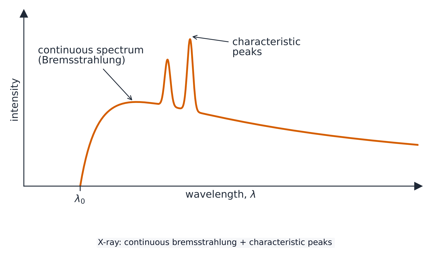
- The spectrum is continuous because each electron may lose any fraction of its energy, in one deceleration or several, so photons of every energy up to the maximum appear.
- It has a sharp cut-off at $\lambda_{\text{min}}$ because no photon can carry more than one electron's energy, $eV$.
- The sharp peaks are characteristic 特征 of the target metal: an incoming electron knocks out an inner electron, an outer one falls into the vacancy, and the photon carries the difference between those two levels.
- So the curve's shape has three separate causes, and a question about it wants all three named.
A cliff, a hump, and two spikes
Lectura del espectro
- El espectro es continuo porque cada electrón puede perder cualquier fracción de su energía, en una desaceleración o varias, por lo que aparecen fotones de todas las energías hasta el máximo.
- Tiene un límite abrupto en $\lambda_{\text{min}}$ porque ningún fotón puede transportar más energía que la de un electrón, $eV$.
- Los picos agudos son característicos del metal objetivo: un electrón entrante arranca un electrón interno, uno externo cae para ocupar la vacancia, y el fotón transporta la diferencia entre esos dos niveles.
- Por tanto, la forma de la curva tiene tres causas separadas, y una pregunta sobre ella pide nombrar las tres.
Un acantilado, un bulto y dos espigas
X-ray production route · Ruta de producción de rayos X
Follow electrons from cathode to X-ray photons. · Seguir a los electrones desde el cátodo hasta los fotones de rayos X.
Why does an X-ray spectrum have the shape it does? Select all · todos that apply. · ¿Por qué el espectro de rayos X tiene esa forma? Selecciona todas las que correspondan.
The peaks belong to the target's energy levels, so only changing the target moves them. The p.d. moves the cut-off. · Los picos pertenecen a los niveles de energía del objetivo, por lo que solo cambiar el objetivo los desplaza. El p.d. desplaza el corte.
Sharpness and contrast are different things
- Sharpness 清晰度 is how well defined the edges are. It improves with a small source spot, a still patient, and the detector close to the patient.
- Contrast 对比度 is the difference in degree of blackening between neighbouring regions, produced by a difference in attenuation 衰减.
- The two are independent: an image can be sharp with poor contrast, or blurred with strong contrast.
- Where two soft tissues attenuate similarly, a contrast medium 造影剂 such as barium is swallowed or injected to outline one of them.
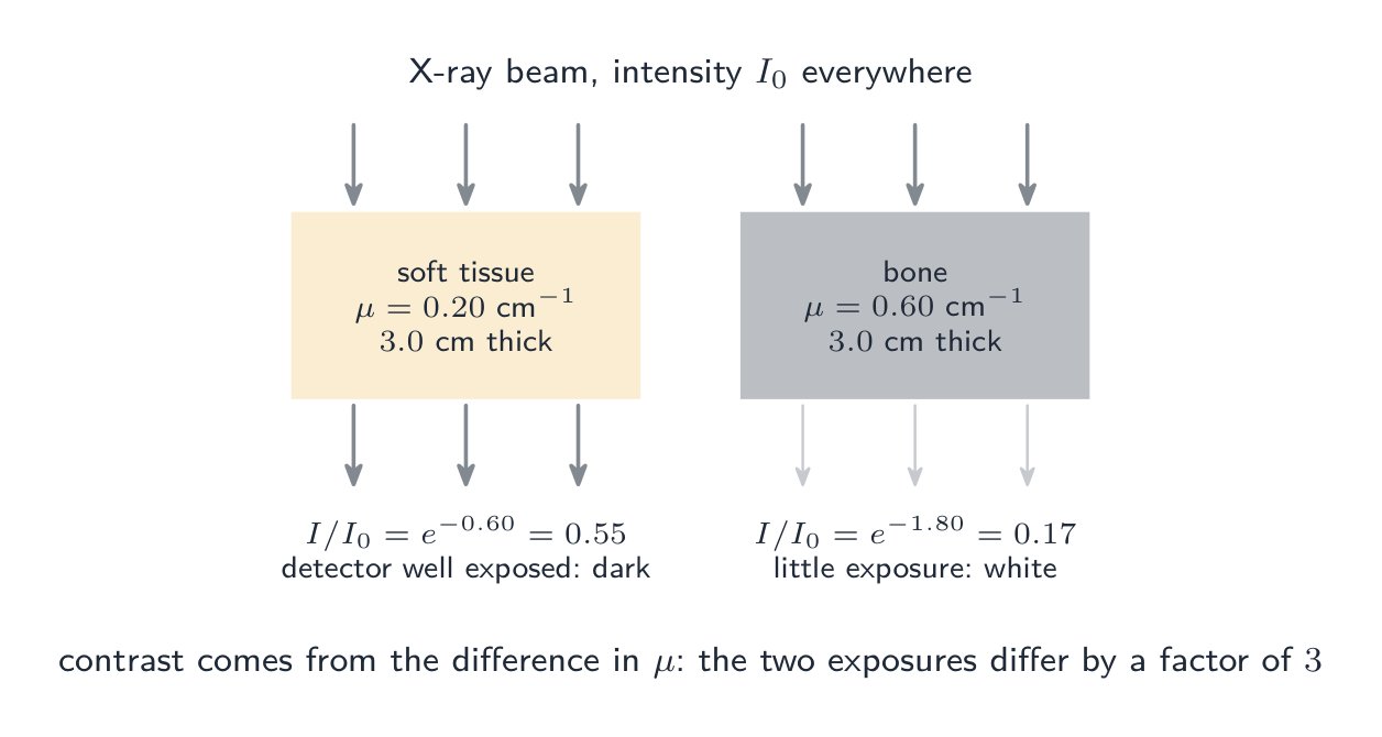
Same beam, very different shadows
La nitidez y el contraste son cosas distintas
- Nitidez es la definición de los bordes. Mejora con un punto focal pequeño, un paciente inmóvil y el detector cerca del paciente.
- Contraste es la diferencia en el grado de oscurecimiento entre regiones vecinas, producida por una diferencia en la atenuación.
- Las dos son independientes: una imagen puede tener buena nitidez pero mal contraste, o estar borrosa pero con fuerte contraste.
- Donde dos tejidos blandos atenúan de manera similar, un medio de contraste como el bario se traga o inyecta para delinear uno de ellos.
Mismo haz, sombras muy distintas
An X-ray image with excellent sharpness must also have good contrast. · Una imagen de rayos X con excelente nitidez también debe tener buen contraste.
They are independent. Sharpness is edge definition, set by the source size and geometry; contrast is a difference in attenuation between tissues. · Son independientes. La nitidez es la definición de bordes, determinada por el tamaño de la fuente y la geometría; el contraste es una diferencia en la atenuación entre tejidos.
Worked example: why bone shows white
- X-rays cross $3.0\ \text{cm}$ of soft tissue ($\mu = 0.20\ \text{cm}^{-1}$) in one region and $3.0\ \text{cm}$ of bone ($\mu = 0.60\ \text{cm}^{-1}$) in another. Compare the transmitted intensities.
- Soft tissue: $I/I_0 = e^{-0.20\times3.0} = e^{-0.60} = 0.55$.
- Bone: $I/I_0 = e^{-0.60\times3.0} = e^{-1.80} = 0.17$.
- The tissue region receives more than three times the exposure, so bone appears white and tissue dark.
- The exposure ratio is $e^{(\mu_{\text{bone}} - \mu_{\text{tissue}})x}$, which grows with thickness: contrast improves for thicker structures, though the total attenuation grows too.
Ejemplo resuelto: por qué el hueso aparece blanco
- Los rayos X atraviesan $3.0\ \text{cm}$ de tejido blando ($\mu = 0.20\ \text{cm}^{-1}$) en una región y $3.0\ \text{cm}$ de hueso ($\mu = 0.60\ \text{cm}^{-1}$) en otra. Compare las intensidades transmitidas.
- Tejido blando: $I/I_0 = e^{-0.20\times3.0} = e^{-0.60} = 0.55$.
- Hueso: $I/I_0 = e^{-0.60\times3.0} = e^{-1.80} = 0.17$.
- La región de tejido recibe más de tres veces la exposición, por lo que el hueso aparece blanco y el tejido oscuro.
- La razón de exposición es $e^{(\mu_{\text{bone}} - \mu_{\text{tissue}})x}$, que crece con el grosor: el contraste mejora para estructuras más gruesas, aunque también aumenta la atenuación total.
The attenuation law
- Higher-energy X-rays penetrate further, meaning a smaller $\mu$; bone has a much larger $\mu$ than soft tissue.
- For a thickness, take logs: $x = \dfrac{1}{\mu}\ln\dfrac{I_0}{I}$. The half-value thickness 半值厚度 is $x_{1/2} = \ln 2/\mu$, exactly like a half-life.
- Through two layers the exponentials multiply, so add the exponents: $I = I_0 e^{-(\mu_1 x_1 + \mu_2 x_2)}$. Never add the thicknesses or the coefficients, and note the order of the layers makes no difference.
- Read the question: "what fraction is absorbed" is $1 - e^{-\mu x}$, not $e^{-\mu x}$.
La ley de atenuación
- Los rayos X de mayor energía penetran más profundamente, lo que significa un $\mu$ menor; el hueso tiene un $\mu$ mucho mayor que el tejido blando.
- Para un grosor dado, tome logaritmos: $x = \dfrac{1}{\mu}\ln\dfrac{I_0}{I}$. El grosor semicorrector es $x_{1/2} = \ln 2/\mu$, exactamente como una vida media.
- Al atravesar dos capas, los exponenciales se multiplican, por lo que se suman los exponentes: $I = I_0 e^{-(\mu_1 x_1 + \mu_2 x_2)}$. Nunca sume los grosores ni los coeficientes, y note que el orden de las capas no importa.
- Lea la pregunta: "¿qué fracción es absorbida" es $1 - e^{-\mu x}$, no $e^{-\mu x}$.
Bone looks white on an X-ray image because it: · El hueso se ve blanco en una imagen de rayos X porque:
Dense, high-$Z$ bone absorbs more ($I = I_0 e^{-\mu x}$ with large $\mu$), casting a stronger shadow. · El hueso denso de alto-$Z$ absorbe más ($I = I_0 e^{-\mu x}$ con gran $\mu$), proyectando una sombra más fuerte.
A beam crosses 2.0 cm of P (mu = 0.35 /cm) then 1.5 cm of Q (mu = 0.90 /cm). What fraction of the intensity is transmitted? · Un haz cruza 2.0 cm de P (mu = 0.35 /cm) luego 1.5 cm de Q (mu = 0.90 /cm). ¿Qué fracción de la intensidad se transmite?
The exponentials multiply, so add the exponents: e^-(0.70 + 1.35) = e^-2.05 = 0.13. Never add the thicknesses or the coefficients, and the order of the layers makes no difference. · Los exponenciales se multiplican, así que se suman los exponentes: e^-(0.70 + 1.35) = e^-2.05 = 0.13. Nunca sumes los grosores ni los coeficientes, y el orden de las capas no importa.
Computed tomography
- A computed tomography 计算机断层扫描 (CT) scan builds a three-dimensional image, and the syllabus wants the construction described in order.
- One. Many X-ray images of the same section are taken from different angles around the patient.
- Two. A computer combines them into one two-dimensional image of that section.
- Three. The process is repeated for many sections along an axis.
- Four. The two-dimensional sections are combined into a three-dimensional image, which can be rotated or sliced on screen. The dose is far higher than a single radiograph, which is the trade-off.
Tomografía computarizada
- Una tomografía computarizada (TC) construye una imagen tridimensional, y el programa exige describir la construcción en orden.
- Uno. Se toman muchas imágenes de rayos X de la misma sección desde diferentes ángulos alrededor del paciente.
- Dos. Una computadora las combina en una sola imagen bidimensional de esa sección.
- Tres. El proceso se repite para muchas secciones a lo largo de un eje.
- Cuatro. Las secciones bidimensionales se combinan en una imagen tridimensional, que puede rotarse o rebanarse en pantalla. La dosis es mucho mayor que en una radiografía simple; ese es el compromiso.
A CT scan takes X-ray images from many angles to build a 3-D image. · Una tomografía computarizada toma imágenes de rayos X desde muchos ángulos para construir una imagen 3-D.
Rotating the tube and detectors and reconstructing the slices separates overlapping tissues into a 3-D picture. · Rotar el tubo y los detectores y reconstruir los cortes separa los tejidos superpuestos en una imagen 3-D.
Put the construction of a CT image in order. · Ordena la construcción de una imagen TC.
Angles first, then sections. The dose is far higher than a single radiograph, which is the trade-off for the 3-D view. · Ángulos primero, luego secciones. La dosis es mucho mayor que en una radiografía simple, que es el compromiso para obtener la vista 3-D.
Marks that slip away
- Filament current sets intensity, accelerating p.d. sets penetrating power. Do not swap them.
- $\lambda_{\text{min}}$ depends only on the p.d., not on the target or the current.
- Sharpness and contrast are independent. A question about blurred edges is not answered by talking about attenuation.
- Two layers: add the exponents $\mu_1 x_1 + \mu_2 x_2$.
- Match the units of $\mu$ and $x$, and check whether the question wants what gets through or what is absorbed.
Puntos que se escapan
- La corriente del filamento establece la intensidad, la d.d.p. de aceleración establece el poder de penetración. No los intercambie.
- $\lambda_{\text{min}}$ depende solo de la d.d.p., no del objetivo ni de la corriente.
- La nitidez y el contraste son independientes. Una pregunta sobre bordes borrosos no se responde hablando de atenuación.
- Dos capas: sume los exponentes $\mu_1 x_1 + \mu_2 x_2$.
- Coincida las unidades de $\mu$ y $x$, y verifique si la pregunta busca lo que transmite o lo que es absorbido.
You've got it
- X-rays are produced when electrons from a heated cathode, accelerated through a high p.d., decelerate rapidly in a metal target, mostly as heat
- $\lambda_{\text{min}} = hc/(eV)$ is the cut-off set by the accelerating p.d. alone, with characteristic peaks from the target metal on a continuous curve
- sharpness is edge definition, contrast is the difference in attenuation, and they are independent
- $I = I_0 e^{-\mu x}$, with half-value thickness $\ln 2/\mu$ and exponents that add through successive layers; CT combines many angles into a section and many sections into a 3-D image
Lo has logrado
- Los rayos X se producen cuando electrones de un cátodo calentado, acelerados a través de una alta d.d.p., se desaceleran rápidamente en un objetivo metálico, principalmente como calor.
- $\lambda_{\text{min}} = hc/(eV)$ es el límite establecido solo por la d.d.p. de aceleración, con picos característicos del metal objetivo sobre una curva continua.
- La nitidez es la definición de los bordes, el contraste es la diferencia en la atenuación, y son independientes.
- $I = I_0 e^{-\mu x}$, con grosor semicorrector $\ln 2/\mu$ y exponentes que se suman a través de capas sucesivas; la TC combina múltiples ángulos en una sección y múltiples secciones en una imagen 3-D.