Why big animals need a pump
- A single cell gets oxygen by diffusion alone: the distance is tiny. A human is about $10^{13}$ cells, and most of them are far from any surface. Diffusion over centimetres would take hours.
- So large animals have a mass transport system: a pump (the heart), tubes (blood vessels) and a carrier (blood) that moves substances in bulk to every cell.
A double circulation
- Mammals have a double circulation: blood passes through the heart twice in each complete circuit. The pulmonary circuit goes heart → lungs → heart; the systemic circuit goes heart → body → heart.
- Why it matters: blood loses pressure in the lung capillaries. Returning to the heart lets it be pumped again at high pressure, so it reaches the body cells quickly. The low-pressure lung circuit also protects the thin lung capillaries.
What is the main advantage of a double circulation?
难度 1/5 · 今日首次完成可获 100 金币及少量额外奖励
Blood loses pressure in the lung capillaries. Returning to the heart lets the left ventricle pump it again at high pressure, so it reaches the body cells quickly.
The heart
- Four chambers: two atria on top receive blood; two ventricles below pump it out. The septum keeps oxygenated blood (left side) apart from deoxygenated blood (right side).
- Atrioventricular valves lie between atria and ventricles; semilunar valves lie at the bases of the aorta and pulmonary artery. Valves only let blood flow one way.
| Chamber | Wall | Why |
|---|---|---|
| atria | thin | push blood only into the ventricles below |
| right ventricle | thick | pumps blood to the nearby lungs, at lower pressure |
| left ventricle | thickest | must make a high pressure to push blood round the whole body |
The cardiac cycle
| Stage | What contracts | Valves |
|---|---|---|
| atrial systole | atria; ventricles fill fully | AV open, semilunar closed |
| ventricular systole | ventricles; blood forced into arteries | AV closed, semilunar open |
| cardiac diastole | nothing; heart fills from the veins | AV open, semilunar closed |
- One rule for every valve: a valve opens when the pressure behind it is higher than the pressure in front of it, and closes when the pressure in front is higher. Use it to read any pressure graph.
Match each stage of the cardiac cycle to the state of the valves.
难度 2/5 · 今日首次完成可获 140 金币及少量额外奖励
Each valve follows the pressure on its two sides. When the ventricles contract, ventricular pressure shuts the AV valves and opens the semilunar valves.
The right ventricle has a thicker wall than the left ventricle because it pumps blood to the lungs.
难度 1/5 · 今日首次完成可获 100 金币及少量额外奖励
The left ventricle is thicker. It must make a high pressure to push blood round the whole body; the right ventricle pumps a short distance to the lungs at lower pressure.
Arteries, veins and capillaries
| Vessel | Structure | How it fits the job |
|---|---|---|
| artery | thick wall: smooth muscle, elastic fibres, collagen; narrow lumen | elastic fibres stretch and recoil, smoothing the high pressure; collagen stops bursting |
| vein | thin wall, wide lumen, valves | blood is at low pressure; valves stop backflow; muscles squeeze veins |
| capillary | wall one endothelial cell thick; very narrow lumen | short diffusion distance; huge total surface area; slow flow gives time for exchange |
- Exam point: for "describe the structure" questions, name the layers (endothelium, smooth muscle and elastic fibres, collagen). For "explain", link each feature to pressure.
Match each feature to its function.
难度 2/5 · 今日首次完成可获 140 金币及少量额外奖励
Link every structure to pressure or to exchange. That is what "explain" questions reward.
Worked example: vessel measurements
A small vein has a total diameter of $46\ \mu\text{m}$ and a wall $4\ \mu\text{m}$ thick. Find the area of its lumen.
- The wall is on both sides, so lumen diameter $= 46 - 2 \times 4 = 38\ \mu\text{m}$; radius $= 19\ \mu\text{m}$.
- Area $= \pi r^2 = \pi \times 19^2 = 1134\ \mu\text{m}^2$.
- Ratios: a vena cava with a $30\ \text{mm}$ lumen and a $500\ \mu\text{m}$ wall has lumen : wall $= 30\,000 : 500 = 60 : 1$. Convert to the same unit first.
An artery has a total diameter of 4.0 mm and a wall 1.0 mm thick. Calculate the area of its lumen in mm², to 2 significant figures.
难度 2/5 · 今日首次完成可获 140 金币及少量额外奖励
Lumen diameter $= 4.0 - 2 \times 1.0 = 2.0$ mm, so $r = 1.0$ mm and area $= \pi \times 1.0^2 = 3.1$ mm².
Haemoglobin carries oxygen
- Haemoglobin is a globular protein with four polypeptide chains, each with a haem group containing an iron ion ($\text{Fe}^{2+}$). Each haem binds one $\text{O}_2$, so one molecule carries four oxygen molecules.
- Its hydrophilic R groups face outwards, so it is soluble in the red blood cell. Carbon dioxide binds to the amino groups of the chains (not to the haem), forming carbaminohaemoglobin.
How many oxygen molecules can one haemoglobin molecule carry when fully saturated?
难度 2/5 · 今日首次完成可获 140 金币及少量额外奖励
Four polypeptide chains, each with one haem group, and each haem binds one $\text{O}_2$.
The dissociation curve
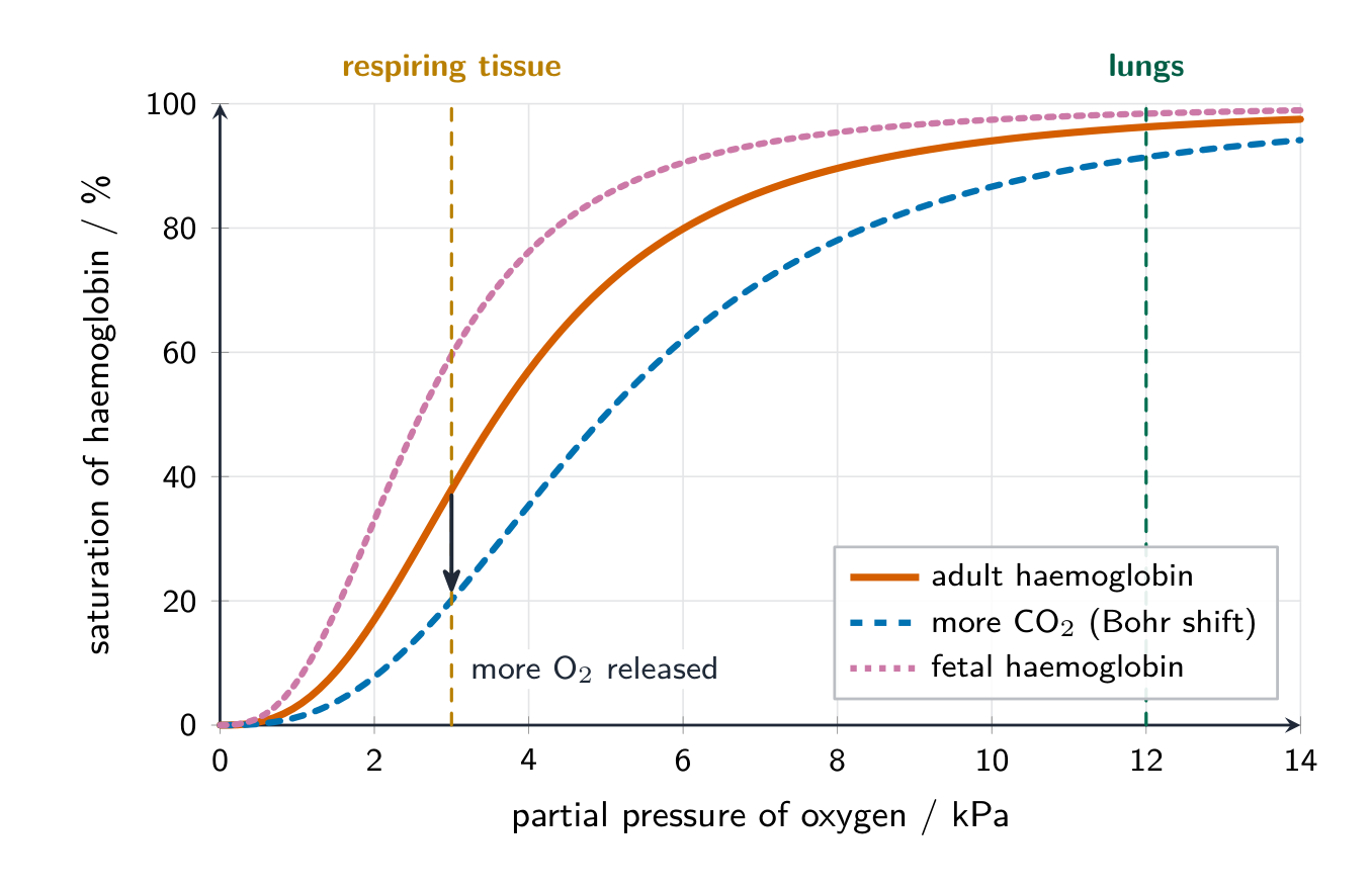
- The curve is S-shaped (sigmoid). When the first $\text{O}_2$ binds, the shape of the molecule changes and the next ones bind more easily (cooperative binding).
- In the lungs (high partial pressure of oxygen) haemoglobin is almost fully saturated. In respiring tissue (low partial pressure) saturation falls steeply, so a lot of oxygen is released exactly where it is needed.
The Bohr effect and fetal haemoglobin
- Bohr effect: respiring tissue makes $\text{CO}_2$, which lowers the pH. This lowers haemoglobin's affinity for oxygen, so the curve shifts right and more oxygen is released in the tissues that need it.
- Fetal haemoglobin has a higher affinity for oxygen (its curve is to the left). At the placenta it can take oxygen from the mother's haemoglobin, which is giving oxygen up at that partial pressure.
During exercise, muscles produce more carbon dioxide. What happens to the dissociation curve, and why does this help?
难度 1/5 · 今日首次完成可获 100 金币及少量额外奖励
This is the Bohr effect. More $\text{CO}_2$ lowers haemoglobin's affinity for oxygen, so at the same partial pressure more oxygen is unloaded where respiration is fastest.
Explain how the structure of haemoglobin is related to its function in transporting oxygen and carbon dioxide. (3 marks)
Each mark links one feature to its job. Never say carbon dioxide binds to the haem group.
Large animals need mass transport. A double circulation lets blood be re-pressurised after the lungs. Valves open and close by pressure differences, which drives the cardiac cycle. Arteries resist and smooth high pressure, veins have valves, capillaries are one cell thick. Haemoglobin's four haem groups load oxygen in the lungs and unload it in tissues; more $\text{CO}_2$ shifts the curve right (Bohr effect), and fetal haemoglobin lies to the left.