| Core | Supplement |
|---|---|
| 1 Describe the circulatory system as a system of blood vessels with a pump and valves to ensure one-way flow of blood | |
| 2 Describe the single circulation of a fish | |
| 3 Describe the double circulation of a mammal | |
| 4 Explain the advantages of a double circulation |
Transport in animals
IGCSE Biology · Topic 9
 8:21
8:21
Transport in Animals
Your heart beats about a hundred thousand times a day, and it has never once stopped to rest. It is not really one pump — it is two, side by side, sharing a…
English narration · English + 中文 subtitles burned in
9.1
The circulatory system

Syllabus
Source: Cambridge International syllabus
The circulatory system 循环系统 carries blood around the body. It has three parts: a pump 泵 (the heart 心脏), a network of blood vessels 血管, and valves 瓣膜 that keep the blood flowing one way only.
Single and double circulation (Supplement)
- A fish has a single circulation 单循环: the blood passes through the heart once on each trip around the body.
- A mammal 哺乳动物 has a double circulation 双循环: the blood passes through the heart twice on each full trip — once on the way to the lungs, and once on the way to the rest of the body.
- The advantage of a double circulation: the blood can be pumped again at high pressure 压力 before going to the body, so it travels faster and delivers oxygen 氧气 quickly.
Explore
Double circulation
Mammals have a double circulation — blood passes through the heart TWICE on each full trip round the body.
Vocabulary
Train
| English |
|---|
| circulatory system/ˌsɜːkjʊˈleɪtəri ˈsɪstəm/ |
| heart/hɑːt/ |
| pump/pʌmp/ |
| blood vessels/blʌd ˈveslz/ |
| valves/vælvz/ |
| single circulation/ˈsɪŋɡl ˌsɜːkjʊˈleɪʃn/ |
| mammal/ˈmæml/ |
| double circulation/ˈdʌbl ˌsɜːkjʊˈleɪʃn/ |
| pressure/ˈpreʃə/ |
| oxygen/ˈɒksɪdʒn/ |
9.2
The heart

Syllabus
| Core | Supplement |
|---|---|
| 1 Identify in diagrams and images the structures of the mammalian heart, limited to: muscular wall, septum, left and right ventricles, left and right atria, one-way valves and coronary arteries | 7 Identify in diagrams and images the atrioventricular and semilunar valves in the mammalian heart |
| 8 Explain the relative thickness of: (a) the muscle walls of the left and right ventricles (b) the muscle walls of the atria compared to those of the ventricles | |
| 9 Explain the importance of the septum in separating oxygenated and deoxygenated blood | |
| 10 Describe the functioning of the heart in terms of the contraction of muscles of the atria and ventricles and the action of the valves | |
| 2 State that blood is pumped away from the heart in arteries and returns to the heart in veins | |
| 3 State that the activity of the heart may be monitored by: ECG, pulse rate and listening to sounds of valves closing | |
| 4 Investigate and describe the effect of physical activity on the heart rate | 11 Explain the effect of physical activity on the heart rate |
| 5 Describe coronary heart disease in terms of the blockage of coronary arteries and state the possible risk factors including: diet, lack of exercise, stress, smoking, genetic predisposition, age and sex | |
| 6 Discuss the roles of diet and exercise in reducing the risk of coronary heart disease |
Source: Cambridge International syllabus
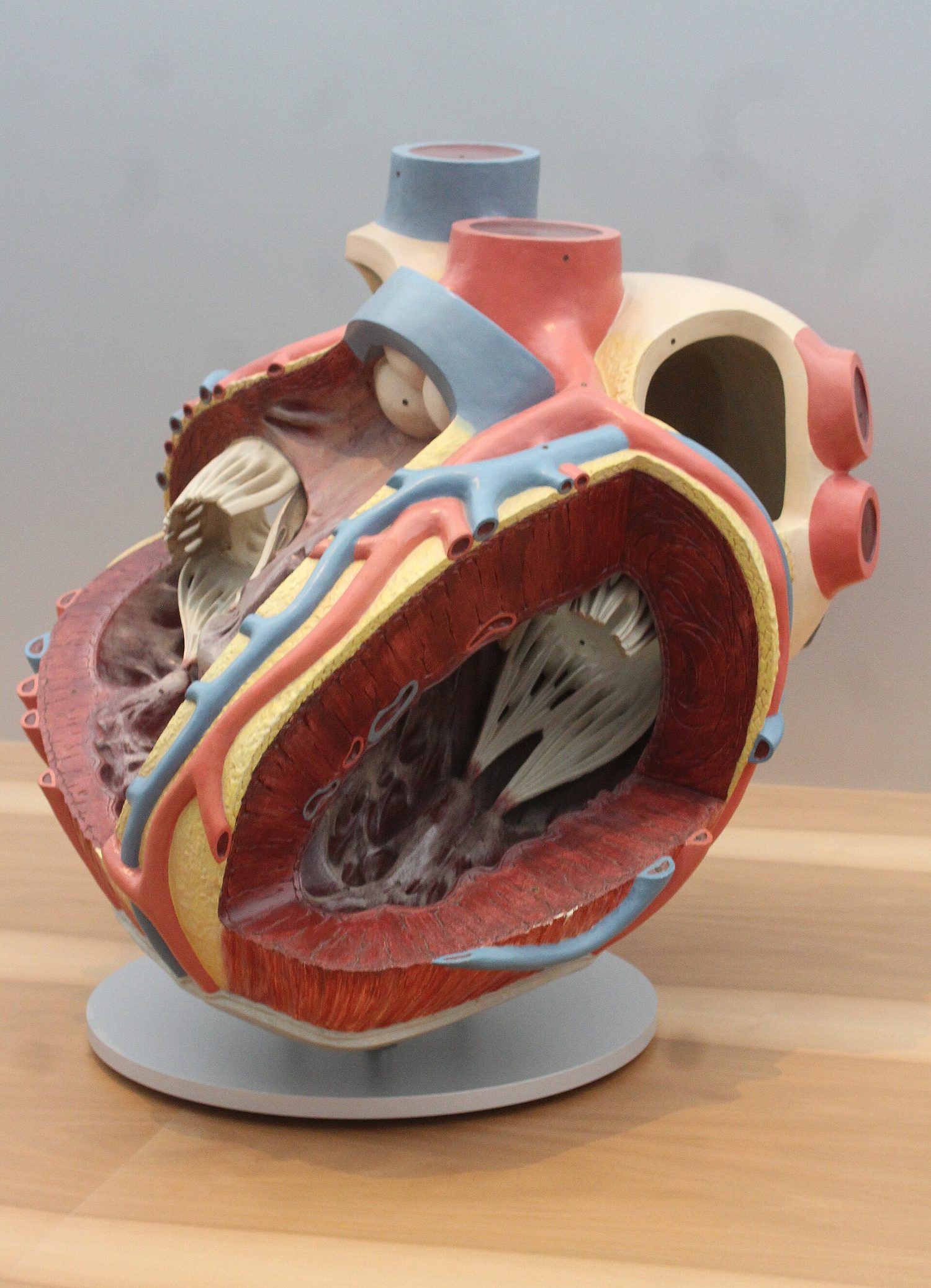
The heart is made of muscle 肌肉. A wall called the septum 隔膜 divides it into a left side and a right side. On each side there is an upper chamber, the atrium 心房, and a lower chamber, the ventricle 心室. Valves between the chambers stop the blood flowing backwards. The heart muscle is fed with blood by the coronary arteries 冠状动脉.
(Supplement) The wall of the left ventricle is thicker than the right, because it must pump blood all the way around the body; the right side only pumps blood to the nearby lungs. The atria have thin walls, as they only push blood into the ventricles just below them.
The septum keeps oxygenated blood 含氧血 on the left side apart from deoxygenated blood 缺氧血 on the right side, so the two never mix.
(Supplement) The valves between each atrium and ventricle are the atrioventricular valves 房室瓣. The valves at the exit of each ventricle are the semilunar valves 半月瓣.
How the heart beats (Supplement)
- The atria contract 收缩 and push blood down into the ventricles.
- The ventricles contract and force blood out into the arteries; the valves snap shut so blood cannot flow back.
- The muscle relaxes and the heart fills with blood again.
Blood is pumped away from the heart in arteries and returns to the heart in veins.
Heart rate and exercise
You can check the heart's activity with an ECG, by feeling the pulse 脉搏 in an artery, or by listening to the valves closing. During exercise the heart rate 心率 goes up, so blood reaches the muscles faster, bringing the extra oxygen and glucose they need.

Worked example. A student counts 18 pulse beats in 15 seconds at rest, and 33 beats in 15 seconds straight after exercise. Give both heart rates in beats per minute. A minute holds 60 ÷ 15 = 4 lots of 15 seconds, so multiply by 4. At rest: 18 × 4 = 72 beats per minute. After exercise: 33 × 4 = 132 beats per minute. The rise delivers more oxygen and glucose to the muscles. Writing the 15-second count straight down as the heart rate is the commonest mistake - always scale it up to a full minute.
Coronary heart disease
Coronary heart disease 冠心病 happens when the coronary arteries become narrow or blocked, so the heart muscle cannot get enough oxygen. Risk factors 风险因素 include a diet high in fat, lack of exercise, stress, smoking, family history (genetics), older age, and being male. A healthy diet and regular exercise lower the risk.

Explore
Blood flow through the heart
The four chambers and their valves keep blood moving one way — deoxygenated to the lungs, oxygenated to the body.
Explore
The heart (double circulation)
Follow the blood: blue comes back from the body, the right side pumps it to the lungs (it turns red), and the left side pumps it out to the body — a double circulation.
Vocabulary
Train
| English |
|---|
| muscle/ˈmʌsl/ |
| septum/ˈseptəm/ |
| atrium/ˈeɪtrɪəm/ |
| ventricle/ˈventrɪkl/ |
| coronary arteries/ˈkɒrənəri ˈɑːtəriz/ |
| oxygenated blood/ˈɒksɪdʒeneɪtɪd blʌd/ |
| deoxygenated blood/diːˈɒksɪdʒeneɪtɪd blʌd/ |
| atrioventricular valves/ˌeɪtrɪˌəʊvənˈtrɪkjʊlə vælvz/ |
| semilunar valves/ˌsemɪˈluːnə vælvz/ |
| contract/ˈkɒntrækt/ |
| pulse/pʌls/ |
| heart rate/hɑːt reɪt/ |
| coronary heart disease/ˈkɒrənəri hɑːt dɪˈziːz/ |
| risk factors/rɪsk ˈfæktəz/ |
9.3
Blood vessels

Syllabus
| Core | Supplement |
|---|---|
| 1 Describe the structure of arteries, veins and capillaries, limited to: relative thickness of wall, diameter of the lumen and the presence of valves in veins | 4 Explain how the structure of arteries and veins is related to the pressure of the blood that they transport |
| 2 State the functions of capillaries | 5 Explain how the structure of capillaries is related to their functions |
| 3 Identify in diagrams and images the main blood vessels to and from the: (a) heart, limited to: vena cava, aorta, pulmonary artery and pulmonary vein (b) lungs, limited to: pulmonary artery and pulmonary vein (c) kidney, limited to: renal artery and renal vein | 6 Identify, in diagrams and images, the main blood vessels to and from the liver as: hepatic artery, hepatic veins and hepatic portal vein |
Source: Cambridge International syllabus
There are three kinds of blood vessel. The space inside a vessel is its lumen 管腔.
| Vessel | Wall | Lumen | Valves? | Job |
|---|---|---|---|---|
| arteries 动脉 | thick, muscular, elastic | narrow | no | carry blood away from the heart, at high pressure |
| veins 静脉 | thin | wide | yes | carry blood back to the heart, at low pressure |
| capillaries 毛细血管 | one cell thick | very narrow | no | let oxygen, glucose and wastes pass between blood and cells |
(Supplement) Arteries have thick elastic walls to cope with the high pressure from the heart. Veins have valves because their blood is at low pressure and could otherwise flow backwards. Capillaries are very thin (one cell thick) and very narrow, giving a large surface area 表面积 and a short distance for fast exchange.
Main blood vessels
| Connects | Artery (carries blood out) | Vein (carries blood in) |
|---|---|---|
| heart ↔ body | aorta 主动脉 | vena cava 腔静脉 |
| heart ↔ lungs | pulmonary artery 肺动脉 | pulmonary vein 肺静脉 |
| ↔ kidney | renal artery 肾动脉 | renal vein 肾静脉 |
(Supplement) The liver is served by the hepatic artery 肝动脉, the hepatic vein 肝静脉, and the hepatic portal vein 肝门静脉, which brings blood from the gut to the liver.
Explore
The three blood vessels
Compare an artery, a vein and a capillary — their walls and lumens suit the pressure and job of each.
Vocabulary
Train
| English |
|---|
| lumen/ˈluːmen/ |
| arteries/ˈɑːtəriz/ |
| veins/veɪnz/ |
| capillaries/ˈkæpɪləriz/ |
| surface area/ˈsɜːfɪs ˈeərɪə/ |
| aorta/eɪˈɔːtə/ |
| vena cava/ˈviːnə ˈkɑːvə/ |
| pulmonary artery/ˈpʌlmənəri ˈɑːtəri/ |
| pulmonary vein/ˈpʌlmənəri veɪn/ |
| renal artery/ˈriːnl ˈɑːtəri/ |
| renal vein/ˈriːnl veɪn/ |
| hepatic artery/heˈpætɪk ˈɑːtəri/ |
| hepatic vein/heˈpætɪk veɪn/ |
| hepatic portal vein/heˈpætɪk ˈpɔːtl veɪn/ |
9.4
Blood

Syllabus
| Core | Supplement |
|---|---|
| 1 List the components of blood as: red blood cells, white blood cells, platelets and plasma | |
| 2 Identify red and white blood cells in photomicrographs and diagrams | 5 Identify lymphocytes and phagocytes in photomicrographs and diagrams |
| 3 State the functions of the following components of blood: (a) red blood cells in transporting oxygen, including the role of haemoglobin (b) white blood cells in phagocytosis and antibody production (c) platelets in clotting (details are not required) (d) plasma in the transport of blood cells, ions, nutrients, urea, hormones and carbon dioxide | 6 State the functions of: (a) lymphocytes – antibody production (b) phagocytes – engulfing pathogens by phagocytosis |
| 4 State the roles of blood clotting as preventing blood loss and the entry of pathogens | 7 Describe the process of clotting as the conversion of fibrinogen to fibrin to form a mesh |
Source: Cambridge International syllabus
Blood has four parts:
| Part | Function |
|---|---|
| red blood cells 红细胞 | carry oxygen, using the red pigment 色素 haemoglobin 血红蛋白 |
| white blood cells 白细胞 | defend the body against disease |
| platelets 血小板 | help the blood to clot 凝血 |
| plasma 血浆 | a liquid that carries blood cells, ions 离子, nutrients 营养物质, urea 尿素, hormones 激素 and carbon dioxide 二氧化碳 |

Defending the body, and clotting
White blood cells fight pathogens 病原体 (the microbes that cause disease) in two ways:
- phagocytes 吞噬细胞 carry out phagocytosis 吞噬作用 — they surround and digest the pathogens.
- lymphocytes 淋巴细胞 make antibodies 抗体, which stick to the pathogens and destroy them.
When you cut yourself, the blood clots to seal the wound. This stops blood loss and stops pathogens getting in. (Supplement) During clotting, a soluble protein called fibrinogen 纤维蛋白原 is changed into threads of fibrin 纤维蛋白. The threads form a net that traps blood cells and makes a solid clot.
Explore
Blood component lab
Classify blood components by the job they do.
Vocabulary
Train
| English |
|---|
| red blood cells/red blʌd selz/ |
| pigment/ˈpɪɡmənt/ |
| haemoglobin/ˌhiːməˈɡləʊbɪn/ |
| white blood cells/waɪt blʌd selz/ |
| platelets/ˈpleɪtlɪts/ |
| clot/klɒt/ |
| plasma/ˈplæzmə/ |
| ions/ˈaɪɒnz/ |
| nutrients/ˈnjuːtrɪənts/ |
| urea/juːˈrɪə/ |
| hormones/ˈhɔːməʊnz/ |
| carbon dioxide/ˈkɑːbən daɪˈɒksaɪd/ |
| pathogens/ˈpæθədʒnz/ |
| phagocytes/ˈfæɡəsaɪts/ |
| phagocytosis/ˌfæɡəsɪˈtəʊsɪs/ |
| lymphocytes/ˈlɪmfəsaɪts/ |
| antibodies/ˈæntɪbɒdiz/ |
| fibrinogen/ˈfɪbrɪnədʒn/ |
| fibrin/ˈfɪbrɪn/ |
9.4
Exam tips
- Circulatory system = pump (heart) + vessels + valves for one-way flow.
- Double circulation (mammals): blood passes through the heart twice. The left ventricle wall is thickest because it pumps to the whole body.
- Arteries carry blood away (thick, high pressure); veins carry it back (valves, low pressure); capillaries are for exchange (one cell thick).
- Blood = red cells (oxygen, haemoglobin) + white cells (defence) + platelets (clotting) + plasma (transport).
- Phagocytes engulf pathogens; lymphocytes make antibodies.
Interactive lessons on this topic
Work through it step by step, with instant-check exercises.